Gastric ulcers are among the most important diseases in the world. The gastric mucosa is continuously exposed to po-tentially injurious agents such as acid, pepsin, bile acids, food ingredients, bacterial products, and drugs.1) These agents have been implicated in the pathogeneses of gastric ulcer, such as an increase in gastric acid and pepsin secre-tion, a decrease in gastric blood flow, the suppression of en-dogenous generation of prostaglandins, inhibition of mucosal growth and cell proliferation, and alteration of gastric mobil-ity.2)
The current therapy for this disease continues to have, as one of its major goals, the control of Helicobacter pylori as well as H1/K1-ATPase, acid secretion and a subsequent
re-versal of mucosal damage and inflammation.3)
Many pharmaceutical products have been employed for the treatment of gastroduodenal ulcer and peptic disease, and for decreasing mortality and morbidity rates, but they are not completely effective and produce many adverse effects. Moreover, these pharmaceutical products are too expensive.4)
Plant extracts are some of the most attractive sources of new drugs and have been shown to produce promising results for the treatment of gastric ulcer.5)
Simaroubaceae is a large botanical family with pantropical distribution; only a few representatives occur in temperate re-gions. Quassia amaraL., a representative of this family, is a
neotropical forest shrub or small tree reputed in traditional medicine to have good effect on stomach diseases, especially gastric ulcers. In Brazil, this species is found in a region from the border with Guiana to the state of Maranhão. It is popularly known in Latin American as “Amargo”, “Hombre Grande”, “Simaruba”, “Pau Quassia”, “Murubá”, “Murupá” and “Quina de Caiena”.6)
This plant is a source of several compounds which include both b-carbonile and cantin-6 alkaloids, as well as, primarily, the bitter components known as quassinoids, a group of sub-stances belonging to the terpens class. Antimalarial, an-tifeedant and antifertility pharmacological activities are atributted to these quassinoids.7)
In considering the use of this plant in Brazilian folk medi-cine and the several chemical substances isolated and identi-fied from Quassia amarabark, we have performed a study of the antiulcerogenic effects for four different extracts of this species using standard rodent models of induced gastric ulcer.
MATERIALS AND METHODS
Drugs and Chemicals Bethanechol, hydrochloric acid (HCl), cimetidine, diazald, ethanol, hexane, indomethacin, lansoprazole, methanol and dichloromethane (Sigma
Chemi-25
∗To whom correspondence should be addressed. e-mail: [email protected] © 2002 Pharmaceutical Society of Japan
Antiulcerogenic Activity of Four Extracts Obtained from the Bark Wood
of
Quassia amara
L. (Simaroubaceae)
Walber TOMA,aJuliano de Souza GRACIOSO,aFábio Donisete Pezzuto de ANDRADE,b Clélia Akiko HIRUMA-LIMA,cWagner VILEGAS,band Alba Regina Monteiro SOUZABRITO*,a
a Departamento de Fisiologia e Biofísica, Instituto de Biologia, Universidade Estadual de Campinas; Campinas, São
Paulo, C.P. 6109, CEP 13083–970 Brazil: bDepartamento de Química Orgânica, Instituto de Química, Universidade Estadual Paulista; Araraquara, São Paulo, Brazil: and cDepartamento de Fisiologia, Instituto de Biociências, Universidade Estadual Paulista; Botucatu, SP, Brazil. Received March 4, 2002; accepted May 16, 2002
Quassia amaraL., a neotropical forest shrub of the Simaroubaceae family, is widely used in Caribbean folk medicine and in some northern states of Brazil for the treatment of gastric ulcers. This plant is a source of nu-merous compounds including both bb-carbonile and cantin-6 alkaloids as well as, primarily, the bitter compounds known as quassinoids. We analyzed the possible antiulcerogenic activities of four extracts of different polarities: 70% ethanol (70% EtOH), 100% EtOH, 100% dichloromethane (DCM), and 100% hexane (HEX) obtained from Quassia amarabark. All extracts, administered at doses of 5000 mg/kg orally and 1000 mg/kg intraperi-toneally, caused neither toxicity or death. In the indomethacin/bethanechol-induced gastric ulcer, 70% EtOH, 100% EtOH, DCM and HEX extracts, 100 mg/kg, p.o., inhibited the gastric ulcer (22.5, 23.4, 50.5, 46.8%, respec-tively). 70% EtOH, 100% EtOH, DCM, and HEX extracts reduced the gastric injury induced by the hypother-mic restraint–stress test in hypother-mice (70.7, 80, 60, 82.7%, respectively). In the pylorus ligature of the mouse stomach, following pre-treatment with a single intraduodenal administration of 100 mg/kg of each extract, only 70% EtOH did not change the biochemical parameters of gastric juice. 100% EtOH, DCM and HEX extracts pre-sented decreased gastric juice content, increased pH values and decreased acid output. We also determined the antiulcerogenic activity on HCl–EtOH-induced gastric ulcers in mice at four doses (25, 50, 75, 100 mg/kg, p.o.), then evaluated the possible dose-dependent relation and calculated the ED50values. Except for 70% EtOH at a dose of 25 mg/kg, the other extracts showed significantly activity (p,0.05). The free mucous amount in the gas-tric stomach content was also evaluated. All extracts showed significant increases (p,0.05) of free mucous. This effect was abolished when the animals were pre-treated with indomethacin. Prostaglandin synthesis was evalu-ated by the administration of HEX extracts by the oral route (100 mg/kg). Prostaglandin synthesis was signifi-cantly, increased by 52.3% (p,0.05), and this effect was abolished with prior administration of indomethacin. We concluded that Quassia amarais a probable source for a new drug to treat gastric ulcers, and the mechanism of its activity relates to cytoprotective factors, such as mucous and prostaglandins, but there is still the possibility that antisecretory activity is involved in its antiulcerogenic effect.
cal Co., St. Louis, MO, U.S.A.) were used in this study. In-domethacin was prepared in sodium bicarbonate (5%), and extracts were dissolved in 12% Tween 80®. All substances
were prepared immediately before use. The reagents used were of analytical grade.
Animals All experiments were performed on male Swiss mice (3065 g) obtained from the Animal House (CEMIB-UNICAMP) in Campinas, SP. The animals were fed a certi-fied Nuvilab CR-a® (Nuvital) diet with free access to tap
water, and were housed in a 12 h light/dark cycle at a humid-ity of 50% and a temperature of 2461 °C. All experiments were done in the morning. The experimental protocols were approved by the Animal Use and Care Committee of Uni-camp. All of the experiments were conducted in accordance with the recommendations of the Canadian Council on Ani-mal Care.8)
Extract Preparation The bark of Quassia amaraL. was collected and identified by botanists from the Enda Caribe Institute, Santo Domingo, Dominican Republic (Exsicata 2114, RD, 84374). The dried bark (5 kg) was powdered in a mill and extracted by maceration for one week with the fol-lowing organic solvents of increasing polarity: HEX, DCM, 100% EtOH and 70% EtOH. After each extraction, the solu-tions were filtered through filter paper and the solvents were evaporated in a rotavapor at reduced pressure in order to sep-arate the raw extract from the solvent. Extractions afforded 8.0 g of HEX, 19.5 g of DCM, 52.5 g of 100% EtOH and 93.5 g of 70% EtOH extracts.
Phytochemical Analysis The Quassia amara extracts were analyzed using thin-layer chromatography (TLC) plates (0.1 mm thick, silica gel G Merck, Art. 7731) eluted with toluene–acetone–acetic acid (70 : 30 : 0.5, v/v/v) or n-butanol acetic-acid water (BAW, 65 : 15 : 25, v/v/v), according to their polarities. The spots were identified under short-wave UV light and plates were sprayed further with iodoplatinate and anisaldehyde–sulfuric-acid reagents or developed with a re-sublimated iodine vapor.
These extracts were also analyzed by HPLC-ES-MS. Twenty milligrams of each extract were dissolved with methanol and filtered through a 0.45mm Millex filter. Twenty microliters of each extract were injected into the HPLC-ES-MS system (RP18, Phenomenex, 25 cm34.6 mm35mm, 1 ml/min, ES positive mode170V).
Pharmacological Assays. Nonsteroidal Antiinflamma-tory Drug (NSAID)-Induced Gastric Ulcers in Choli-nomimetic-Treated Mice The experiment was performed by the method of Rainsford, 1978.9) In this model, gastric
ulcer was induced using indomethacin (30 mg/kg, s.c.) and bethanechol (5 mg/kg, i.p.), administered to mice after a 24 h fast. The extracts 70% EtOH, 100% EtOH, DCM and HEX from Quassia amara(100 mg/kg), cimetidine (100 mg/kg) or Tween were administered orally 30 min before the induction of gastric ulcer. The animals were killed by cervical disloca-tion 4 h after treatment with the ulcerogenic agents; the stom-achs were removed and inflated with 4% formalin in buffered saline, and the gastric damage was determined by the methodology of Szelenyi and Thiemer, 1978.10)
Hypothermic Restraint-Stress Ulcer The experiment was performed by the method of Levine (1971),11)with some
modifications. After 24 h of starvation, the animals received an oral administration of 70% EtOH, 100% EtOH, DCM and
HEX extracts from Quassia amara (100 mg/kg), cimetidine (100 mg/kg) or Tween (10 ml/kg). One hour after treatment, mice were immobilized in a restraint cage at 4 °C for 3 h to induce gastric ulcer. The animals were killed and the stom-achs removed and opened along the greater curvature to de-termine the lesion index.
Determination of Gastric Secretion The assay was per-formed by the method of Shay (1945)12)with a few
modifica-tions. All groups of mice were fasted for 24 h, with free ac-cess to water. Immediately after pylorus ligature, 70% EtOH, 100% EtOH, DCM and HEX extracts from Quassia amara (100 mg/kg), cimetidine (100 mg/kg) as positive control, or the vehicle, Tween, were administered intraduodenally. The animals were killed 4 h later by cervical dislocation; the ab-domen was opened and another ligature was placed around the esophagus, close to the diaphragm. The stomachs were removed and the gastric content collected to determine the total amount of gastric-juice acid (ml) and pH values. Dis-tilled water (5 ml) was added, and the resultant solution was centrifuged at 3000 rpm for 10 min. Total acid in the gastric secretion was determined in the supernatant volume by titra-tion to pH 7.0 with 0.01NNaOH.
HCl/Ethanol-Induced Ulcer The antiulcerogenic activ-ity of the four extracts obtained from Quassia amarain this model was assessed in mice, as described by Mizui and Doteuchi (1983).13)Mice were divided into 6 groups which were fasted for 24 h prior to oral dosing with the vehicle, 12% Tween 80® (10 ml/kg), lansoprazole (30 mg/kg), 70%
EtOH, 100% EtOH, DCM or HEX (25, 50, 75 and 100 mg/kg). Fifty minutes after the treatments, all animals re-ceived orally 0.2 ml of a 0.3MHCl/60% EtOH solution.
Ani-mals were killed 1 h after the administration of HCl/EtOH solution; the stomachs were excised, inflated by an injection of saline (2 ml) and opened along the greater curvature. Then the stomachs were fixed in 5% formalin for 30 min and the ulcerative lesion index (ULI) was calculated. The ED50 val-ues were also calculated by linear regression.
Determination of Mucous in Gastric Content This assay was performed according to the methodology de-scribed previously by Sun et al.,14)with some modifications.
Mice were fasted for 24 h, under anesthesia, the abdomen in-cised and the pylorus ligated. The 70% EtOH, 100% EtOH, DCM and HEX extracts (100 mg/kg body weight (kg)) of Quassia amaraor vehicle were administered intraduodenally after the pylorus ligature. The animals were killed by cervical dislocation 4 h after the drug treatments. The stomach con-tent was immersed in 10 ml 0.02% alcian blue in 0.16M
su-crose/0.05Msodium acetate, pH 5.8, and incubated for 24 h
at 20 °C. The alcian blue binding extract was centrifuged at 3000 rpm for 10 min. The absorbance of the supernatant was measured at 615 nm using a light spectrophotometer U/2000 (Hitachi, Japan). The free mucous in the gastric content was calculated from the amount of alcian blue binding (mg/wet tissue (g)).
Determination of Prostaglandin Synthesis The assay was performed by the method of Curtis et al.15)Thirty
experi-mental group. Samples of the corpus (full thickness) were excised, weighed and suspended in 1 ml of 10 mM sodium
phosphate buffer, pH 7.4. The tissue was minced finely with scissors, then incubated at 37 °C for 20 min. Prostaglandin in the buffer was measured using an enzyme immunoassay (RPN222-Amersham).
Statistical Analysis The results are presented as the mean6standard error of the mean (S.E.M.), and were com-pared using one-way analysis of variance (ANOVA), fol-lowed by Dunnet’s pairwise test. pvalues of less than 0.05 were considered significant.
RESULTS AND DISCUSSION
Traditionally, medicinal plants have been used in folk medicine throughout the world to treat various diseases, es-pecially gastric ulcer. There are several types of experimental models for evaluating anti-ulcer drugs.
We evaluated the preventive effects of 70% EtOH, 100% EtOH, DCM and HEX extracts obtained from Quassia amarabark in mice using the different standard experimental models of induced gastric ulcer.
It is already known that the suppression of prostaglandin synthesis by NSAIDs, such as indomethacin, results in in-creased susceptibility to mucosal injury and causes gastro-duodenal ulcerations.16) The co-administration of
choli-nomimetic agents, such as bethanechol, promotes a syner-gism with NSAIDs in the gastric lesion induced by the in-creased secretion of acid and pepsin in the stomach.17)
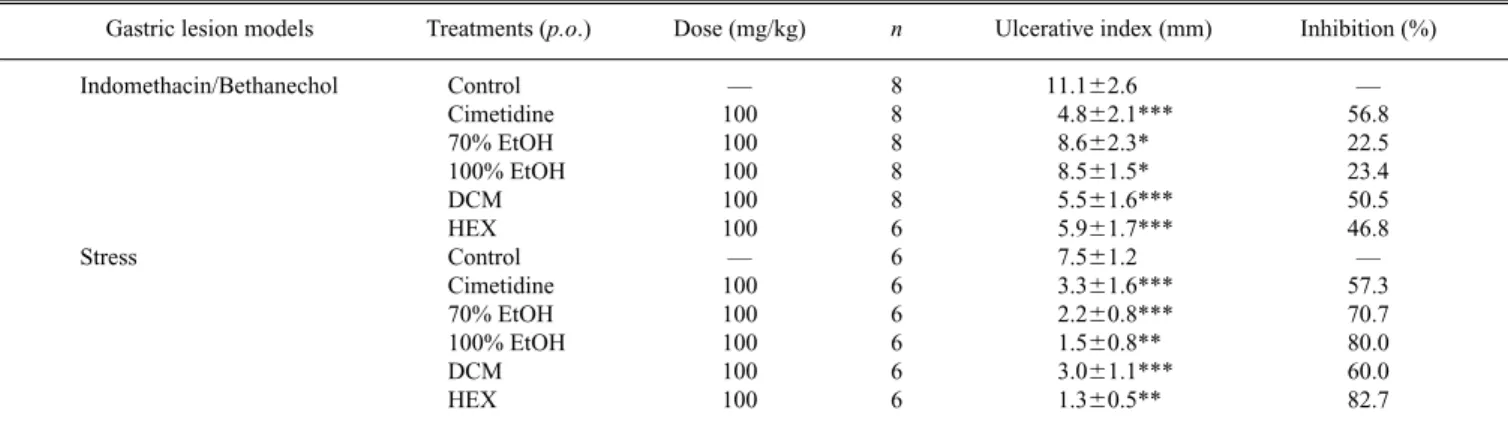
The antiulcerogenic effect of these extracts in the NSAID/ Cholinomimetic-induced lesion model (Table 1), mainly the
DCM and HEX, demonstrated significantly reduced damage to these gastric lesions (p,0.05) compared with the respec-tive control and, suggested the probable involvement of these extracts with the cytoprotective mechanism by increasing mucous and/or prostaglandin synthesis.
Gastric stress ulceration is a serious complication which may occur in patients with burns, surgery or central-nervous-system trauma. Stress-induced ulcers are probably mediated by histamine release, with an enhancement of acid secretion and a reduction in mucous production. Moreover, vagal over-activity has been suggested as the principal factor in stress-induced ulceration.2)
In the gastric ulcer induced by hypothermic-restraint stress, again all the extracts showed significant activity (p,0.05), and the obtained inhibitions index for extracts were more effective than those obtained for the positive con-trol, cimetidine (70.7, 80, 60, 82.7, respectively), considering the fact that both extracts and cimetidine were used at same dose, 100 mg/kg p.o., (Table 1). Antisecretory mechanisms, such as reduced histamine secretion and increased mucous synthesis, are responsible for this activity.
In the next step we showed the biochemical effects of Quassia amaraextracts on gastric-juice parameters obtained after submitting the mice to pylorus ligature using all of the treatments, extracts and positive and negative control admin-istered intraduodenally (Table 2). We observed that 100% EtOH, DCM and HEX, as well as the cimetidine positive control, significantly decreased the gastric acid secretion (p,0.05), increased the pH values and promoted reduced acid output. The 70% EtOH did not show significant results in the parameters evaluated.
Table 1. Effects of Quassia amaraExtracts and Cimetidine on Indomethacin/Bethanecol and Hypothermic Restraint Stress Induced Gastric Ulcer in Mice
Gastric lesion models Treatments (p.o.) Dose (mg/kg) n Ulcerative index (mm) Inhibition (%)
Indomethacin/Bethanechol Control — 8 11.162.6 —
Cimetidine 100 8 4.862.1*** 56.8
70% EtOH 100 8 8.662.3* 22.5
100% EtOH 100 8 8.561.5* 23.4
DCM 100 8 5.561.6*** 50.5
HEX 100 6 5.961.7*** 46.8
Stress Control — 6 7.561.2 —
Cimetidine 100 6 3.361.6*** 57.3
70% EtOH 100 6 2.260.8*** 70.7
100% EtOH 100 6 1.560.8** 80.0
DCM 100 6 3.061.1*** 60.0
HEX 100 6 1.360.5** 82.7
The results are the mean6S.E.M. ANOVA: F(5,42)511.739 (p,0.05) for indomethacin/bethanecol and F(5,30)527.097 (p,0.05) for hypothermic restraint stress model.
Dunnett’s test: ∗p,0.05, ∗∗p,0.01, ∗∗∗p,0.0001.
Table 2. Effects of Quassia amaraExtracts and Cimetidine Administered Intraduodenally (i.d.) on the Biochemical Parameters of Gastric Juice Obtained from Pylorus–Ligature Mice
Treatments (i.d.) Dose (mg/kg) n pH (Units) Volume gastric juice (ml) Total gastric acid (mEq/4h)
Control — 20 3.161.0 469.56230.9 11.263.9
Cimetidine 100 15 5.861.1*** 257.86150.9** 7.565.4**
70% EtOH 100 18 3.861.4 454.66227.6 8.763.4
100% EtOH 100 18 4.861.4*** 302.06134.0* 7.063.3**
DCM 100 17 4.761.7** 300.66225.6* 7.863.2*
HEX 100 18 5.061.5*** 292.26175.2* 6.562.8***
Robert et al.,18) showed that E series prostaglandins
(PGEs) inhibit gastric acid secretion in dogs and rats. Subse-quent studies confirmed this inhibitory effect of PGE2and its analogs in both monkeys and man. In addition, PGI2, PGF2a,
and their analogs, were shown to have similar acid-suppres-sant properties.16) Probably, the activities exhibited by these
extracts are responsible for the synthesis of mucous, phos-pholipid, bicarbonate and prostaglandins, consequently pro-moting the inhibition of gastric-acid secretion. Moreover, it is important to remember that the compound(s) present in the extracts of Quassia amara by intraduodenal administration were absorbed and then arrived at the stomach to promote ef-fects.
Considering the efficacy of these extracts led us to perform another HCl–EtOH-induced gastric-ulcer experiment using four dose levels (25, 50, 75, 100 mg/kg) for each extract, all of them administered orally. In this experiment we tried to establish the ED50 value for each extract and the possible dose-dependent relation of the obtained effect. As shown in Table 3, only 70% EtOH at a dose of 25 mg/kg was not sig-nificant in the inhibition of gastric ulcer. The 70% EtOH, 100% EtOH and HEX were dose-dependent (p,0.05), and the ED50values were, respectively, 7.0, 7.2 and 5.3 mg/kg.
The balance of defensive and aggressive factors is thought to be important in maintaining gastric mucosal integrity.19)
The formation of gastric mucosal lesions by necrotizing agents such as HCl and EtOH has been reported to involve the depression of these gastric defensive mechanisms.20) HCl–EtOH-induced gastric ulcers also promote stasis in gas-tric blood flow which contributes to the development of the hemorrhagic and necrotic aspects of tissue injury.2)
It has been found that EtOH-induced ulcers are not ited by antisecretory agents such as cimetidine, but are inhib-ited by agents that enhance mucosal defensive factors such as prostaglandins.21)These results show that
Quassia amara ex-tracts have an antiulcerogenic effect related to cytoprotection activity.
In looking for a possible mechanism for the increase in mucosal protective factors, we also investigated free mucous
production after the administration of the 70% EtOH, 100% EtOH, DCM and HEX extracts (100 mg/kg, p.o.), and prostaglandin synthesis with an HEX extract of Quassia amara. We observed that all of the extracts significantly in-creased the “free” mucous in the gastric juice (14.8, 14.3, 27.1, 52.4%, respectively) (Table 4). When indomethacin was subcutaneously administered 30 min before treatment with the extracts or controls, the increase of this mucous was halted.
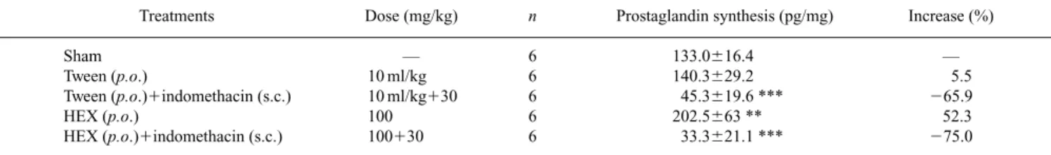
As shown in Table 5, HEX extract increased the synthesis of prostaglandin levels (52.3%). However, it was not signifi-cantly different from the sham group or negative control.
Several compounds, such as b-carbonile alkaloids, indole alkaloids and steroids, are presented in the extracts obtained from Quassia amara,22)moreover, the bitter principle
quassi-noids are the most interesting group of substances present in these extracts. Our phytochemical analyses by TLC and HPLC-ES-MS confirmed the presence of these quassinoids (mainly quassin and neoquassin) in all of the extracts (data not shown).
These quassinoids probably are responsible for the ob-served antiulcerogenic activity. Raji and Bolarinwa,23)
showed that orally administered quassin reduced the plasma level of testosterone hormone and consequently promoted antifertility action in animals. Aston et al.,24) showed that
sexual hormones such as testosterone, when exogenously ad-ministered, aggravated the gastroduodenal ulcer induced by cysteamine in rats. In addition, Adeniyi,25)observed that in orchidoctemized rats, with testosterone synthesis inhibited, a reduction of gastric-acid secretion occurred compared with that of normal rats. Montoneri & Drago,26) in contrast,
showed that the elevation of endogenous progesterone levels and the administration of exogenous progesterone increased the layer of mucous in the stomach and duodenum, and pro-tected the cysteamine-induced ulcer.
There are many additional works relating exogenous testosterone with an improvement in the ability to increase the number of receptors of endogenous vasopressin, an im-portant aggressive factor of gastrointestinal mucosa,27,28) Table 3. Effects of Quassia amaraExtracts and Lansoprazole on HCl/EtOH-Induced Gastric Ulcer in Mice
Treatments (p.o.) Dose (mg/kg) n Ulcerative index (mm) Inhibition (%) r ED50(mg/kg) p
Control — 8 35.465.2 — — — —
Lansoprazole 30 8 7.461.1*** 79.1 — — —
70% EtOH 25 8 33.366.6 5.9
50 8 26.564.4*** 25.1 **0.98 7.0 **0.003
75 8 16.462.9*** 53.7
100 8 12.662.1*** 64.4
100% EtOH 25 8 22.862.7*** 35.6
50 8 16.863.1*** 52.5 *0.92 7.2 *0.03
75 8 15.061.7*** 57.6
100 8 12.164.2*** 65.8
DCM 25 8 16.161.6*** 54.5
50 8 16.062.8*** 54.8 0.50 — 0.4
75 8 5.662.0*** 83.8 — — —
100 8 5.662.0*** 83.8 — — —
HEX 25 8 13.563.0*** 61.9
50 8 13.661.7*** 61.6 *0.92 5.3 *0.03
75 8 6.062.1*** 83.1
100 8 2.561.9*** 92.9
while orchidectomy decreases these receptors as well as the vasopressin levels in the plasma.29)It is probable that
quassi-noids (quassin and neoquassin) are the compounds responsi-ble for antiulcerogenic activities exhibited in all of the per-formed experiments.
We conclude that 70% EtOH, 100% EtOH, DCM and HEX extracts obtained from Quassia amara provide an ex-cellent preventive effect in gastric ulcer models. The main compounds are the quassinoids, and the mechanism of this effect is probably related to prostaglandins and mucous syn-thesis. However, the antisecretory mechanism is still possible in this activity. The fact that low doses obtain the best effects, with no toxicity at a dose of 5000 mg/kg, is crucial, demon-strating the high efficacy and security of these extracts.
Acknowledgements We thank Fundação de Amparo à Pesquisa do Estado de São Paulo (FAPESP) for financial sup-port and Ana Claudia B. de Paula, Ana Beatriz A. de Almeida, Fernanda Rocha Soares, Leônia M. Baptista and Luciana de P. Magri for technical assistance.
REFERENCES
1) Peskar B. M., Maricic N., Dig. Dis. Sci., 43(Suppl. S), 23S—29S (1998).
2) Konturek P. C., Brzowski T., Sliwowski Z., Pajido R., Stachura J., Hahn E. G., Konturek S. J., Scand. J. Gastroenterol., 33, 691—700 (1998).
3) Woo T. W., Chang M. S., Chung M. S., Kim K. B., Sohn S. K., Kim S. G., Choi W. S., Biol. Pharm. Bull., 21, 449—455 (1998).
4) Germano M. P., Sanago R., Guglielmo M., De Pasquale R., Crisafi G., Bisignano G., J. Ethnopharmacology, 59, 167—178 (1998).
5) Alkofahi A., Atta A. H., J. Ethnopharmacology, 67, 341—345 (1999). 6) Corrêa, M. P. “Dicionário das Plantas Úteis do Brasil e das Exóticas
Cultivadas” Vol. V, Imprensa Nacional, R. J., 1984, pp. 556—557. 7) Ajaiyeoba E. O., Abalogu U. I., Krebs H. C., Oduola A. M. J., J.
Ethnopharmacology, 67, 321—325 (1999). 8) Zimmerman M., Pain, 16, 109—110 (1983).
9) Rainsford K. D., Biochem. Pharmacol., 27, 1281—1289 (1978). 10) Szelenyi I., Thiemer K., Arch. Toxicol., 41, 99—105 (1978).
11) Levine R. J., “Peptic Ulcer,” ed. by Pfeiffer C. J., Munksgarrd, Copen-hagen, 1971, pp. 92—97.
12) Shay H., Komarov S. A., Fels S. S., Meranze D., Gruestein M., Siplet H., Gastroenterol., 5, 43—61 (1945).
13) Mizui T., Doteuchi M., Jpn. J. Pharmacol., 33, 939—945 (1983). 14) Sun S. B., Matsumoto T., Yamada H., Jpn. J. Pharmacol., 43, 699—
704 (1991).
15) Curtis G. H., Macnaughton W. K., Gall D. G., Wallace J. L., Can. J. Physiol. Pharmacol., 73, 130—134 (1995).
16) Atay S., Tarnawski A. S., Dubois A., Prostag. Lipid. Med., 61, 105— 124 (2000).
17) Rainsford K. D., Biochem. Pharmacol., 27, 1281—1289 (1978). 18) Robert A., Nezamis J. E., Phillips J. P., Am. J. Dig. Dis., 12, 1073—
1076 (1967).
19) Sun D. C. H., “Gastroenterology,” 3rd ed., Vol. 1, ed. by Bochus H. L., Saunders W. B., Philadelphia, 1974, pp. 579—610.
20) Kinoshita M., Tsunehisa N., Tamaki H., Biol. Pharm. Bull., 18, 223— 226 (1995).
21) Morimoto Y., Shimohara K., Oshima S., Sukamoto T., Jpn. J. Pharma-col., 57, 595—605 (1991).
22) Germonsén-Robineau L., “Farmacopea Caribeña,” Enda Caribe, Tramil-9, 1998, pp. 1—9.
23) Raji Y., Bolarinwa A. F., Life Sci., 61, 1067—1074 (1997).
24) Aston N. O., Kalaichadran S., Carr J. V., Can. J. Sur., 34, 482—483 (1991).
25) Adeniyi K. O., Gastroenterol., 101, 66—69 (1991).
26) Montoneri C., Drago F., Dig. Dis. Sci., 42, 2572—2575 (1997). 27) László F., Karácsony G., Pávo J., Varga Cs., Rojik I., László F. A., Eur.
J. Pharmacol., 258, 15—22 (1994).
28) László F., Szepes Z., Varga Cs., László F. A., Scand. J. Gastroenterol.,
33 (Suppl. 228), 62—67 (1998).
29) Pávo I., Varga Cs., Szucs M., László F., Szécsi M., Gardi J., László F. A., Life Sci., 56, 1215—1222 (1995).
Table 4. Effect of Quassia amaraExtracts Administered by the Intraduodenal Route on Alcian Blue Binding to Free Gastric Mucous from Pylorus–Liga-ture Mice
Treatments Dose (mg/kg) n Alcian blue bound [mg/wet tissue (g)] Alcian blue bound (%)
Sham – 6 2.160.1 —
Sham1indomethacin 10 ml/kg130 6 2.060.1* 24.8
Tween 10 ml/kg 6 2.160.09 —
Tween1indomethacin 10 ml/kg130 6 2.060.1 24.8
70% EtOH 100 6 2.460.07* 14.8
70% EtOH1indomethacin 100130 6 2.160.1 —
100% EtOH 100 6 2.460.08* 14.3
100% EtOH1indomethacin 100130 6 2.060.06 —
DCM 100 6 2.760.4*** 27.1
DCM1indomethacin 100130 6 1.960.2*** 29.5
HEX 100 6 3.260.4*** 52.4
HEX1indomethacin 100130 6 2.060.1 —
The results are the mean6S.E.M. ANOVA following Fvalues: F(11,60)520.748 (p,0.05) for alcian blue bound. Dunnett’s test: ∗p,0.05; ∗∗∗p,0.0001.
Table 5. Effects of HEX Extract from Quassia amaraon Prostaglandin Synthesis by the Gastric Mucosa of Rats
Treatments Dose (mg/kg) n Prostaglandin synthesis (pg/mg) Increase (%)
Sham — 6 133.0616.4 —
Tween (p.o.) 10 ml/kg 6 140.3629.2 5.5
Tween (p.o.)1indomethacin (s.c.) 10 ml/kg130 6 45.3619.6 *** 265.9
HEX (p.o.) 100 6 202.5663 ** 52.3
HEX (p.o.)1indomethacin (s.c.) 100130 6 33.3621.1 *** 275.0