ORIGINAL ARTICLE
Bacteria and bacterial DNA in atherosclerotic plaque and
aneurysmal wall biopsies from patients with and without
periodontitis
Zahra Armingohar
1*, Jørgen J. Jørgensen
2, Anne Karin Kristoffersen
1,
Emnet Abesha-Belay
1and Ingar Olsen
1#1Department of Oral Biology, Faculty of Dentistry, University of Oslo, Oslo, Norway;2Department of
Vascular Surgery, Oslo University Hospital, Aker and University of Oslo, Oslo, Norway
Background: Several studies have reported an association between chronic periodontitis (CP) and cardio-vascular diseases. Detection of periodontopathogens, including red complex bacteria (RCB), in cardio-vascular lesions has suggested these bacteria to be involved in the pathogenesis of atherosclerosis and abdominal aortic aneurysms.
Objective: In this study, we investigate bacteria and their DNA in vascular biopsies from patients with vascular diseases (VD; i.e. abdominal aortic aneurysms, atherosclerotic carotid, and common femoral arteries), with and without CP.
Methods: DNA was extracted from vascular biopsies selected from 40 VD patients: 30 with CP and 10 without CP. The V3-V5 region of the 16S rDNA (V3-V5) was polymerase chain reaction (PCR)-amplified,
and the amplicons were cloned into Escherichia coli, sequenced, and classified (GenBank and the Human
Oral Microbiome database). Species-specific primers were used for the detection ofPorphyromonas gingivalis.
In addition, 10 randomly selected vascular biopsies from the CP group were subjected to scanning electron
microscopy (SEM) for visualization of bacteria. Checkerboard DNADNA hybridization was performed to
assess the presence of RCB in 10 randomly selected subgingival plaque samples from CP patients.
Results: A higher load and mean diversity of bacteria were detected in vascular biopsies from VD patients
with CP compared to those without CP. Enterobacteriaceaewere frequently detected in vascular biopsies
together with cultivable, commensal oral, and not-yet-cultured bacterial species. While 70% of the subgingival
plaque samples from CP patients showed presence of RCB, onlyP. gingivaliswas detected in one vascular
biopsy. Bacterial cells were seen in all 10 vascular biopsies examined by SEM.
Conclusions: A higher bacterial load and more diverse colonization were detected in VD lesions of CP patients as compared to patients without CP. This indicated that a multitude of bacterial species both from the gut and the oral cavity, rather than exclusively periodontopathogens, may be involved as additional risk factors in the pathogenesis of VD.
Keywords: chronic periodontitis;vascular disease;16S rDNA; Porphyromonas gingivalis
Responsible Editor: Bruce J. Paster, Forsyth Institute and Harvard School of Dental Medicine, United States.
*Correspondence to: Zahra Armingohar, Department of Oral Biology, Faculty of Dentistry, University of Oslo, P.B.1052, Blindern, NO-0316 Oslo, Norway, Email: [email protected]
To access the supplementary material for this article, please see Supplementary Files under Article Tools online.
Received: 2 February 2014; Revised: 23 April 2014; Accepted: 23 April 2014; Published: 15 May 2014
C
hronic periodontitis (CP) and atherosclerotic and abdominal aortic aneurysmal vascular disease (VD) are highly prevalent, chronic inflammatory conditions with complex and multifactorial etiology. CP is initiated by the accumulation of dental plaque with a subsequent gingival inflammatory immune response that may progress to the destruction of connective tissue andalveolar bone (1). Atherosclerosis is characterized by initial endothelial injury followed by invasion of macro-phages, accumulation of lipids, and calcification of the subendothelial layer, and can cause disabling and life-threatening conditions such as myocardial infarction and stroke (2). The mechanisms causing the initiation and progression of the abdominal aortic aneurysm are
#Ingar Olsen, the chief editor, has not had any part in the review and decision process for this paper.
icrobiology i
i
Journal of Oral Microbiology 2014.#2014 Zahra Armingohar et al. This is an Open Access article distributed under the terms of the Creative Commons Attribution-Noncommercial 3.0 Unported License (http://creativecommons.org/licenses/by-nc/3.0/), permitting all non-commercial use, distribution, and
poorly understood. Inflammation, matrix degradation, and thrombosis formation are important features of the disease, which is a significant cause of morbidity and mortality in the adult population (3).
Traditional risk factors such as smoking, hypercholes-terolemia, hypertension, and diabetes mellitus cannot fully account for the overall incidence of VD (4). Chronic bacterial infections such as periodontitis are suggested to be additional risk factors (58).
The gingival epithelium is part of the innate immune system acting as a physical barrier against invasion of oral bacteria. In the oral cavity, approximately 700 dif-ferent major bacterial species have been identified by 16S rDNA sequencing, and more than 400 species are identified from periodontal pockets (9). In sites with active periodontal disease, the epithelial barrier is ulcer-ated and discontinuous and may allow dissemination of oral bacteria into the underlying connective tissue and capillaries, and eventually into the systemic circulation. Transient bacteremias in patients with periodontitis have been detected after regular activity such as tooth brush-ing and followbrush-ing periodontal treatment (1012). Direct hematogenous spreading of oral bacteria and invasion of endothelial and smooth muscle cells are considered to be the main mechanisms of the association between period-ontitis and cardiovascular diseases. In addition, oral bacteria may also reach the arterial walls by invading phagocytic cells (6, 13).
Several studies have identified periodontopathogens such asPorphyromonas gingivalisandTreponema denticola in atherosclerotic lesions (1417).In vitrostudies and in vivo animal studies have shown that bacteria associated with periodontal disease, such asP. gingivalis, may con-tribute to vascular inflammation by several mechanisms such as activation of toll-like receptors, increasing the production of pro-inflammatory mediators, and the expression of cell-surface adhesion molecules, causing apoptosis of vascular cells, and triggering of the coagula-tion cascade (6, 13). Using quantitative polymerase chain reaction (PCR), Gaetti-Jardim et al. demonstrated that periodontopathic bacterial DNA represented 47.3% of the total bacterial DNA found in atheromatous samples from patients with periodontitis and 7.2% of the total bacterial DNA detected in atheromas from periodontally healthy subjects (18). Ott et al. (19) reported an overall high bacterial diversity with more than 50 bacterial species in the coronary atherosclerotic tissue using 16S rDNA clone libraries. These and other studies suggest that a multitude of bacterial species may contribute to atherogenesis and abdominal aortic aneurysm formation (5, 20, 21).
The objective of this case-control study was to investigate the bacterial diversity in atherosclerotic pla-que and aneurysmal wall biopsies from patients with and without CP by sequencing 16S rDNA clone libraries.
In addition, the presence of P. gingivalisin VD biopsies from CP patients was investigated using species-specific PCR and subsequent sequencing. Scanning electron micro-scopy (SEM) was used to visualize bacterial cells in VD lesions.
Material and methods
Participants
This study included a total of 77 patients with VD who were recruited at the Department of Vascular Surgery, Oslo University Hospital, Aker, Oslo, Norway. They were scheduled for vascular surgery, including abdominal aortic aneurysm repair and carotid or femoral arterial endarterectomy. Clinical oral examinations were per-formed at the hospital ward to determine the periodontal status of the patient.
The patients were categorized into two groups accord-ing to their clinical periodontal status. The first group (VD with CP,n30) were patients undergoing treatment for VD and who were diagnosed with CP. The second group (VD without CP,n47) were patients undergoing treat-ment for VD and who were assessed without periodontitis. The patients’ medical records were obtained for general health information. Questions on ethnicity and smok-ing behavior were self-reported. Written informed con-sent was obtained from all participants. This study was approved by the Regional Ethical Committee (REK Sør, NO. 08/322b) and was in accordance with the Helsinki declaration of 1975, as revised in 1983. This study has been registered at ClinicalTrials.gov (ID. NCT01358630).
Inclusion criteria
The patients in both groups were diagnosed and treated according to standard procedures at the Department of Vascular Surgery, Oslo University Hospital, Aker. Diag-nosis of CP was based on the classification system of the American Academy of Periodontology, established in 1999 at the International Workshop for Classification of Periodontal Diseases and Conditions (22). Clinical per-iodontal status was assessed by perper-iodontal pocket probing depth and bleeding on probing. The measure-ments were done at the mesial, buccal, distal, lingual, and palatinal surfaces of all teeth. Subjects who had at least four sites with a probing depth ]5 mm and bleeding on probing were categorized as having CP. All periodontal examinations were done by an experienced dentist. No subject had received periodontal treatment within the last 6 months or taken antibiotics within the past month.
Sample collection and DNA extraction
treatment from the walls of aneurysms and during exci-sion of intravascular plaques in carotid or common femo-ral arteries. The biopsies were immediately transferred from bedside to a sterilized container that was brought on ice to the laboratory and stored at808C. The collec-tion of the biopsies and further processing in the labo-ratory were performed under strict aseptic conditions.
Extraction of genomic DNA was done by using the Masterpure Complete DNA Purification Kit (Epicentre Biotechnologies, Madison, WI), according to the manu-facturer’s extraction protocol for tissue samples with some modifications.
In order to make a representable selection of the biop-sies for DNA extraction and assist cell lysis, the vascular biopsies were mechanically homogenized. Proteinase K (100 mg) was used for cell lysis treatment. After the samples were treated with RNase A (5mg) and protein-precipitated, they were kept in 0.5 M NaCl. Further, they were purified with (1:1 v/v) phenolchloroform (VWR International AS, Oslo, Norway) and centrifuged in Phase Lock Gel tubes (5Prime, Gaithersburg, MD). The aqueous phase was treated twice with chloroform iso-amyl alcohol (1:1 v/v) (AppliChem GmbH, Darmstadt, Germany). DNA was precipitated with 100% ethanol (Kemetyl, Vestby, Norway) in 0.3 M sodium acetate (AppliChem GmbH). The pellet was washed twice with 75% ethanol, resuspended in 1TE buffer (10 mM Tris, 1 mM EDTA, pH 8.0) and stored at208C.
Negative and positive control reactions were per-formed for the tissue homogenization and DNA extrac-tion methods.
The subgingival plaque samples were collected from four periodontal sites for each subject. In patients with CP, the deepest pockets with bleeding on probing were chosen, while in patients without CP, four sites were chosen randomly. After removal of supragingival plaque, isolation, and drying of sample sites with cotton rolls, subgingival plaque was collected by using sterile Gracey curettes. Samples were pooled and transferred to tubes containing 300ml TE buffer.
PCR amplification of bacterial DNA
Universal bacterial primers were used for 16S rDNA ampli-fication under standardized conditions with forward primer: E334F 5? -CCAGACTCCTACGGGAGGCAGC-3? and reverse primer: E939R 5?-CTTGTGCGGGCCC CCGTCAATTC-3?(23). These primers cover the hyper-variable region V3-V5 of the 16S rRNA gene, have high specificity to bacterial sequences, and low match to Eukarya and Archaea sequences (23). PCRs were performed with Accuprime Supermix II (Invitrogen, Carlsbad, CA) at an annealing temperature of 698C and a total of 32 cycles.
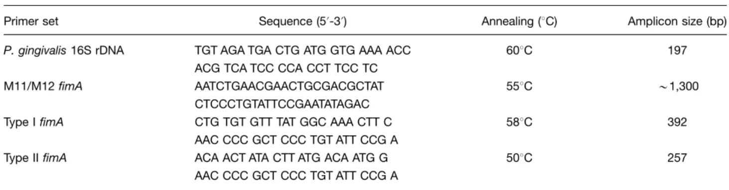
Species-specific PCR assays andfimAgenotyping were applied for the identification of the periodontopathogen P. gingivalis in vascular biopsies from patients with CP. The previously reported primers for P. gingivalis 16S rRNA andfimAgene (24, 25) were used (Table 1). PCRs were performed with OneTaq 2X Master Mix with standard buffer (New England Biolabs, Beverly, MA) and annealing temperatures as given in Table 1.
Each set of experiments included negative controls with sterile molecular grade water instead of template DNA and positive controls containing purified DNA fromVeillonella dispar,P. gingivalisstrains forfimAtype I (ATCC 33277T) andfimAtype II (A7A1-28). The PCR products were detected by 1% agarose gel electrophoresis. Gels were stained with ethidium bromide, using 1Kb Plus DNA Ladder (Invitrogen).
Cloning and sequencing
The universal 16S rDNA PCR amplicons (primers E334/ E939) were ligated into the pCR4-TOPO vector using the TOPO TA cloning kit (Invitrogen) and transformed into competent Escherichia coli TOP10 cells according to the manufacturer’s instructions. Ninety-six colonies were collected from each sample to make a representative library of the PCR products, which were stored in the TE buffer at 208C until further processing. After PCR amplification of the inserts with M13 forward primer, 5? -GTA AAA CGA CGG CCA G-3?, and M13 reverse primer, 5?-CAG GAA ACA GCT ATG AC-3?, the PCR
Table 1. Porphyromonas gingivalis16S rDNA andfimAgene-specific primers used in this study
Primer set Sequence (5?-3?) Annealing (8C) Amplicon size (bp)
P. gingivalis16S rDNA TGT AGA TGA CTG ATG GTG AAA ACC ACG TCA TCC CCA CCT TCC TC
608C 197
M11/M12fimA AATCTGAACGAACTGCGACGCTAT CTCCCTGTATTCCGAATATAGAC
558C 1,300
Type IfimA CTG TGT GTT TAT GGC AAA CTT C AAC CCC GCT CCC TGT ATT CCG A
588C 392
Type IIfimA ACA ACT ATA CTT ATG ACA ATG G AAC CCC GCT CCC TGT ATT CCG A
products were purified by Exosap-IT (Affymetrix, Santa Clara, CA) according to the manufacturer’s protocol. Purified amplicons were sequenced using the BigDye Terminator v1.1 Cycle Sequencing Kit (Applied Biosys-tems, Foster City, CA) and the M13 forward primer.
The P. gingivalis-specific PCR amplicons were se-quenced using the BigDye terminator v1.1 Cycle Sequen-cing Kit according to the manufacturer’s instructions and the respective forward primers (Table 1). Sequence reactions were run on an ABI Prism 3,730 DNA analyzer (Applied Biosystems).
Data analysis of the sequences
Sequence trimming and quality check were performed with the Sequencher 5.0 program (Gene Codes Corp., Ann Arbor, MI). The UCHIME program (26) at the mothur platform (27) was used for chimeric check. After the elimination of suspected chimeric sequences, the 16S rDNA sequences (500 bp) were used to determine bacterial identity. The sequences were aligned by using the Molecular Evolutionary Genetics Analysis (MEGA) software version 5 (28).
For identification of closest relatives, the consensus sequences were compared with known sequences in the GenBank databases using the NCBI BLAST search tool (http://www.ncbi.nlm.nih.gov/BLAST/) and Human Oral Microbiome Database (HOMD; http://www.homd.org/) (29).
Checkerboard DNADNA hybridization
Ten randomly selected subgingival plaque samples from VD patients with CP were assessed for the presences of red complex bacteria (RCB) using checkerboard DNA DNA hybridization. NaOH (100ml of 0.5 M) was added to 100 ml of subgingival plaque samples in TE buffer and vortexed. After boiling for 10 min, 800 ml of 5 M ammonium acetate was added to neutralize the samples. The DNA was then applied into the extended slots of a Minislot-30 apparatus (Immunetics, Cambridge, MA) with a positively charged nylon membrane (Boehringer Mannheim, Indianapolis, IN). The membrane had two lanes with DNA standards equivalent to 105106cells per target species tested. The membranes were hybridized with 20 digoxigenin labeled whole genome probes of species significant for the subgingival microbiota, includ-ing RCB (Table 2) (30). DNA from the type species was used as probes.
Scanning electron microscopy
Ten randomly selected vascular biopsies from VD patients with periodontitis were selected at random for SEM. The biopsies were cut into blocks of approximately 55 mm. Tissue blocks were fixed in 2.5% glutaralde-hyde or 0.1 mol/L Sorensen phosphate buffer, and stored at 48C until processing. After dehydration in ethanol, the blocks were critically point-dried with carbon dioxide.
The specimens were finally sputter-coated with gold or palladium in a vacuum evaporator and examined in a scanning electron microscope (XL 30 ESEM; Philips, Eidenhoven, The Netherlands) (31).
Statistical analyses
The statistical analyses were performed using the SPSS software (IBM SPSS Statistic version19). Independent samples t-tests were used for comparisons of species diversity and bacterial load between the two groups. For comparisons of demographic data between the two groups, independent t-test and Fisher’s exact test were performed. A statistically significant difference was de-fined withpB0.05.
Results
This study included a total of 77 patients with VD. They were categorized into two groups according to their clinical periodontal status. The first group (VD with CP) were patients undergoing treatment for VD and who Table 2. Bacterial species detected by DNADNA hybridi-zation (checkerboard analysis) in subgingival dental plaque samples from patients with atherosclerotic and abdominal aortic aneurysmal vascular disease and chronic periodontitis
Detection frequency in subgingival dental plaque
samples (n10)
Fusobacterium nucleatumssp. vincentii
1.0
Eubacterium saburreum 1.0 Prevotella nigrescens 0.9 Actinomyces viscosus 0.8 Actinomyces gerencseriae 0.8 Actinomyces israelii 0.7 Streptococcus intermedius 0.7 Porphyromonas gingivalis 0.7 Fusobacterium nucleatumssp.
polymorphum
0.6
Fusobacterium nucleatumssp. nucleatum
0.6
Tannerella forsythia 0.6 Aggregatibacter
actinomycetemcomitans
0.4
Treponema denticola 0.4 Porphyromonas endodontalis 0.4 Campylobacter rectus 0.2 Treponema socranskiissp.
socranskii
0.2
were diagnosed with CP (n30, mean age66.9, stan-dard deviation (SD)7.9, range: 51.385.0, male25/30 (83%)). The second group (VD without CP) were patients undergoing treatment for VD and who were assessed without periodontitis (n47, mean age70.9, SD9.7, range: 45.784.8, male37/47 (79%)). Vascular biopsies from all 30 VD patients with CP and 10 randomly selected VD patients without CP were further analyzed for detection of 16S rDNA sequences. In total, 27 biop-sies from abdominal aortic aneurysmal walls, 9 from carotid, and 4 from common femoral atherosclerotic plaque were examined. Demographic data of the two study groups are presented in Table 3. The patients with CP had significantly higher number of periodontal pockets with probing depth]5 mm (pB0.01). They also had a significantly higher pack-year smoking history (pB0.01). There was no significant difference in terms of age, gender, diabetes, body mass index, and number of teeth between the two groups.
After discarding incomplete sequences, a total of 2,638 sequences from VD patients with CP and 859 sequences from VD patients without CP were analyzed. A sequence similarity threshold of ]97 and ]99% were applied for identification at the genus and species level, respectively
(32). Sequences with similarity lower than 97% were ranked as unclassified and were excluded (listed accord-ing to the closest relative in Supplementary fileSA). After optimization of the PCR, weak PCR bands were detected in the negative PCR controls (50%) and were subse-quently sequenced. These background sequences (Sup-plementary file SB) as well as eukaryote and chimeric sequences were also discarded from the sequences ob-tained from the vascular samples and further analyzed. A final total of 38 out of 40 (95%) vascular samples with bacterial DNA were subjected to further analysis.
After discarding unwanted sequences, the final fraction of bacterial sequences, reflecting overall bacterial load, obtained from the vascular biopsies in patients with CP was in average 68923% (range: 098%) at an individual level. In patients without CP, this fraction was signifi-cantly lower with an average of 25926% (range: 084%; pB0.01). No significant difference in bacterial load was found comparing intravascular plaque versus aneurysmal vascular biopsies in any group.
Eighty-four different bacterial taxa were detected from a total of 1,808 sequences from the VD patients with CP (Table 4). From the VD patients without CP, 18 dif-ferent taxa were identified from a total of 210 sequences
Table 3. Demographic data of the study groups with atherosclerotic and abdominal aortic aneurysmal vascular disease (VD), with and without chronic periodontitis (CP)
VD with CP VD without CP
n30 n10 p
Age
Mean9SD 66.997.9 68.4910.2 0.66
Median (range) 66.6 (51.385.0) 69.1 (48.983.6)
Gender (n) 1.00*
Male 25 8
Female 5 2
Teeth (n)
Mean9SD 19.197.0 19.699.2 0.87
Median (range) 20 (427) 23.5 (228)
Periodontal pocket probing depth (n]5 mm)
Mean9SD 17.9911.8 0.590.97 B0.01
Median (range) 15.5 (455) 0 (03)
Diabetes (n) 0.15*
1 2
Smoking (n)
Non-smokers 1 3
Current or previous smokers 29 7
Pack-year Mean9SD 36.4920.3 19.4911.4 B0.01
Pack-year Median (range) 34.2 (4.988.0) 17.3 (5.251.3) BMI (kg/m2)
Mean9SD 26.494.2 27.794.2 0.43
Median (range) 26.4 (14.235.3) 26.3 (22.637.0)
Table 4. Bacterial taxa identified in atherosclerotic plaque and abdominal aortic aneurysmal wall biopsies from patients with chronic periodontitis
16S rDNA sequence frequency (totaln1,808)
nvascular biopsies positive (totaln30)
Closest relative n %
1. Serratiasp.* 251 13.9 15 2. Propionibacterium acnes*** 229 12.7 21 3. Pseudomonassp.** 179 9.9 11 4. Acinetobactersp.** 108 6.0 11 5. Phenylobacteriumsp. 79 4.4 8
6.UnculturedFirmicutesbacterium 79 4.4 10
7. Flavobacteriumsp. 60 3.3 6
8. Uncultured unclassified bacterium**** 44 2.4 7
9.UnculturedActinobacterium 31 1.7 3
10. Bacillussp.*** 30 1.7 2 11. Staphylococcus petenkoferri** 28 1.5 1 12. Chitinophagaceaebacterium 28 1.5 1 13. Corynebacteriumsp.** 27 1.5 5 14. Klebsiellasp.* 27 1.5 5 15. Streptococcussp.** 25 1.4 4 16. Dietziasp. 24 1.3 3 17. Bradyrhizobiumsp. 23 1.3 3 18. Enterococcussp. 22 1.2 3
19.UnculturedRhodobacteraceaebacterium 22 1.2 3
20. Microbacteriumsp. 21 1.2 4 21. Moraxella osloensis 21 1.2 2
22.UnculturedGeobactersp. 20 1.1 1
23. Staphylococcus epidermidis** 19 1.1 2 24. Ralstonia picketti** 18 1.0 2 25. Burkholderiasp.** 14 0.8 5 26. Pedobactersp. 14 0.8 2 27. Alishewanellasp. 13 0.7 2 28. Stenotrophomonas maltophilia 13 0.7 2 29. Kytococcus sedentarius 12 0.7 1 30. Oxalobacteraceaebacterium 12 0.7 1 31. Sediminibacteriumsp. 12 0.7 1 32. Acinetobacter bereziniae** 11 0.6 2 33. Afipiasp. 11 0.6 2 34. Alloprevotellasp. 11 0.6 1 35. Capnocytophaga sputigena 11 0.6 1 36. Micrococcussp. 11 0.6 1
37.UnculturedFlavobacteriumsp 11 0.6 1
38. Hymenobacter roseosalivarius** 10 0.6 1 39. Rhizobiumsp. 10 0.6 1
40.UnculturedOxalabacteriacaebacterium 10 0.6 1
(Table 5). The relative abundance of the different taxa detected in vascular biopsies from patients with and without CP is presented in Tables 4 and 5, respectively. The mean diversity of bacterial taxa detected from the vascular biopsies was significantly higher in patients with CP (n30), average of 7.193.2 (range: 012), compared to those without CP (n10), average of 2.891.6 (range:
05;pB0.01). No significant difference in mean bacterial diversity was found comparing intravascular plaque versus aneurysmal vascular biopsies in any group.
Patients with CP displayed a wide variety of bacteria identified at the genus level from their vascular biop-sies, including some known oral bacterial taxa (e.g. Stre-ptococcus spp., Prevotella sp., andCapnocytophaga sp.). Table 4(Continued)
16S rDNA sequence frequency (totaln1,808)
nvascular biopsies positive (totaln30)
Closest relative n %
49. Ornithinimicrobiumsp. 7 0.4 1 50. Uncultured Gallionellasp. 7 0.4 1 51. Acidovoraxsp. 6 0.3 3 52. Actinomyces oris 6 0.3 2 53. Carnobacteriumsp. 6 0.3 1 54. Peptoniphilus tyrrelliae 6 0.3 1 55. Psychrobactersp. 6 0.3 2 56. Rhodoplanessp. 6 0.3 1 57. Roseinatronobactersp. 6 0.3 1
58.Uncultured betaProteobacteria 6 0.3 1
59. Brevundimonassp. 5 0.3 1 60. Comamonassp. 5 0.3 1 61. Facklamia tabacinasalis 5 0.3 2 62. Thermoanaerobacteriumsp. 5 0.3 1 63. Veillonella dispar 5 0.3 1
64.UnculturedChitinophagasp. 5 0.3 1
65.UnculturedElusimicrobiumsp. 5 0.3 1
66. Aerococcussp. 4 0.2 1 67. Porphyromonas gingivalis 4 0.2 1 68. Rhodococcussp. 4 0.2 1 69. Tepidimonassp. 4 0.2 1
70.Uncultured deltaProteobacterium 4 0.2 1
71. Agrococcus terreus 2 0.1 1 72. Alphaproteobacteria 2 0.1 1 73. Aquabacteriumsp. 2 0.1 1 74. Delftia acidovorans 2 0.1 1 75. Lysinibacillussp. 2 0.1 1 76. Massilia haematophila 2 0.1 1 77. Roseococcussp. 2 0.1 1 78. Roseomonas aquatica 2 0.1 1 79. Staphylococcus aureus** 2 0.1 1 80. Cellulomonassp. 1 0.1 1 81. Corynebacterium afermentans** 1 0.1 1 82. Streptococcus sanguinis** 1 0.1 1 83. Streptococcus salivarius** 1 0.1 1 84. Undibacteriumsp. 1 0.1 1
The different taxa were identified by NCBI BLAST analysis using a similarity threshold of ]97% for identification
Possible overlap with bacterial taxa identified in vascular biopsies from VD patients without CP at *family, **genus, ***species, and ****unranked levels.
Members of theEnterobacteriaceaefamily (e.g.Serratiasp. andKlebsiellasp.) were also frequently identified in this group (Table 4).Pseudomonassp. andPropionibacterium acnes were abundant in both groups of patients. Stre-ptococcus, Staphylococcus, and Acinetobacter spp. were also detected in both groups and were among the most prevalent species in the VD group with CP. In total, 15% and 4.3% of the sequences were identified as not-yet-cultured bacterial spp. in the VD group with and without CP, respectively. Environmental species, e.g. Phenylobacterium and Bradyrhizobium spp., were also identified.
Using universal bacterial 16S rDNA PCR,P. gingivalis was detected in the vascular biopsy from only one VD patients with CP. No other members of the RCB were detected from the vascular biopsies in either group. None of the 30 vascular biopsies from VD patients with CP were positive for P. gingivalis using the specific primers listed in Table 1. Weak PCR bands were detected in the vascular samples using thefimAII primer set. Sequencing these bands showed non-specific binding of primers to human DNA. However, checkerboard analysis revealed presence of one to three members of the RCB species in
7 out of 10 subgingival plaque samples from the CP patients (Table 2).
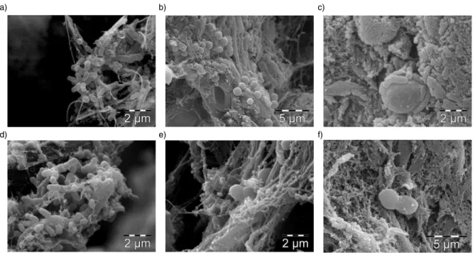
Ten vascular biopsies from VD patients with CP were randomly selected for SEM. Cocci and rod-shaped bacterial cells of different sizes were seen in all biopsies (Fig. 1af). Bacteria were observed at the surface of the intravascular plaque lining the arterial lumen entangled in a meshwork of delicate fibers and plaque remnants. Several preparations from each biopsy were inspected in order to visualize bacterial cells. Most often, the bacterial cells were found coaggregated as micro-colonies at a distance from each other. The coaggregated bacteria in each location were most often of uniform morphology. Apparent bacterial cell division with invagination of the cell membrane was seen in some of the SEM images.
Discussion
Bacterial DNA was detected in 95% of the vascular biopsies, and most biopsies contained DNA from multi-ple bacterial species, pointing to a less specific relation-ship between single species and VD. These findings are supported by recent studies suggesting multiple bacterial species being involved in cardiovascular diseases (5, 21). Table 5. Bacterial taxa identified in atherosclerotic plaque and abdominal aortic aneurysmal wall biopsies from patients without chronic periodontitis
16S rDNA sequence frequency
(Totaln210) nvascular biopsies positive
Closest relative* n % (Totaln10)
1.Bacillus idriensis 44 21.0 1 2.Pseudomonassp. 35 16.7 5 3.Delftiasp. 20 9.5 1 4.Ralstoniasp. 18 8.6 2 5.Corynebacteriumsp. 15 7.1 1 6.Propionibacterium acnes 15 7.1 3 7.Mycobacteriumsp. 12 5.7 1 8.Salinicoccussp. 10 4.8 1 9.Burkholderiasp. 9 4.3 2 10.Hymenobactersp. 6 2.9 2 11.Uncultured unclassified bacteria 6 2.9 1 12.Streptococcussp. 5 2.4 2 13.Staphylococcussp. 4 1.9 1
14. UnculturedBacteroidalesbacterium 3 1.4 1
We detected a significantly higher mean diversity and a higher load of bacteria in the vascular biopsies from the CP group as compared to the group without CP. In patients with CP, the defective epithelial barrier of the periodontal pockets together with a high load and diversity of bacteria in the subgingival plaque make it conceivable that the oral ‘gateway’ is contributing to the bacterial composition detected in VD biopsies. Bacterial translocation and transient bacteremia are caused by dental procedures, oral infections, and may even follow tooth brushing and chewing (10). Blood-borne micro-organisms are usually cleared by the immune system within minutes. However, favorable conditions may allow colonization at a given site, as seen in cases of infective endocarditis (33).
Even though RCBs were detected at high frequencies in the subgingival plaque samples, only P. gingivalis was identified from only one vascular biopsy from a patient with CP. In addition, we used P. gingivalis-specific primers; however, none of these primers yielded positive detection of P. gingivalis in our VD biopsies. Similarly, several other studies, using species-specific PCR, nested PCR and hybridization detection techniques, also re-ported no detection of RCB in atherosclerotic carotid lesions, despite the presence of one or more period-ontopathogens in the patients’ subgingival plaque
sam-ples (3436). A possible explanation to the negative or sparse detection of RCB in VD biopsies may be that, in contrast to commensal bacteria, ‘more antigenic’ mi-crobes such as the RCB are readily recognized by the immune system and eliminated from the circulation. In a study by Nakano et al. (37),Streptococcus mutansstrains serotype k survived in blood for a longer time due to lower antigenicity as compared to more cariogenic strains. In addition, a variation in ability to invade cardiovascular cells has been demonstrated among differentP. gingivalis strains (38). Similar invasive heterogeneity is reported for Aggregatibacter actinomycetemcomitans and S. mutans strains (39, 40). Individual differences in harboring invasive strains may be an alternative explanation of the discrepancies in the detection of RCB in vascular tissue reported in the literature (11).
Nevertheless, many studies have reported the presence of periodontopathogens in atherosclerotic lesions from aorta, coronary, carotid, and femoral arteries (14, 16, 17, 41), although with variable frequency (11, 36). These variations may be due to methodological differences, e.g. origin of specimens, DNA extraction techniques, the use of specific or universal primers, and PCR conditions (14, 35). However, even by using similar PCR-based techni-ques with the same specific primers, various detection rates for RCB (032% for P. gingivalis) in peripheral
a) b) c)
d) e) f)
atherosclerotic biopsies from patient with CP have been reported (41, 42). With theP. gingivalis fimAII primer set previously used for P. gingivalis detection in cardiovas-cular specimen (43), weak PCR bands were detected in some of our vascular samples. Sequencing of these bands showed non-specific binding of primers to human DNA. These findings together with the human sequences identified, using bacterial-specific 16S rDNA primers, emphasize the importance of identifying PCR products by sequencing in studies involving specimens where bacterial DNA is sparse. Using nested PCR and sequen-cing, Fiehn et al. (44) reported a detection rate of 4.17% for P. gingivalis in carotid and femoral atherosclerotic biopsies. None of the other members of the RCB were detected. Ott et al. (19) found an overall high bacterial diversity with more than 50 bacterial species in coronary atherosclerotic tissue using 16S rDNA clone libraries; however, they did not detect any members of RCB. By using 454 high-throughput sequencing, Koren et al. (45) also could not confirm the presence of DNA from periodontopathogens in carotid artery biopsies. Sparse finding of RCB in abdominal aneurysmal samples (Tannerella forsythia in 1 out of 10 samples) was also reported in a study by Marques da Silva et al. using 16S rRNA PCR and sequencing (31). DNA from none of the other RCB was detected. However, the DNA of period-ontopathogens was detected in the atheromatous plaques from coronary arteries in a Chinese population with CP (46). The prevalence of T. forsythia, Fusobacterium nucleatum,Prevotella intermedia, andP. gingivaliswas 31, 12, 18, and 33%, respectively. The same specific primers forT. forsythia,F. nucleatum, andP. intermediawere used by Cairo et al. (35) for investigating carotid atheromatous plaque for bacterial DNA. They were not able to detect DNA from these species, even though dental plaque samples were positive in 79, 63, and 53% of the cases, respectively.
Although we were able to detect bacterial DNA in most VD biopsies, the bacterial load was generally sparse as reflected by the amount of PCR products obtained. Also, with SEM several preparations from each biopsy had to be inspected in order to visualize bacterial cells. Most often the bacterial cells were found coaggregated in intravascular plaque lining the arterial lumen. Due to scant amounts of PCR products, several rounds of cloning and sequencing had to be performed to obtain 96 clone libraries with sequences of adequate quality and size, especially when processing VD biopsies from patients without periodontitis. In these patients, the final fraction of bacterial sequences averaged 25%, and the majority of the excluded sequences were background sequences and eukaryote sequences (Supplementary file SB). Thus, the number of vascular biopsies examined in this group was limited to 10 randomly selected biopsies. Renko et al. (47) using 16S rDNA sequencing, reported
the average number of different bacterial sequences from atherosclerotic lesions in patients without signs of clinical infections to be 2.291.2, which is comparable to our patients without CP (2.891.6).
In subjects with CP, several common oral taxa were detected in the vascular biopsies, i.e. Streptococcus, Prevotella, Capnocytophaga, Veillonella, and Porphyro-monas spp., while in patients without CP, only one common oral taxon was found (Streptococcussp.). These species and other possible oral taxa detected in the vascular biopsies are highlighted in Tables 4 and 5. Although these taxa represent only a minor proportion of the oral bacterial microbiota, the data suggest that the oral cavity may be a potential source for bacterial dissemination to vascular tissue. Due to the near absence of RCB in vascular biopsies, we wanted to confirm their presence in the subgingival plaque in CP patients by using checkerboard DNADNA hybridization analysis. Although this method cannot be compared to 16S rDNA sequencing, it is an efficient and sensitive detection method for this purpose (30). The checkerboard analysis revealed presence of one to three members of the RCB in 7 out of 10 subgingival plaque samples from the CP patients (Table 2). Further, only 3 out of 19 bacterial spp. found in the subgingival plaque by checkerboard analysis were detected in the vascular biopsies in the CP group (i.e. P. gingivalis, Actinomyces sp., and Streptococcus spp.), suggesting dissimilar bacterial composition in the subgingival plaque and vascular biopsies. In a previous study, 16S rDNA-based PCR was positive for bacterial detection, whereas checkerboard DNADNA hybridiza-tion methods were negative. This indicated that the number of RCB-positive subgingival plaque samples in our study could possibly be even higher (48).
Similarly, Nakano et al. (49) showed that the bacterial composition in dental plaque was different from what was found in cardiovascular specimens. The presence of periodontopathogens and other oral bacterial species were sparse in cardiovascular tissue, and only a few species, including S. mutans, may have originated from the oral cavity.
dissemination of gut bacteria into the blood stream have been described (52). Koren et al. (45) found several operational taxonomic units shared between the gut and atherosclerotic plaque suggesting that bacteria present in the atherosclerotic plaque could have been derived from the gut. In a study by Hietbrink et al. (53), systemic inflammation induced by intravenously administratedE. coli lipopolysaccharide increased the intestinal perme-ability which may facilitate bacterial translocation. Ani-mal studies have shown inflammation to induce and sustain increased intestinal permeability (54, 55). We may hypothesize that low-grade chronic systemic inflamma-tion caused by periodontal disease (56) may affect intes-tinal permeability and thereby translocation of enteric bacteria, dissemination to the bloodstream, and further colonization of vascular lesions, as seen in our vascular biopsies from patients with CP.
We detected DNA from as-yet-uncultured bacterial taxa in both groups. Much of the human microbiota is not yet cultivated (57), and the presence of different not-yet-cultured bacterial taxa is to be expected. Their significance for oral and cardiovascular disease remains to be elucidated.
Several environmental bacterial species with unde-fined pathogenicity were detected in the VD biopsies, such as members of the families Chitinophagaceae and Rhodobacteraceae, Geobacter sp., and Alishewanella sp. Through the skin and mucous membrane linings of the respiratory, gastrointestinal, and genitourinary tracts, the human body is constantly exposed to environmental species. Some of the environmental bacteria detected in the VD biopsies may be part of our transient microbiota. The inclusion of negative controls and the high level of interindividual differences in detection of environmental species make it unlikely that these bacterial taxa are introduced by technical contamination. Several other studies have reported the presence of environmental species in vascular lesions and clinical samples (47, 58); however, the pathogenicity and clinical relevance of most environmental bacterial taxa in patient samples are yet to be determined.
Bacterial DNA belonging to Pseudomonas sp. and Propionibacteriumacnes were frequently detected in both groups of patients. Pseudomonas is an opportunistic human pathogen of clinical relevance, and has been con-sidered by some to be associated with periodontal disease (59).P. acnesis part of the commensal microbiota of skin and respiratory and digestive mucosa. This species is a common blood contaminant and can be the cause of postoperative infections, and has been associated with different cases of apical periodontitis and endocarditis (60, 61).
Acinetobacter sp. was found in both groups and was among the most prevalent in the group with CP. Acinetobacterhas been isolated from periodontal pockets
(50), and is known to be a frequent cause of nosocomial infections, especially pneumonias, as well as bloodstream and urinary tract infections (62, 63). Streptococcus and Staphylococcus spp. were detected in both groups. Non-pathogenic streptococcal spp. are part of the commensal microbiota of the mouth, skin, intestines, and upper res-piratory tract. However, they can cause serious conditions such as bacteremia, meningitis, and pneumonia, and have been associated with endocarditis (64). Staphylococcus spp., such asS. epidermidis andS. aureus, is part of the commensal skin and respiratory tract microflora. Similar to streptococci they can cause cardiovascular conditions such as endocarditis (64).
By SEM, bacterial cells with different morphology were detected in all examined specimens from CP patients. Bacteria seemingly going through cell division were also observed. These findings emphasize the pre-sence of bacterial cells, not only bacterial DNA, in the VD biopsies. Viable invasive bacteria such asP. gingivalis andA. actinomycetemcomitanshave been cultivated from human atherosclerotic plaque (65, 66). Others have suggested that bacteria from the circulation might get caught in the vascular lesions as ‘innocent bystanders’ (67). However, animal studies have shown that bacterial taxa such asChlamydia pneumoniaandP. gingivalismay invade atherosclerotic plaque, alter inflammatory path-ways, and cause molecular mimicry, and thereby may contribute to the progression of atherogenesis (6, 13).
In conclusion, our results indicate that the cumulative effect of different infectious agents may be associated with the pathophysiology of atherosclerotic and abdom-inal aortic aneurysmal vascular diseases. The infectious burden seems to be associated with chronic periodontal disease. However, periodontopathogens were rarely iden-tified in the vascular biopsies, whilst gut bacteria were detected more frequently. A possible association between chronic periodontal disease, systemic inflammation, in-testinal permeability, and enterobacterial colonization of atherosclerotic and aneurysmal tissue should be studied further.
Acknowledgements
We thank the staff at the Department of Vascular Surgery, Oslo University Hospital, Aker, for their kind help with the patients and the vascular biopsies. We also wish to thank Steinar Stølen for skillful help with SEM, Ibrahimu Mdala for his statistical advice, and Morten Enersen for his advice regarding P. gingivalis fimA genotyping.
Conflict of interest and funding
References
1. Kornman KS. Mapping the pathogenesis of periodontitis: a new look. J Periodontol 2008; 79: 15608.
2. Ross R. Atherosclerosis an inflammatory disease. N Engl J Med 1999; 340: 11526.
3. Golledge J, Norman PE. Atherosclerosis and abdominal aortic aneurysm: cause, response, or common risk factors? Arterioscler Thromb Vasc Biol 2010; 30: 10757.
4. Tonetti MS. Periodontitis and risk for atherosclerosis: an update on intervention trials. J Clin Periodontol 2009; 36(Suppl 10): 1519.
5. Elkind MS. Infectious burden: a new risk factor and treatment target for atherosclerosis. Infect Disord Drug Targets 2010; 10: 8490.
6. Kebschull M, Demmer RT, Papapanou PN. ‘‘Gum bug, leave my heart alone!’’ epidemiologic and mechanistic evidence linking periodontal infections and atherosclerosis. J Dent Res 2010; 89: 879902.
7. Desvarieux M, Demmer RT, Jacobs DR, Papapanou PN, Sacco RL, Rundek T. Changes in clinical and microbiological period-ontal profiles relate to progression of carotid intima-media thickness: the oral infections and vascular disease epidemiology study. J Am Heart Assoc 2013; 2: e000254.
8. Tonetti MS, Van Dyke TE, Working group 1 of the joint EFPAAPw. Periodontitis and atherosclerotic cardiovascular disease: consensus report of the Joint EFP/AAP Workshop on Periodontitis and Systemic Diseases. J Clin Periodontol2013; 40(Suppl 14): S249.
9. Dewhirst FE, Chen T, Izard J, Paster BJ, Tanner AC, Yu WH, et al. The human oral microbiome. J Bacteriol 2010; 192: 500217.
10. Forner L, Larsen T, Kilian M, Holmstrup P. Incidence of bacteremia after chewing, tooth brushing and scaling in individuals with periodontal inflammation. J Clin Periodontol 2006; 33: 4017.
11. Reyes L, Herrera D, Kozarov E, Roldan S, Progulske-Fox A. Periodontal bacterial invasion and infection: contribution to atherosclerotic pathology. J Clin Periodontol 2013; 40(Suppl 14): S3050.
12. Tomas I, Diz P, Tobias A, Scully C, Donos N. Periodontal health status and bacteraemia from daily oral activities: systematic review/meta-analysis. J Clin Periodontol 2012; 39: 21328.
13. Kozarov E. Bacterial invasion of vascular cell types: vascular infectology and atherogenesis. Future Cardiol 2012; 8: 12338. 14. Kozarov E, Sweier D, Shelburne C, Progulske-Fox A, Lopatin D. Detection of bacterial DNA in atheromatous plaques by quantitative PCR. Microbes Infect 2006; 8: 68793.
15. Cavrini F, Sambri V, Moter A, Servidio D, Marangoni A, Montebugnoli L, et al. Molecular detection of Treponema denticolaand Porphyromonas gingivalis in carotid and aortic atheromatous plaques by FISH: report of two cases. J Med Microbiol 2005; 54: 936.
16. Okuda K, Ishihara K, Nakagawa T, Hirayama A, Inayama Y, Okuda K. Detection ofTreponema denticolain atherosclerotic lesions. J Clin Microbiol 2001; 39: 111417.
17. Stelzel M, Conrads G, Pankuweit S, Maisch B, Vogt S, Moosdorf R, et al. Detection of Porphyromonas gingivalis DNA in aortic tissue by PCR. J Periodontol 2002; 73: 86870. 18. Gaetti-Jardim E, Jr., Marcelino SL, Feitosa AC, Romito GA, Avila-Campos MJ. Quantitative detection of periodontopathic bacteria in atherosclerotic plaques from coronary arteries. J Med Microbiol 2009; 58: 156875.
19. Ott SJ, El Mokhtari NE, Musfeldt M, Hellmig S, Freitag S, Rehman A, et al. Detection of diverse bacterial signatures in
atherosclerotic lesions of patients with coronary heart disease. Circulation 2006; 113: 92937.
20. Marques da Silva R, Caugant DA, Eribe ER, Aas JA, Lingaas PS, Geiran O, et al. Bacterial diversity in aortic aneurysms determined by 16S ribosomal RNA gene analysis. J Vasc Surg 2006; 44: 105560.
21. Lund Haheim L, Olsen I, Nafstad P, Schwarze P, Ronningen KS. Antibody levels to single bacteria or in combination evaluated against myocardial infarction. J Clin Periodontol 2008; 35: 4738.
22. Armitage GC. Periodontal diagnoses and classification of periodontal diseases. Periodontol 2000 2004; 34: 921. 23. Baker GC, Smith JJ, Cowan DA. Review and re-analysis of
domain-specific 16S primers. J Microbiol Methods 2003; 55: 54155.
24. Amano A, Nakagawa I, Kataoka K, Morisaki I, Hamada S. Distribution of Porphyromonas gingivalis strains with fimA genotypes in periodontitis patients. J Clin Microbiol 1999; 37: 142630.
25. Nakagawa I, Amano A, Kimura RK, Nakamura T, Kawabata S, Hamada S. Distribution and molecular characterization of Porphyromonas gingivalis carrying a new type of fimA gene. J Clin Microbiol 2000; 38: 190914.
26. Edgar RC, Haas BJ, Clemente JC, Quince C, Knight R. UCHIME improves sensitivity and speed of chimera detection. Bioinformatics 2011; 27: 2194200.
27. Schloss PD, Westcott SL, Ryabin T, Hall JR, Hartmann M, Hollister EB, et al. ntroducing mothur: open-source, platform-independent, community-supported software for describing and comparing microbial communities. Appl Environ Microbiol 2009; 75: 753741.
28. Tamura K, Peterson D, Peterson N, Stecher G, Nei M, Kumar S. MEGA5: molecular evolutionary genetics analysis using maximum likelihood, evolutionary distance, and maximum parsimony methods. Mol Biol Evol 2011; 28: 27319. 29. Chen T, Yu WH, Izard J, Baranova OV, Lakshmanan A,
Dewhirst FE. The Human Oral Microbiome Database: a web accessible resource for investigating oral microbe taxonomic and genomic information. Database (Oxford) 2010; baq013. doi: 10.1093/database/baq013.
30. Socransky SS, Haffajee AD, Smith C, Martin L, Haffajee JA, Uzel NG, et al. Use of checkerboard DNA-DNA hybridiza-tion to study complex microbial ecosystems. Oral Microbiol Immunol 2004; 19: 35262.
31. Marques da Silva R, Lingaas PS, Geiran O, Tronstad L, Olsen I. Multiple bacteria in aortic aneurysms. J Vasc Surg 2003; 38: 13849.
32. Drancourt M, Bollet C, Carlioz A, Martelin R, Gayral JP, Raoult D. 16S ribosomal DNA sequence analysis of a large collection of environmental and clinical unidentifiable bacterial isolates. J Clin Microbiol 2000; 38: 362330.
33. Moreillon P, Que YA, Bayer AS. Pathogenesis of streptococcal and staphylococcal endocarditis. Infect Dis Clin North Am 2002; 16: 297318.
34. Romano F, Barbui A, Aimetti M. Periodontal pathogens in periodontal pockets and in carotid atheromatous plaques. Minerva Stomatol 2007; 56: 16979.
35. Cairo F, Gaeta C, Dorigo W, Oggioni MR, Pratesi C, Pini Prato GP, et al. Periodontal pathogens in atheromatous plaques. A controlled clinical and laboratory trial. J Periodontal Res 2004; 39: 4426.
37. Nakano K, Nomura R, Matsumoto M, Ooshima T. Roles of oral bacteria in cardiovascular diseases from molecular mechanisms to clinical cases: Cell-surface structures of novel serotype kStreptococcus mutansstrains and their correlation to virulence. J Pharmacol Sci 2010; 113: 1205.
38. Dorn BR, Burks JN, Seifert KN, Progulske-Fox A. Invasion of endothelial and epithelial cells by strains of Porphyromonas gingivalis. FEMS Microbiol Lett 2000; 187: 13944.
39. Brissette CA, Fives-Taylor PM. Actinobacillus actinomy-cetemcomitans may utilize either dependent or actin-independent mechanisms of invasion. Oral Microbiol Immunol 1999; 14: 13742.
40. Kesavalu L, Lucas AR, Verma RK, Liu L, Dai E, Sampson E, et al. Increased atherogenesis during Streptococcus mutans infection in ApoE-null mice. J Dent Res 2012; 91: 25560. 41. Chen YW, Umeda M, Nagasawa T, Takeuchi Y, Huang Y,
Inoue Y, et al. Periodontitis may increase the risk of peripheral arterial disease. Eur J Vasc Endovasc Surg 2008; 35: 1538. 42. Padilla C, Lobos O, Hubert E, Gonza´lez C, Matus S, Pereira M,
et al. Periodontal pathogens in atheromatous plaques isolated from patients with chronic periodontitis. J Periodontal Res 2006; 41: 3503.
43. Nakano K, Inaba H, Nomura R, Nemoto H, Takeuchi H, Yoshioka H, et al. Distribution of Porphyromonas gingivalis fimA genotypes in cardiovascular specimens from Japanese patients. Oral Microbiol Immunol 2008; 23: 1702.
44. Fiehn NE, Larsen T, Christiansen N, Holmstrup P, Schroeder TV. Identification of periodontal pathogens in atherosclerotic vessels. J Periodontol 2005; 76: 7316.
45. Koren O, Spor A, Felin J, Fa˚k F, Stombaugh J, Tremaroli V, et al. Human oral, gut, and plaque microbiota in patients with atherosclerosis. Proc Natl Acad Sci U S A 2011; 108(Suppl 1): 45928.
46. Zhang YM, Zhong LJ, Liang P, Liu H, Mu LT, Ai SK. Relationship between microorganisms in coronary atheroma-tous plaques and periodontal pathogenic bacteria. Chin Med J 2008; 121: 15957.
47. Renko J, Lepp PW, Oksala N, Nikkari S, Nikkari ST. Bacterial signatures in atherosclerotic lesions represent human commen-sals and pathogens. Atherosclerosis 2008; 201: 1927. 48. Siqueira JF, Rocas IN, De Uzeda M, Colombo AP, Santos KR.
Comparison of 16S rbased PCR and checkerboard DNA-DNA hybridisation for detection of selected endodontic patho-gens. J Med Microbiol 2002; 51: 10906.
49. Nakano K, Inaba H, Nomura R, Nemoto H, Takeda M, Yoshioka H, et al. Detection of cariogenicStreptococcus mutans in extirpated heart valve and atheromatous plaque specimens. J Clin Microbiol 2006; 44: 331317.
50. Slots J, Feik D, Rams TE. Prevalence and antimicrobial susceptibility of Enterobacteriaceae, Pseudomonadaceae and Acinetobacterin human periodontitis. Oral Microbiol Immunol 1990; 5: 14954.
51. Barbosa FC, Irino K, Carbonell GV, Mayer MP. Characteriza-tion ofSerratia marcescens isolates from subgingival biofilm, extraoral infections and environment by prodigiosin produc-tion, serotyping, and genotyping. Oral Microbiol Immunol 2006; 21: 5360.
52. Barnes PD, Bergman MA, Mecsas J, Isberg RR. Yersinia pseudotuberculosis disseminates directly from a replicating bacterial pool in the intestine. J Exp Med 2006; 203: 1591601. 53. Hietbrink F, Besselink MG, Renooij W, de Smet MB, Draisma A, van der Hoeven H, et al. Systemic inflammation increases intestinal permeability during experimental human endotoxe-mia. Shock 2009; 32: 3748.
54. Yang R, Han X, Uchiyama T, Watkins SK, Yaguchi A, Delude RL, et al. IL-6 is essential for development of gut barrier dysfunction after hemorrhagic shock and resuscitation in mice. Am J Physiol Gastrointest Liver Physiol 2003; 285: G6219. 55. Han X, Fink MP, Delude RL. Proinflammatory cytokines cause
NO*-dependent and -independent changes in expression and localization of tight junction proteins in intestinal epithelial cells. Shock 2003; 19: 22937.
56. Dave S, Van Dyke T. The link between periodontal disease and cardiovascular disease is probably inflammation. Oral Dis 2008; 14: 95101.
57. Paster BJ, Boches SK, Galvin JL, Ericson RE, Lau CN, Levanos VA, et al. Bacterial diversity in human subgingival plaque. J Bacteriol 2001; 183: 377083.
58. Sontakke S, Cadenas MB, Maggi RG, Diniz PP, Breitschwerdt EB. Use of broad range 16S rDNA PCR in clinical microbiology. J Microbiol Methods 2009; 76: 21725.
59. da Silva-Boghossian CM, do Souto RM, Luiz RR, Colombo AP. Association of red complex,A. actinomycetemcomitansand non-oral bacteria with periodontal diseases. Arch Oral Biol 2011; 56: 899906.
60. Haidar R, Najjar M, Der Boghossian A, Tabbarah Z. Propionibacterium acnes causing delayed postoperative spine infection: review. Scand J Infect Dis 2010; 42: 40511. 61. Debelian GJ, Eribe ER, Olsen I, Tronstad L. Ribotyping of
bacteria from root canal and blood of patients receiving endodontic therapy. Anaerobe 1997; 3: 23743.
62. Antunes LC, Imperi F, Carattoli A, Visca P. Deciphering the multifactorial nature ofAcinetobacter baumanniipathogenicity. PLoS One 2011; 6: e22674.
63. Bergogne-Berezin E, Towner KJ.Acinetobacterspp. as nosoco-mial pathogens: microbiological, clinical, and epidemiological features. Clin Microbiol Rev 1996; 9: 14865.
64. Pierce D, Calkins BC, Thornton K. Infectious endocarditis: diagnosis and treatment. Am Fam Physician 2012; 85: 9816. 65. Kozarov EV, Dorn BR, Shelburne CE, Dunn WA, Jr.,
Progulske-Fox A. Human atherosclerotic plaque contains viable invasive Actinobacillus actinomycetemcomitans and Porphyromonas gingivalis. Arterioscler Thromb Vasc Biol 2005; 25: e1718.
66. Rafferty B, Jonsson D, Kalachikov S, Demmer RT, Nowygrod R, Elkind MS, et al. Impact of monocytic cells on recovery of uncultivable bacteria from atherosclerotic lesions. J Intern Med 2011; 270: 27380.