Molecular Analysis of Oral Bacteria in Heart Valve of
Patients With Cardiovascular Disease by Real-Time
Polymerase Chain Reaction
Francisco Artur Forte Oliveira, DDS, MSc, Clarissa Pessoa Fernandes Forte, DDS, MSc,
Paulo Goberlaˆnio de Barros Silva, DDS, MSc, Camile B. Lopes, DDS, MSc,
Raquel Carvalho Montenegro, PhD, Aˆndrea Kely Campos Ribeiro dos Santos, PhD,
Carlos Roberto Martins Rodrigues Sobrinho, MD, PhD, Ma´rio Roge´rio Lima Mota, DDS, PhD,
Fabrı´cio Bitu Sousa, DDS, PhD, and Ana Paula Negreiros Nunes Alves, DDS, PhD
Abstract:Structural deficiencies and functional abnormalities of heart valves represent an important cause of cardiovascular morbidity and mortality, and a number of diseases, such as aortic stenosis, have been recently associated with infectious agents. This study aimed to analyze oral bacteria in dental plaque, saliva, and cardiac valves of patients with cardiovascular disease. Samples of supragingival plaque, subgingival plaque, saliva, and cardiac valve tissue were collected from 42 patients with heart valve disease. Molecular analysis ofStreptococcus mutans, Prevotella intermedia,Porphyromonas gingivalis,andTreponema denti-colawas performed through real-time PCR. The micro-organism most frequently detected in heart valve samples was theS. mutans(89.3%), followed byP. intermedia(19.1%),P. gingivalis(4.2%), andT. denticola (2.1%). The mean decayed, missing, filled teeth (DMFT) was 26.46.9 (meanSD), and according to the highest score of periodontal disease observed for each patient, periodontal pockets>4 mm and dental calculus were detected in 43.4% and 34.7% of patients, respectively. In conclusion, oral bacteria, especiallyS. mutans, were found in the cardiac valve samples of patients with a high rate of caries and gingivitis/periodontitis.
(Medicine94(47):e2067)
Abbreviations: CTAB = cetyltrimethylammonium bromide, CVD = cardiovascular disease, CVDs = cardiovascular diseases, DMFT = decayed, missing, and filled teeth, DNA = deoxyribonucleic acid, IPC = internal PCR control, P. gingivalis = Porphyromonas gingivalis, P. intermedia = Prevotella intermedia, PBS = phosphate-buffered saline, PCR = polymerase chain reaction, PSR = periodontal screening and recording, S. mutans = Streptococcus mutans, SD = standard deviation, SEM = standard error of the mean,T. denticola=Treponema denticola.
INTRODUCTION
S
tructural deficiencies and functional abnormalities of heart valves are conditions that need cardiovascular surgery. These abnormalities may be caused by congenital diseases or by a variety of acquired diseases that result in valvular stenosis, valvular insufficiency, or both.1The rheumatic heart disease (RHD), the most common heart valve disease in underdeveloped countries, is a condition that causes damage to the valve function, due to an abnormal immune response to group A streptococcal infection, especially during infancy.2Patients with RHD have valvular lesion caused
by the rheumatic valve involvement (stenosis and/or regurgita-tion) or secondary to ventricular dilatation, leading to mitral or tricuspid insufficiency.3
The aortic stenosis, the most common valve disease in industrialized countries, had been considered, for many years, a degenerative disease that would appear with aging, and that was caused by the passive accumulation of calcium on the surface of the valve leaflet. Recent studies, however, have demonstrated that this disease represents an active process that may be divided into 2 distinct phases: an early initiation phase, similar to atherosclerosis, and a later progression phase that involves pro-calcifying and pro-osteogenic factors.4 –6
In the last few decades, these conditions have been the object of great attention in the cardiology field, mainly due to changes in their presentation profile and treatment. These changes came as a result of a significant reduction in the incidence and sequelae of rheumatic fever, increase in life expectancy, technological advancement, and the discovery of new causes for valve diseases, including the alleged role of infectious agents in their pathogenesis.7
Infectious agents are well known to cause infective endo-carditis, another major cause of valve replacement, but there are recent evidence also connecting these pathogens to valvular stenosis and/or regurgitation. In this context, an association betweenChlamydophila pneumoniainfection and aortic stenosis Editor: Manal Elshmaa.
Received: June 30, 2015; revised: October 16, 2015; accepted: October 23, 2015.
From the Department of Stomatology and Oral Pathology, School of Dentistry, Federal University of Ceara, Fortaleza, Ceara´ (FAFO, CPF, PGBS, MRLM, FBS); Department of Oral Pathology, School of Dentistry, Federal University of Ceara, Fortaleza, Ceara´ (APNNA); Department of Clinical Medicine, School of Medicine, Federal University of Ceara, Fortaleza, Ceara´ (CRMRS); Laboratory of Human and Medical Genetics, Institute of Biological Sciences, Federal University of Para´, Bele´m, Para´, Brazil (CBL, AKCRS); Human Cytogenetics Laboratory, Institute of Biological Sciences, Federal University of Para´, Bele´m, Para´, Brazil (RCM).
Correspondence: Francisco Artur Forte Oliveira, Department of Clinical Dentistry, Division of Oral Pathology, School of Dentistry, Federal University of Ceara´. Address: Rua: Monsenhor Furtado, S/N, Rodolfo Teofilo, 60430-350, Fortaleza, Ceara, Brazil (e-mail: arturforte@ymail. com).
Funding: the authors also thank CAPES (Coordenac¸a˜o de Aperfeic¸oamento de Pessoal de Nı´vel Superior) for financial support with the scholarships of postgraduate students. Otherwise, no outside funding sources sup-ported this work.
The authors declare no potential conflicts of interest with respect to author-ship and/or publication of this article.
Copyright#2015 Wolters Kluwer Health, Inc. All rights reserved. This is an open access article distributed under the Creative Commons Attribution-NonCommercial-NoDerivatives License 4.0, where it is permissible to download, share and reproduce the work in any medium, provided it is properly cited. The work cannot be changed in any way or used commercially.
ISSN: 0025-7974
DOI: 10.1097/MD.0000000000002067
has been suggested by Nystro¨m-Rosander et al8and Turgeman et al.9Conversely, Kaden et al10did not find this association.
Oral micro-organisms have already been indirectly associ-ated with other cardiovascular diseases (CVD), such as athero-sclerosis. Oral bacteria involved in the etiopathogenesis of chronic periodontitis are known to cause an immune and moderate systemic inflammatory response, elevating the serum concentration of multiple cytokines and inflammatory markers that are abundantly produced in pathological periodontal tissues and strongly associated with the pathogenesis of some CVDs.11 The presence of oral bacteria in valvular tissue with or without clinical endocarditis have been investigated through sensitive molecular exams, such as the polymerase chain reaction (PCR), in order to investigate the mechanisms that can link oral infections to CVD.12–14In addition to identifying oral bacteria DNA in valvular tissue, Nomura et al15 have reported the mechanisms that led cariogenic bacteria to colonize heart valves. Considering the recent findings, the aim of this study is to identify cariogenic and periodontopathogenic micro-organisms in the dental plaque, saliva, and cardiac valves of patients undergoing valve replacement surgery, regardless of the dis-ease, in order to contribute with epidemiologic data regarding the presence of oral bacteria in cardiac valves. Detailed evalu-ation of oral health status of all patients, including presentevalu-ation of caries and periodontal disease history, by dental examination, will also be reported. This data could provide evidence of a connection between oral diseases and the frequency of oral micro-organisms in valve samples.
MATERIAL AND METHODS
Calibration, Oral Examination, and Collection of Samples
Patients admitted to Hospital Universita´rio Walter Cantı´-dio and Hospital de Messejana Dr. Carlos Alberto Studart Gomes (Fortaleza, Ceara´, Brazil) who were consecutively scheduled for cardiovascular surgery of valve replacement between March 2012 and September 2012 were enrolled in this study, which accounted for a convenience sample of 42 patients. All participants gave their informed consent and this study was approved by the Ethics Committee of the Hospitals where the research was performed.
Two examiners (F.A.F.O and C.P.F) were calibrated to evaluate teeth condition and current periodontal status of the patients, using the indexes DMFT (decayed, missing, and filled teeth) and PSR (periodontal screening and recording), respect-ively. For inter-examiner calibration, both examiners evaluated the same 10 volunteers that were not enrolled in the study. After 1 week, the same patients were re-evaluated to assess intra-exam-iner calibration. The Kappa values ranged from 0.80 to 0.97.16
Before the cardiovascular surgery, the previously cali-brated examiners performed bedside oral examination. Infor-mation regarding smoking history, hypertension, diabetes mellitus, dyslipidemia, and other comorbidities were obtained through medical charts and anamnesis. Only 1 patient could not undergo the periodontal examination due to his poor physical condition.
For dentate patients, during oral examination, supragingi-val and subgingisupragingi-val dental plaque were collected.17 Only for
edentulous patients, saliva samples were collected by rubbing sterilized absorbent paper points in the oral mucosa and on the alveolar ridge, palate, and tongue. The oral samples were stored in a sterile vial containing phosphate-buffered saline (PBS) at 20oC for subsequent molecular analysis.
A total of 47 cardiac valves were collected, aseptically, during valve replacement surgeries. A fragment of each sample was stored in a sterile container containing PBS at20oC for further analysis using real-time PCR.
DNA Extraction and Real-Time PCR
DNA extraction and molecular analysis through real-time PCR were carried out, respectively, at the Laboratory of Human Cytogenetics and Laboratory of Human and Medical Genetics of the Federal University of Para´ (Bele´m, Brazil).
A total of 114 samples of supragingival and subgingival dental plaque, saliva, and cardiac valves (30 mg) were trans-ferred to sterile plastic tubes of 2 mL containing a buffer, maintaining aseptic handling, and posteriorly homogenized. DNA extraction proceeded according to a standard protocol that used proteinase K and cetyltrimethylammonium bromide (CTAB) to remove complex polysaccharide.18
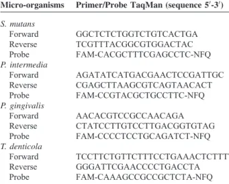
The samples of extracted DNA were subjected to real-time PCR for the detection of DNA from 4 different bacterial species:S. mutans, P. gingivalis, P. intermedia,andT. denticola. TaqMan probes to 16S bacterial ribosomal DNA (Table 1) were specifically designed for this study (Life Technologies1). For real-time PCR, 1mL of genomic DNA (5 ng) was added to 8mL
of TaqMan1 Universal PCR Master Mix (Applied Biosys-tems1), 0.35mL of probe, and 3.15mL of water. The final
volume of 14mL was obtained by adding IPC (1mL) and IPC
DNA (0.5mL), which was used as an internal control for the
reaction. Amplification was carried out in a Thermal Cycler 7500 real-time PCR System (Applied Biosystems1). Bacterial DNA was replaced by water as a negative control for the reaction. The real-time PCR protocol consisted of an initial step of denaturation at 95oC for 10 min, followed by 40 cycles at
95oC for 15 s, and 60oC for 1 min.
Statistical Analysis
The distribution pattern of the quantitative data of the sample was evaluated through the Shapiro–Wilk test. Poster-iorly the quantitative data were submitted to the Studentttest
TABLE 1. List of Primers and Probes Designed for the Identi-fication of Different Cariogenic and Periodontopathogenic Micro-organisms
Micro-organisms Primer/Probe TaqMan (sequence 50-30)
S. mutans
Forward GGCTCTCTGGTCTGTCACTGA
Reverse TCGTTTACGGCGTGGACTAC
Probe FAM-CACGCTTTCGAGCCTC-NFQ
P. intermedia
Forward AGATATCATGACGAACTCCGATTGC
Reverse CGAGCTTAAGCGTCAGTAACACT
Probe FAM-CCGTACGCTGCCTTC-NFQ
P. gingivalis
Forward AACACGTCCGCCAACAGA
Reverse CTATCCTTGTCCTTGACGGTGTAG
Probe FAM-CCCCTCCTGCAGATCT-NFQ
T. denticola
Forward TCCTTCTGTTCTTTCCTGAAACTCTTTT
Reverse GGGATTCGAACCCCTGACCTA
and expressed as meanSEM. The quantitative demographic variables were expressed as the meanSD.
Qualitative nominal variables were expressed as absolute frequency (relative frequency) and analyzed by the Fisher exact test or the chi-square test with Bonferroni correction. When possible, the prevalence ratio expressed as the prevalence ratio (confidence interval minimummaximum) was calculated.
The statistical software EpiInfo 3.5.1 for Windows (CDC Atlanta) was used and a significance level of P<0.05 was
established for all analyses.
RESULTS
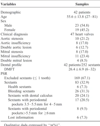
For the present study, a total of 114 oral samples (supra-gingival plaque, sub(supra-gingival plaque, and saliva) and cardiovas-cular samples (heart valves) were collected from 42 patients with a mean age of 55.613.8 years. Regarding the medical conditions that led to valve replacement surgery, mitral regur-gitation (23.4%) and aortic stenosis (21.2%) were the most common (Table 2).
Detailed oral examination for the investigation of dental caries and periodontitis was carried out in all 42 individuals. The mean number of teeth missing due to caries was 23.529.41 per patient, and all patients have already had a previous experience of caries resulting in tooth loss. According to the highest degree of periodontal disease observed in the individual, excluding edentulous patients (44.0%), periodontal pockets >4 mm (43.4%), and dental calculus (34.7%) were
present in a greater number of patients. Other demographic,
medical, and dental characteristics of the patients are summar-ized in Table 2.
Molecular analysis of oral samples revealed high fre-quency ofS. mutansandP. intermediain supragingival plaque, saliva, and subgingival plaque of dentate and edentulous patients (ranging from 60.0% to 100.0%), whereasP. gingivalis andT. denticolawere present in fewer oral samples (ranging from 17.6% to 64.0%) (Fig. 1). Distribution profile of oral bacteria between dentate and edentulous patients revealed significant differences for P. gingivalis (P¼0.024) and T. denticola (P¼0.037). According to the probing depth, P. gingivalis(P¼0.002) andT. denticola(P¼0.044) were found with a higher frequency in patients with periodontal pockets
>4 mm, with a rate>75.0%.
Regarding the presence of oral bacteria in the heart valves, all 4 micro-organisms were found in at least 1 sample, and only 5 valves (10.6%) were not infected with cariogenic or period-ontopathic bacteria. The micro-organism most frequently found in the valve samples was theS. mutans(89.3%), followed byP. intermedia (19.1%), P. gingivalis (4.2%), and T. denticola (2.1%) (Fig. 1). Significant difference was observed between the frequency ofS. mutans and the studied periodontopathic bacteria in the valve tissue (P<0.001) (Fig. 1). Likewise,P. intermediawas significantly more present in the valve tissue compared to P. gingivalis (P¼0.025) and T. denticola (P¼0.007). Differing from other bacteria (P. intermedia, P<0.001; P. gingivalis, P<0.001; T. denticola, P<0.001),
there was no significant difference between the presence ofS. mutansin heart valve and dental plaque or saliva (P¼0.060) (Fig. 1). There was no significant difference between the frequency of oral bacteria in the heart valves regarding dental condition (dentate, edentulous) (P¼0.504), anatomical location (aortic or mitral) (P¼0.596), and clinical diagnosis (stenosis, insufficiency, or both) (P¼0.256).
DISCUSSION
For many years, studies have been developed with an aim to investigate the possible connection between periodontal disease and cardiovascular disease, through the evaluation of inflammatory markers that are common to both pathologies. However, recent studies, such as the study of Nakano et al,12 attempted to elucidate the direct mechanisms that link oral diseases to CVDs, and, according to this author, the presence of oral bacteria in the bloodstream (bacteremia) is probably one of the initiators of biological events that justify this association. TABLE 2. Demographic, Clinic, and Dental Characteristics of
Patients With Heart Valve Diseases
Variables Samples
Demographic 42 patients
Age 55.613.8 (27–81)
Sex
Male 23 (54.8)
Female 19 (45.2)
Clinical diagnosis 47 heart valves
Aortic stenosis 10 (21.2)
Aortic insufficiency 8 (17.0)
Double aortic lesion 6 (12.7)
Mitral stenosis 8 (17.0)
Mitral insufficiency 11 (23.4)
Double mitral lesion 4 (8.5)
Dental profile 42 patients/252 sextants
DMFT 26.46.9 (6–32)
PSR
Excluded sextants (1 tooth) 169 (67.1)
Sextants 83 (32.9)
Health sextants 6 (7.3)
Bleeding sextants 26 (31.3)
Sextants with dental calculus 20 (24.1)
Sextants with periodontal
pockets 3.5–5.5 mm for 4–5 mm
17 (20.5)
Sextants with periodontal
pockets>5.5 mm for6 mm
8 (9.5)
Lost information 6 (7.3)
Qualitative dada expressed by ‘‘n(%)’’.
FIGURE 1. Percentage distribution of cariogenic and periodonto-pathic bacteria in dental plaque, saliva, and heart valve samples.
P<0.05 versus valve sample of Pi, Pg, and Td.yP<0.05 versus supragingival dental plaque, subgingival dental plaque, and saliva sample for each oral bacteria.z
The involvement of cariogenic bacteria in the pathogenesis of some CVDs has been studied, and the reported detection rate ofS. mutansin cardiovascular samples has been superior to the detection rate of periodontopathic bacteria.12 In the present study, a high detection rate of S. mutansin the heart valve samples, as well as dental plaque and saliva samples was observed. The higher frequency ofS. mutansin the heart valves compared to other studies may be related to the great previous experience of dental caries among the participants of the present study. It was also observed a high percentage of tooth loss due to caries, and epidemiological studies have demonstrated that the absence of teeth may be a risk factor associated with many cardiovascular diseases, including aortic stenosis.19 In our research, the high detection rate ofS. mutansin dental plaque samples and saliva samples may suggest that the S. mutans found in the valve samples was originated from the oral cavity. In the present study, edentulous patients also presented a high frequency of the cariogenic bacterium in saliva and cardiac valve samples. These bacteria can be found in the oral cavity even after complete tooth loss, adhered to soft tissue or den-tures.20The colonization ofS. mutansin the cardiac tissue of these patients may have happened previously to complete tooth loss. Another theory is that these bacteria may have entered the bloodstream after complete tooth loss through soft tissue trauma. The possible occurrence of bacteremia in edentulous patients needs further investigation.
Statistically significant difference was found between the frequency ofS. mutansand the other investigated micro-organ-isms in the heart valves. In the past few years,in vitroandin vivo experimental studies have reported thatS. mutanshas the ability to modify the expression of certain genes influenced by plasma components, in order to obtain advantages while inside the bloodstream.21These findings may justify this bacterium’s high ability to settle in the heart valve tissue.
Jung et al22reported the role of AtlA, a recent discovered fibronectin binding protein, in the S. mutans resistance to fagocitosis and in its ability to survive in the bloodstream. Its maturation is enhanced by physiological serum calcium concentration. Fibronectin, elastin, laminin, and collagen are considered the most common extracellular matrix components and may serve as receptors forS. mutans.23
The discovery of bacterial surface structures such as the proteins P1, WapA, GtfB, GtfC, GtfD, Cnm, and Cbm, has been important to understand the mechanisms of attachment of these pathogens to the cardiovascular tissue, with subsequent inva-sion of endothelial cells, induction of inflammation with the production of cytokines, platelet aggregation, and foam cell formation.24 –27
Jung et al28,29and Matsumoto-Nakano et al24have already reported the important interaction between this bacterium and the platelets in heart valve tissue through experimental studies, where they show that platelet aggregation is stimulated by this micro-organism.
Pariodontophatic bacteria were found less frequently in heart valve tissue, when compared to the gram-positive bacter-iumS. mutans,corroborating the findings of Nakano et al.14 Great variability is reported in the literature regarding the prevalence of these bacteria in heart valve tissues and athero-sclerotic plaques, and the reported frequency is either superior or inferior to that observed in the present study.12,14,30–32Many
authors that found a higher frequency of these periodontal bacteria in cardiovascular tissues, compared to the present study, have included in their studies only dentate patients with moderate to severe periodontitis.
In the present study, the majority of the dentate patients presented periodontal disease, exhibiting dental calculus and periodontal pockets>3.5 mm for>4 mm. This fact was
deter-minant for the periodontal bacteria of the orange complex (P. intermedia) and red complex (P. gingivalis,T. denticola) to be detected at a high frequency in the dental plaque, specially subgingival plaque removed from periodontal pockets>6 mm,
corroborating previously reported findings.20,33
Among the gram-negative micro-organisms,P. intermedia was significantly more detected in the heart valve tissue (P<0.05). The high detection rate of this micro-organism in
the oral samples of dentate (supragingival and subgingival dental plaque) and edentulous patients (saliva) probably explains its higher frequency in the heart valve samples, com-pared to the other periodontopathic bacteria. The hypothesis that periodontopathic bacteria can infect cardiovascular samples prior to complete tooth loss of a patient that had periodontal disease may be considered, as suggested by some authors, such as Zaremba et al.34
The frequency of periodontopathic bacteria in the heart valve samples was considered low, when compared to their frequency in the oral samples, which may suggest that these micro-organisms have greater difficulty in surviving inside the bloodstream and adhering to heart valve tissue, compared toS. mutans.
P. gingivalis has received special attention among the periodontopathic bacteria regarding its association with CVDs, although it was found at a low frequency in the present study. This bacterium is detected at a high frequency in studies that investigate its presence in atherosclerotic plaques, andin vitro and in vivo experimental studies have demonstrated that P. gingivalis accelerates the process of atherosclerotic plaque formation through different mechanisms.32,35,36
In the present study, real-time PCR was used because it has previously demonstrated high sensitivity and efficacy in detect-ing bacterial DNA in valve tissue and oral samples, even at low levels.12,37 Nevertheless, the polymerase chain reaction may also detect DNA of dead bacteria, which poses a doubt regard-ing the role of the detected bacteria in the pathogenesis of the disease. It is unlikely that the bacteria present in the heart valve tissue are innocuous, as recent laboratorial studies have given support to the idea that these bacteria may have a direct influence in the initiation and progression of the disease. However, new studies are essential to confirm this association. The evaluation of the expression of different biomarkers (inflammatory, thrombotic, and osteogenic) in human heart valves affected with chronic diseases and its correlation with the presence and frequency of oral bacteria in those samples may provide a positive association between the infection and the intensity of the local inflammatory process. This would be an important finding to confirm an active role of the oral bacteria in the progression of these diseases.
In summary, cariogenic and periodontopathic bacteria were found in dental plaque, saliva, and cardiac valve samples of patients with cardiovascular disease. The detection rate of S. mutansin cardiovascular samples was superior to the detection rate of periodontopathic bacteria. This finding may be related to the great previous experience of dental caries among the partici-pants of the present study andS. mutanspossible ability to survive in the bloodstream and attach to extracellular matrix components.
ACKNOWLEDGMENTS
Gomes, Human Cytogenetics Laboratory of the Federal Uni-versity of Para´ (UFPA), Laboratory of Human and Medical Genetics (UFPA), for their assistance during the development of this study.
REFERENCES
1. Fernandes AMS, Bitencourt LS, Lessa IN, et al. Impact of socio-economic profile on the prosthesis type choice used on heart surgery. Rev Bras Cir Cardiovasc.2012;27:211–216.
2. Marijon E, Mirabel M, Celermajer DS, et al. Rheumatic heart disease.Lancet.2012;379:953–964.
3. Fernandes AM, Andrade GM, Oliveira RM, et al. Evaluation of variables responsible for hospital mortality in patients with rheumatic heart disease undergoing double valve replacement.Rev Bras Cir Cardiovasc.2014;29:537–542.
4. Pawade TA, Newby DE, Dweck MR. Calcification in aortic stenosis: the skeleton key.J Am Coll Cardiol.2015;66:561–577.
5. Dweck MR, Boon NA, Newby DE. Calcific aortic stenosis: a disease of the valve and the myocardium.J Am Coll Cardiol.2012;60:1854– 1863.
6. Ginghin? C, Florian A, Beladan C, et al. Calcific aortic valve disease and aortic atherosclerosis—two faces of the same disease? Rom J Intern Med.2009;47:319–329.
7. Boudulas H. Etiology of valvular heart disease in the 21st century. Hellenic J Cardiol.2002;43:183–188.
8. Nystro¨m-Rosander C, Lindh U, Ilba¨ck NG, et al. Interactions between chlamydia pneumoniae and trace elements: a possible link to aortic valve sclerosis.Biol Trace Elem Res.2003;91:97–110.
9. Turgeman Y, Levahar P, Lavi I, et al. Adult calcific aortic stenosis and chlamydia pneumoniae: the role of chlamydia infection in valvular calcification.Isr Med Assoc J.2006;8:464–468.
10. Kaden JJ, Bickelhaupt S, Grobholz R, et al. Pathogenetic role of chlamydia pneumoniae in calcific aortic stenosis: immunohistochem-istry study and review of the literature.J Heart Valve Dis. 2003;12:447–453.
11. D’Aiuto F, Parkar M, Andreou G, et al. Periodontitis and systemic inflammation: control of the local infection is associated with a reduction in serum inflammatory markers.J Dent Res.2004;83: 156–160.
12. Nakano K, Inaba H, Nomura R, et al. Detection of cariogenic streptococcus mutans in extirpated heart valve and atheromatous plaque specimens.J Clin Microbiol.2006;44:3313–3317.
13. Nakano K, Nemoto H, Nomura R, et al. Serotype distribution of streptococcus mutans a pathogen of dental caries in cardiovascular specimens from Japanese patients.J Med Microbiol.2007;56:551–556.
14. Nakano K, Nemoto H, Nomura R, et al. Detection of oral bacteria in cardiovascular specimens.Oral Microbiol Immunol.2009;24:64–68.
15. Nomura R, Otsugu M, Naka S, et al. Contribution of the interaction of streptococcus mutans serotype k strains with fibrinogen to the pathogenicity of infective endocarditis.Infect Immun.2014;82:5223– 5234.
16. WHO (World Health Organization). Calibration of Examiners for Oral Health Epidemiology Surveys. Technical Report. Geneva: WHO; 1993.
17. Cairo F, Gaeta C, Dorigo W, et al. Periodontal pathogens in atheromatous plaques. A controlled clinical and laboratory trial. J Periodontal Res.2004;39:442–446.
18. Wilson K. Preparation of genomic DNA from bacteria.Curr Protoc Molecul Biol.2001;2-4:.
19. Vo¨lzke H, Schwahn C, Hummel A, et al. Tooth loss is independently associated with the risk of acquired aortic valve sclerosis.Am Heart J.2005;150:1198–1203.
20. Socransky SS, Haffajee AD. Periodontal microbial ecology. Period-ontol 2000.2005;38:135–187.
21. Negrini TC, Duque C, Vizoto NL, et al. Influence of VicRK and CovR on the interactions ofStreptococcus mutanswith phagocytes. Oral Dis.2012;18:485–493.
22. Jung CJ, Zheng QH, Shieh YH, et al. Streptococcus mutans autolysin AtlA is a fibronectin-binding protein and contributes to bacterial survival in the bloodstream and virulence for infective endocarditis.Mol Microbiol.2009;74:888–902.
23. Chia JS, Yeh CY, Chen JY. Identification of a fibronectin binding protein from streptococcus mutans.Infect Immun.2000;68:1864– 1870.
24. Matsumoto-Nakano M, Tsuji M, Inagaki S, et al. Contribution of cell surface protein antigen c ofStreptococcus mutansto platelet aggregation.Oral Microbiol Immunol.2009;24:427–430.
25. Nagata E, de Toledo A, Oho T. Invasion of human aortic endothelial cells by oral viridans group streptococci and induction of inflamma-tory cytokine production.Mol Oral Microbiol.2011;26:78–88.
26. Abranches J, Zeng L, Be´langer M, et al. Invasion of human coronary artery endothelial cells by streptococcus mutans OMZ175.Oral Microbiol Immunol.2009;24:141–145.
27. Nomura R, Nakano K, Naka S, et al. Identification and characteriza-tion of a collagen-binding protein, Cbm, inStreptococcus mutans. Mol Oral Microbiol.2012;27:308–323.
28. Jung CJ, Yeh CY, Shun CT, et al. Platelets enhance biofilm formation and resistance of endocarditis-inducing streptococci on the injured heart valve.J Infect Dis.2012;205:1066–1075.
29. Jung CJ, Yeh CY, Hsu RB, et al. Endocarditis pathogen promotes vegetation formation by inducing intravascular neutrophil extracellu-lar traps through activated platelets.Circulation.2015;131:571–581.
30. Haraszthy VI, Zambon JJ, Trevisan M, et al. Identification of periodontal pathogens in atheromatous plaques.J Periodontol. 2000;71:1554–1560.
31. Gaetti-Jardim E, Marcelino SL, Feitosa AC, et al. Quantitative detection of periodontopathic bacteria in atherosclerotic plaques from coronary arteries.J Med Microbiol.2009;58:1568–1575.
32. Figuero E, Sa´nchez-Beltra´n M, Cuesta-Frechoso S, et al. Detection of periodontal bacteria in atheromatous plaque by nested polymerase chain reaction.J Periodontol.2011;82:1469–1477.
33. Stingu CS, Jentsch H, Eick S, et al. Microbial profile of patients with periodontitis compared with healthy subjects.Quintessence Int. 2012;43:e23–31.
34. Zaremba M, Go´rska R, Suwalski P, et al. Evaluation of the incidence of periodontitis-associated bacteria in the atherosclerotic plaque of coronary blood vessels.J Periodontol.2007;78:322–327.
35. Takahashi Y, Davey M, Yumoto H, et al. Fimbria-dependent activation of pro-inflammatory molecules in porphyromonas gingiva-lis infected human aortic endothelial cells.Cell Microbiol. 2006;8:738–757.
36. Khalaf H, Bengtsson T. Altered t-cell responses by the periodontal pathogen porphyromonas gingivalis.PLoS One.2012;7:e45192.