Citation:Luo R, Jin Z, Deng Y, Strokes N, Piao X (2012) Disease-Associated Mutations Prevent GPR56-Collagen III Interaction. PLoS ONE 7(1): e29818. doi:10.1371/ journal.pone.0029818
Editor:Lin Mei, Medical College of Georgia, United States of America
ReceivedSeptember 14, 2011;AcceptedDecember 6, 2011;PublishedJanuary 4, 2012
Copyright:ß2012 Luo et al. This is an open-access article distributed under the terms of the Creative Commons Attribution License, which permits unrestricted use, distribution, and reproduction in any medium, provided the original author and source are credited.
Funding:This research was supported in part by National Institute of Neurological Disorders and Stroke (NINDS) grant R01 NS057536 (X.P.), a Flight Attendant Medical Research Institute Young Clinical Scientist Award (Z.J.), and the Leonard and Isabelle Goldenson Research Fellowship (R.L.). The funders had no role in study design, data collection and analysis, decision to publish, or preparation of the manuscript.
Competing Interests:The authors have declared that no competing interests exist.
* E-mail: [email protected]
.These authors contributed equally to this work.
Introduction
Adhesion GPCRs are a relatively new family of GPCRs that have a very long N-terminal extracellular domain. In humans, there are a total of 33 members of adhesion GPCRs that are thought to mediate cell-cell and cell-extracellular matrix interaction, with GPR56 as the first one linked to a human developmental malformation [1,2]. Mutations in GPR56 cause BFPP, a specific human brain malformation [3,4]. To date, a total of fourteen BFPP-associated mutations have been identi-fied, including one deletion, two splicing, and eleven missense mutations [2,5]. Based on the fact that all of the missense mutations render an identical clinical phenotype as the deletion mutation, it is presumed that all missense mutations result in a null allele [4]. Previously, we have shown that all of the missense mutations affect GPR56 protein trafficking and cell surface expression to variable degrees [6]. However, we can only say for certain that the two mutations in the GPCR proteolytic site (GPS) domain, C346S and W349S, cause a brain malformation through trapping the mutated proteins in the endoplasmic reticulum [6].
Most recently, we have discovered that the ligand of GPR56 is collagen III [7]. In this report, we aim to identify and characterize the binding domain of GPR56 to collagen III. Although the N-terminal extracellular domain of GPR56, GPR56N, is heavily glycosylated, we provide strong evidence that glycosylation is not required for ligand binding. Moreover, disease-associated muta-tions in the ligand binding domain completely eliminate its ligand binding ability, revealing the molecular mechanism for GPR56-related brain malformation.
Results
GPR56Nbinds specifically to collagen III in the developing brain
We have previously shown that GPR56Nbinds a putative ligand in the meninges and pial basement membrane and subsequently demonstrated that the ligand of GPR56 is collagen III in the developing brain [7,8]. To examine the binding specificity of GPR56Nto collagen III, we performed a putative ligand binding assay in wild type, Col3a12/2, and Gpr562/2 brains. Both Col3a12/2 and Gpr562/2 mice are true null allele mutants
(Fig. 1B) [8]. Deletion ofGpr56has no effect on the expression of collagen III protein and GPR56Nbinding patterns (Fig. 1C and F), whereas loss of Col3a1 completely removed the binding of GPR56N (Fig. 1E). Taken together, our data supports that GPR56Nbinds specifically to collagen III in the developing brain.
The ligand binding domain of GPR56 is within aa 27–160
ones containing aa 27–142 and aa 49–160 (Fig. 2C–J). The shortest fragment that has full ligand binding capacity was the fusion protein containing aa 27–160 (Fig. 2H).
To further verify the putative ligand binding data, we carried out a series of co-immunoprecipitation (co-IP) experiments using meningeal fibroblasts as the ligand source. We failed to detect
Figure 1. GPR56Nbinds specifically to collagen III in the developing brain.(A–C) Immunostaining of collagen III in E14.5 mouse brain sections of wild type,Col3a12/2, andGpr562/2revealed an expression of collagen III (red) in the meninges and pial basement membrane of wild type
andGpr562/2brains. Deletion ofCol3a1completely abolished the expression of collagen III. Scale bar, 100mm. (D–F) Strong GPR56Nbinding was detected in meninges and pial basement membrane of wild type and Gpr562/2, but not Col3a12/2 mouse brains. Nuclear counterstain was
performed with Hoechst 33342 (blue). Scale bar, 200mm.
doi:10.1371/journal.pone.0029818.g001
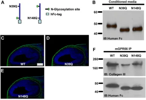
Figure 2. The ligand binding domain of GPR56.(A) The truncated GPR56N-hFc fusion protein constructs are schematically shown, as well as the full length GPR56 protein with its identified N-glycosylation sites. (B) The fusion constructs were transfected into HEK-293T cells. Secreted proteins in the conditioned media were collected, concentrated, and verified by western blot. (C–J) Putative ligand binding on E14.5 mouse cortex. The shortest fragment with a specific binding pattern (green) is the truncated GPR56N-hFc fusion protein containing aa 27–160 (H). Nuclear counterstain was performed by Hoechst 33342 (blue). Scale bar, 200mm. (K) The binding of collagen III and various truncated GPR56N-hFc was confirmed by co-IP.
(Fig. 3B). The mutant fusion proteins were concentrated from the conditioned media and used for both the putative ligand binding assay and co-IP experiments. Both mutants retained a ligand binding capacity comparable to the wild-type protein, suggesting glycosylation of GPR56 is not necessary for ligand binding (Fig. 3C–F).
Disease-associated mutations abolish ligand binding
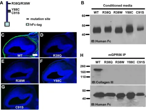
The four reported disease-associated mutations in the ligand binding domain are R38Q, R38W, Y88C, and C91S. We have previously shown that these four mutations decreased mutant proteins expression on the cell surface and its secretion into the conditioned media [6]. It is not clear, however, how each of the four missense mutations completely demolishes the receptor function. To study whether these mutations also affect ligand binding, we generated individual mutant ligand binding domains by site-directed mutagenesis in the pFUSE-hFc2 construct containing aa 27–160 of GPR56 (Fig. 4A). We were able to detect the fusion mutant proteins in the conditioned media of the transfected cells (Fig. 4B). The mutant fusion proteins were
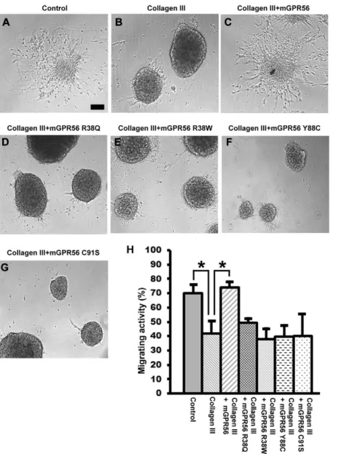
the impact of BFPP-associated mutations on the receptor function, we performed a neurosphere migration assay to examine whether the four BFPP-associated mutations - R38Q, R38W, Y88C, and C91S - functionally abolish receptor-ligand interaction. As expected, in contrast to the wild type GPR56N, none of the four mutant proteins were able to reverse the collagen III-mediated neural migration inhibition (Fig. 5).
Wild type human GPR56Nbinds to both human and
mouse collagen III, whereas mutant GPR56 proteins lose their ligand binding ability
We have thus demonstrated that mouse GPR56 binds to both mouse and human collagen III. To study whether the interaction can extend to the human GPR56 protein, we constructed a human GPR56N-hFc fusion protein (hGPR56N-hFc) and the correspond-ing BFPP-associated mutants: R38Q, Y88C, and C91S. We first performed co-IP experiments using purified human collagen III. As expected, we can only detect collagen III in wild type hGPR56N but not in the mutant hGPR56N co-IP protein complexes (Fig. 6B). We next examined the tissue binding activity
Figure 3. Glycosylation mutations of GPR56 did not affect its ligand binding.(A) The schematic representation of two separate N-glycosylation mutations within the GPR56 ligand binding domain is shown. (B) Secreted fusion proteins in the conditioned media were collected, concentrated, and verified by western blot. (C–E) Putative ligand binding on E14.5 mouse cortex. The N-glycosylation mutation did not impair GPR56 ligand binding (green). Nuclear counterstain was performed by Hoechst 33342 (blue). Scale bar, 200mm. (F) The binding ability of the two
on E14.5 mouse brain sections. As illustrated in Figure 6 C–F, wild type hGPR56N binds to mouse collagen III whereas mutant hGPR56N fails to exhibit any binding activity on mouse embryonic brains. Taken together, our data suggests that there is no species restriction between mouse and human in regards to the interaction of GPR56 and collagen III.
Discussion
GPR56 is a particularly distinct member of the adhesion GPCRs since its mutations are associated with a devastating human brain malformation called BFPP [3,9,10]. There are a total of eleven reported null allele missense mutations associated with BFPP, two of which reside in the GPS domain and cause brain malformation by demolishing the autoproteolytic process, thus trapping the mutant proteins in the endoplasmic reticulum [5,10]. A recent study by Lin’s group indicates that mutations in the tip of GPR56Nlikely render the receptor null status by affecting their
binding to a putative cellular ligand in HT1080 cells [11]. Here, we show four mutations in the tip of GPR56Nimpair the receptor function by aborting its ligand binding ability, in addition to affecting the receptor cell surface expression.
GPR56 is cleaved at aa 382 via a GPS-mediated autoproteolytic process [6,12,13]. Although the cleaved GPR56N lacks a transmembrane sequence, it is expressed on the cell surface via an unknown mechanism [6]. In addition, GPR56Nis secreted into the conditioned media [6,13]. We previously showed that the four missense mutations on the tip of GPR56N(R38Q, R38W, Y88C, and C91S) affect the secretion of GPR56Ninto the conditioned media [6]. In this study, we were able to detect secreted mutant proteins in their short forms. The possible explanations could be either that the pFUSE-hFc2 vector was more efficient in driving protein expression/secretion since it uses the IL2 signal peptide
sequence or that replacing aa 160–382 with the large hFc fragment facilitated the shedding of the fusion protein.
GPR56 functions in a capacity to mediate cell-extracellular matrix interaction. Our recent finding demonstrates that collagen III is the ligand of GPR56 in the developing brain [7]. Collagen III is a major collagen in the connective tissues with integrina1b1 and
a2b1 both serving as it’s receptors [14,15]. In this report, we identified the binding domain of GPR56 to collagen III. Since there is no consensus amino acid sequence between integrin
a1b1/a2b1 and GPR56, it is likely that GPR56 binds to a different region of collagen III.
In addition to its vital function in brain development, GPR56 also plays an important role in tumor growth and metastasis. GPR56 was originally cloned by two independent groups in 1999, one using a degenerative PCR approach and the other through differential display in high and low metastatic melanoma cell lines [16,17]. The latter group revealed that GPR56 is significantly down regulated in high metastatic melanoma cell lines, indicating a possible role of GPR56 in tumor metastasis [17]. It was subsequently demonstrated that GPR56 inhibits tumor growth/metastasis, likely by regulating VEGF production and tumor angiogenesis [13,18]. In addition, they showed that GPR56 binds to TG2, a major crosslinking enzyme in the extracellular matrix, at aa 108–177 [13,18]. Although knockdown of TG2 did not lead to an increase of VEGF, deleting the TG2 binding domain led to enhanced angiogenesis and tumor growth [18]. There is some overlap between the collagen III and TG2 binding domains on GPR56. It is highly desirable to reveal the crystal structure of these two binding domains in order to fully understand how GPR56 dynamically interacts with both collagen III and TG2 in both the developing brain as well as tumor growth and metastasis.
Figure 4. Disease-associated GPR56 abolished its ligand binding.(A) GPR56N-hFc aa 27–160 schematic. The positions of the disease-associated mutations within the ligand binding domain are shown. (B) Secreted fusion proteins in the conditioned media were collected, concentrated, and verified by a western blot. (C–G) Putative ligand binding on E14.5 mouse cortex. Each of the four disease-associated mutations completely killed the receptor-ligand binding ability in contrast to the strong binding signal (green) displayed by the wild type fusion protein (C). Nuclear counterstain was performed with Hoechst 33342 (blue). Scale bar, 200mm. (H) The impaired binding of disease-associated mutants to
Materials and Methods
Ethics Statement
Experiments were performed in accordance with National Institutes of Health guidelines for the care and use of laboratory animals, and with the approval of the Animal Care and Use Committee of Children’s Hospital Boston.
Mice
Col3a1 knockout mice were obtained from the Jackson Laboratory with the strain name C.129S4 (B6)-Col3a1tm1Jae/J in a BALB/c background. The Gpr56 knockout mice, kindly provided by Genentech, were produced in collaboration between Genentech and Lexicon Genetics to analyze the function of,500
secreted and transmembrane proteins. All animals were treated according to the guidelines of the Animal Care and Use Committee of Children’s Hospital Boston (approval ID: A3303-01).
Histology and Immunohistochemistry
Histology analysis was carried out as previously described [7,8]. Frozen sections were collected on a cryostat. Sections were incubated with rabbit anti-human collagen III antibody (Lifespan Biosciences). Primary antibodies were visualized by appropriate fluorophore-conjugated secondary antibodies. Images were cap-tured using a Nikon 80i upright microscope. Representative photographs were obtained with the same exposure setting for both the control and mutant.
Figure 5. Mutant mouse GPR56 proteins failed to rescue collagen III-mediated neural migration inhibition.(A–G) Neurospheres were generated and plated on PDL-coated dishes in regular neurosphere culture medium. After 20 h, the cultures were changed to experimental medium containing collagen III (84 nM) with or without wild type or mutant GPR56 proteins (90 nM), or carrier solution (acetic acid). Representative images are shown. Scale bar, 100mm. (H) The degree of collagen III-mediated migration inhibition was quantified as a percentage of the migrating
Plasmid Constructions
The human IgG Fc-tagged mouse GPR56N(mGPR56N-hFc) and its truncated fragments were constructed in pcDNA3.1-zeo vector (Invitrogen). The hFc tag was cloned into the Xho I/Apa I sites of the vector. Mouse GPR56Nand its truncated fragments, including the GPR56 signal peptide sequence, aa 1–26, were generated by PCR using primers listed in Table S1 and subsequently fused to hFc by inserting into the Nhe I/Xho I sites. Our previous data showed that BFPP-associated mutations affect protein intracellular trafficking and secretion [6]. To increase protein secretion, we cloned the mouse GPR56 ligand binding domain, aa 27–160, into the pFUSE-hFc2 vector (Invivogen) that contains the IL2 signal peptide sequence. N-glycosylation mutations and BFPP-associated mutations were created by site-directed mutagenesis using the QuikChange II XL Site-Directed Mutagen-esis kit (Stratagene), as previously described [6]. The primers used for the site-directed mutagenesis are listed in Table S2.
The human IgG Fc-tagged human GPR56N(hGPR56N-hFc) was also constructed in pFUSE-hFc2 vector (Invivogen) between Nco1 and BglII sites. The primers used for human GPR56NPCR are listed in Table S3. BFPP-associated mutations were created by the same approach described above using the primers listed in Table S4.
Generation of hFc fusion protein and putative ligand binding assay
Each of the above GPR56N-hFc expression constructs was transiently transfected into HEK-293T cells (obtained from ATCC). The culture media was changed to serum-reduced
OPTI-MEM 24 hours after transfection. The conditioned media was harvested 48–72 hours later, and concentrated as previously described [8]. For the neurosphere migration assay, the mouse GPR56 27–160 hFc wild type and mutant proteins were purified through protein A column (GE Healthcare).
Time pregnant wild type mice in the CD-1 background were ordered from Charles River. Embryonic day (E) 14.5 wild type mouse brain sections were obtained at 12mm in thickness on a cryostat (Leica). Equivalent amounts of fusion proteins were used as probes to examine their binding ability to mouse brain sections. The localization of GPR56N-hFc proteins were visualized by fluorescein-conjugated rabbit anti-human IgG antibody (Thermo Scientific).
Co-immunoprecipitation (co-IP) and western blot analysis
Co-IP for mouse GPR56Nand collagen III and immunoblotting were done as previously described, using meningeal fibroblasts as the ligand source [7]. Purified human collagen III (Abcam) was used in co-IP to test the interaction of human GPR56N and collagen III. Protein-G beads were used to pull-down the GPR56N-hFc protein complex. Immunocomplexes were subjected to SDS-PAGE and western blot using rabbit anti-human collagen III antibody (Lifespan Biosciences), and rabbit anti-human IgG Fc antibody (Thermo Scientific) following standard protocols.
Migration assay
Neurosphere generation and migration assay were done as described previously [7]. The neurospheres were plated in a 48-well dish precoated with 100mg/ml poly-D-lysine (PDL) and
cultured in neuron culture medium overnight. After the neurospheres adhered to PDL substrate, the culture medium was then replaced with one of the experimental mediums: neuron culture medium with 84 nM purified human collagen III (Abcam) with or without 90 nM wild type or mutant GPR56N, or with carrier solution (acetic acid) as
control. The neurospheres were imaged and the number of migrating neurospheres was quantified after a 2 day culture. It is worth noting that Abcam only guarantees the quality of purified or recombinant human collagen III by western blot analysis. We prescreened the biological function of each lot by a neural migration assay prior to a bulk order, since there is a significant variation between each lot.
Supporting Information
Table S1 Primers for mouse truncated GPR56N-hFc cloning.
(DOC)
Table S2 Primers for mouse GPR56N-hFc site-directed muta-genesis.
(DOC)
Table S3 Primers for human GPR56N-hFc cloning. (DOC)
Table S4 Primers for human GPR56N-hFc site-directed
muta-genesis. (DOC)
Acknowledgments
We thank Genentech for kindly providing theGpr56knockout mice.
Author Contributions
Conceived and designed the experiments: XP RL ZJ. Performed the experiments: RL ZJ YD NS. Analyzed the data: XP RL ZJ YD NS. Contributed reagents/materials/analysis tools: XP RL ZJ. Wrote the paper: XP.
Figure 6. Human GPR56 binds to both mouse and human collagen III and disease-associated GPR56 mutations abolish their ligand binding ability. (A) Human GPR56N-hFc aa 27–382 schematic. The positions of the disease-associated mutations are shown. (B) Co-IP of human GPR56N and purified human collagen III. Collagen III was detected in GPR56 IP complex, but not in mutant GPR56 complexes. Anti-hFc immunoblot served as a loading control. (C–F) Putative ligand binding of human GPR56N-hFc on E14.5 mouse cortex. Strong binding signal was detected (green) with wild type human GPR56N-hFc protein staining, whereas a loss of signal occurred in mutant proteins. Nuclear counterstain was performed with Hoechst 33342 (blue). Scale bar, 200mm.
lamination. Proc Natl Acad Sci U S A 108: 12925–12930.
8. Li S, Jin Z, Koirala S, Bu L, Xu L, et al. (2008) GPR56 regulates pial basement membrane integrity and cortical lamination. J Neurosci 28: 5817–5826. 9. Piao X, Basel-Vanagaite L, Straussberg R, Grant PE, Pugh EW, et al. (2002) An
autosomal recessive form of bilateral frontoparietal polymicrogyria maps to chromosome 16q12.2-21. Am J Hum Genet 70: 1028–1033.
10. Jin Z, Luo R, Piao X (2009) Chapter 1 GPR56 and Its Related Diseases. Prog Mol Biol Transl Sci 89: 1–13.
296–305.
17. Zendman AJ, Cornelissen IM, Weidle UH, Ruiter DJ, van Muijen GN (1999) TM7XN1, a novel human EGF-TM7-like cDNA, detected with mRNA differential display using human melanoma cell lines with different metastatic potential. FEBS Lett 446: 292–298.