Listeria monocytogenes
Fitness Towards Life within the
Host versus Environmental Survival
Joseph C. Bruno Jr.1, Nancy E. Freitag1,2*
1Department of Global Health, University of Washington, Seattle, Washington, United States of America,2Department of Microbiology and Immunology, University of Illinois at Chicago, Chicago, Illinois, United States of America
Abstract
PrfA is a key regulator ofListeria monocytogenes pathogenesis and induces the expression of multiple virulence factors within the infected host. PrfA is post-translationally regulated such that the protein becomes activated upon bacterial entry into the cell cytosol. The signal that triggers PrfA activation remains unknown, however mutations have been identified (prfA*mutations) that lock the protein into a high activity state. In this report we examine the consequences of constitutive PrfA activation onL. monocytogenesfitness both in vitroandin vivo. WhereasprfA*mutants were hyper-virulent during animal infection, the mutants were compromised for fitness in broth culture and under conditions of stress. Broth culture
prfA*-associated fitness defects were alleviated when glycerol was provided as the principal carbon source; under these conditionsprfA*mutants exhibited a competitive advantage over wild type strains. Glycerol and other three carbon sugars have been reported to serve as primary carbon sources forL. monocytogenesduring cytosolic growth, thusprfA*mutants are metabolically-primed for replication within eukaryotic cells. These results indicate the critical need for environment-appropriate regulation of PrfA activity to enableL. monocytogenesto optimize bacterial fitness inside and outside of host cells.
Citation:Bruno JC Jr, Freitag NE (2010) Constitutive Activation of PrfA Tilts the Balance ofListeria monocytogenesFitness Towards Life within the Host versus Environmental Survival. PLoS ONE 5(12): e15138. doi:10.1371/journal.pone.0015138
Editor:Roy Martin Roop II, East Carolina University School of Medicine, United States of America
ReceivedSeptember 5, 2010;AcceptedOctober 25, 2010;PublishedDecember 7, 2010
Copyright:ß2010 Bruno Jr., Freitag. This is an open-access article distributed under the terms of the Creative Commons Attribution License, which permits unrestricted use, distribution, and reproduction in any medium, provided the original author and source are credited.
Funding:This work was supported by Public health service grant AI41816 (N.E.F) from NIAID. The contents of the article are solely the responsibility of the authors and do not necessarily represent the official views of the funding sources. The funder had no role in study design, data collection and analysis, decision to publish, or preparation of the manuscript.
Competing Interests:The authors have declared that no competing interests exist.
* E-mail: nfreitag@uic.edu
Introduction
The environmental bacterial pathogenListeria monocytogenesis an intriguing example of a microorganism that has become well adapted to life in the soil as well as to life within the cytosol of mammalian host cells. This bacterium is widespread in the environment where it is believed to live as a saprophyte on decaying plant material [1]. Upon ingestion by a susceptible mammalian host, L. monocytogenes transitions into a physiological state that facilitates bacterial survival and replication within host cells [2,3]. While disease caused by L. monocytogenes in healthy individuals is usually restricted to a self-limiting gastroenteritis, in immunocompromised individuals and pregnant womenL. mono-cytogenes is capable of causing systemic infections that lead to meningitis, encephalitis, and in the case of pregnant women, infection of the developing fetus leading to abortion, stillbirth, or neonatal infections [4,5]. L. monocytogenes contamination of food products has resulted in some of the most expensive food recalls in U.S. history [2,6–12] and this is thought to reflect the bacterium’s widespread environmental distribution and its ability to withstand a variety of stress conditions [13–16].
A significant amount of research has focused on the mechanisms used by L. monocytogenes to establish its replication niche within mammalian host cells.L. monocytogenesinvades a wide variety of cell types and is capable of escaping from the phagosome following cell
entry, of replicating within the cytosol, and of utilizing host cell actin polymerization machinery to propel itself through the cytosol and into neighboring cells [3,5,17]. To survive and flourish within eukaryotic cells the bacterium requires the regulated expression of a number of secreted virulence factors, and the expression of most of these gene products is regulated by a transcriptional regulator known as PrfA [18]. PrfA is an essential regulator ofL. monocytogenes pathogenesis, and bacterial mutants that lack functional PrfA are severely attenuated in animal infection models [19,20].
indicate that PrfA activity must be carefully modulated in response to environmental signals so as to enableL. monocytogenesto optimize bacterial fitness both inside and outside of the infected host.
Results
Constitutive activation of PrfA reduces the fitness ofL. monocytogenesin nutrient-rich broth
Until recently, it has proven difficult to construct isogenic L. monocytogenes prfA*mutant strains containing the alleles that confer the highest PrfA activity by standard methods. As a result, these high activityprfA*mutations have been introduced intoDprfAstrains on plasmids [30,31,34,40,43,44]. While these approaches have been informative, there are associated caveats that include multicopy plasmid effects or altered gene expression profiles resulting from the use of integrated plasmids in ectopic locations. We recently reported the successful construction of high activityprfA*isogenic mutants in strains containing promoterless copies of the genes encoding b -glucuronidase (gus) and neomycin resistance (neo) located in the chromosome downstream of the PrfA-dependent gene actA [41]. IsogenicprfA*mutants constructed via allelic exchange were isolated based on the PrfA*-dependent increase inactA-gus-neoexpression that enabled selection for prfA* colonies on selective media containing neomycin and 5-bromo-4-chloro-3-indolyl-b-D-glucuronic acid (x-gluc), a substrate for GUS activity. This approach now enables the direct comparison of independently isolated L. monocytogenes prfA* mutants with strains containing the wild type allele.
To assess if the constitutive activation of PrfA influences the fitness of L. monocytogenes outside of host cells, strains containing mid-level (prfAG155S) or high-level (prfAG145S andprfAL140F) prfA* activity mutations [41] were compared with a wild type strain for growth in BHI broth. Consistent with previous reports, prfA* mutations conferred high levels of PrfA activity in broth culture as indicated byactAexpression levels (Fig. 1A). Expression from the actA promoter for the prfA G155S and prfA G145S mutants was 230-fold and 1870-fold higher respectively than the levels observed for wild typeprfAstrains after 24 hours of growth in BHI (Fig. 1A). Overall growth of theprfA*mutants was very similar to that of the wild type strain, although the doubling times of the prfA* mutants during logarithmic growth were slightly longer (Table S1) and the final bacterial cell densities at stationary phase were slightly lower in the prfA*monocultures than in the wild type monocultures (Fig. 1B).
In contrast to monoculture growth, pronounced fitness effects were observed for high activity prfA* strains when the mutants were mixed and grown with wild type bacteria in BHI. EachprfA* mutant exhibited a competitive defect when cultures were inoculated in equal numbers with wild type bacteria and grown to stationary phase with subsequent cycles of dilution and outgrowth (Fig. 1C). After nine sequential cycles of overnight growth and dilution, wild type bacteria were observed in two-fold
Bruno, unpublished), indicating that there is no apparent inhibitory substance produced by wild type bacteria that compromised mutant growth.
To determine if the competitive defects exhibited by theprfA* mutants occurred during logarithmic growth or whether the defects were associated with entry into or survival during stationary phase, mixed cultures were diluted into fresh BHI upon reaching late-logarithmic phase (OD600of 0.8–1.0), prior to bacterial entry into stationary phase. When mixed cultures of wild type andprfAG145S bacteria were grown under these conditions, the resulting competitive defect was essentially identical to the competitive defect observed for mixed cultures grown to stationary phase (Fig. 1D). This indicates that constitutive activation of PrfA impairs the competitive fitness ofL. monocytogenesin broth culture during logarithmic growth.
The presence of glucose exacerbates the competitive defect exhibited by L. monocytogenes prfA* strains. It has been previously reported that multicopy plasmid-based over-expression of constitutively activated PrfA (prfAG145S) interferes with bacterial utilization of glucose as a carbon source [43,44]. To examine if isogenicprfA*mutants exhibited a fitness defect in the presence of glucose, the prfA G145S mutant was grown in LB buffered to pH 7.4 and supplemented with 55 mM of glucose. LB was selected for monitoring growth asL. monocytogenesrequires an added carbon source for optimal growth in this medium. Similar to the observations made forprfA*monocultures in BHI, cultures grown in LB and glucose-supplemented LB resembled the wild type strain with only subtle growth differences, indicating that the isogenic prfA* strains were able to efficiently use glucose as a carbon source (Fig. 2A and Table S1). However, whenprfAG145S cultures were mixed with the wild type strain and grown in LB or in LB supplemented with glucose, the competitive defect exhibited by the prfA G145S mutant in LB with glucose was of greater magnitude than that exhibited in LB alone (Fig. 2B). After seven cycles of dilution and outgrowth, wild type bacteria outnumbered the prfAG145S mutants by 30-fold and 170-fold in LB and in glucose-supplemented LB, respectively (Fig. 2B). The presence of glucose thus exacerbated the competitive defect associated with PrfA activation in broth culture.
Constitutive activation of PrfA increases the fitness ofL. monocytogenesin the presence of glycerol
Consistent with this growth advantage, the prfA G145S mutant exhibited a competitive advantage when it was mixed and grown with wild typeL. monocytogenesin glycerol-supplemented LB. After seven cycles of dilution and outgrowth,prfAG145S outnumbered wild type bacteria by more than 20-fold (Fig. 2B). These findings indicate that constitutive activation of PrfA increases the fitness ofL. monocytogenes in the presence of glycerol. The findings further indicate that competitive defects associated with theprfA*strains in other media cannot simply be attributed to the metabolic burden of increased PrfA-dependent gene product expression, as high expression levels are maintained byprfA* strains in the presence of glycerol ([44,45] and J. Bruno, unpublished).
Constitutive activation of PrfA increases the sensitivity of
L. monocytogenesto osmotic stress and acid stress
The ability ofL. monocytogenesto withstand a variety of stresses is vital for its survival and replication in disparate environments
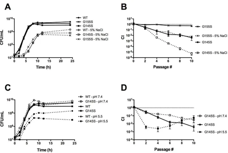
[5,13,46,47], including food processing facilities [9,48] and the gastrointestinal tract [3,17,49,50]. To determine if constitutive activation of PrfA influences the ability of L. monocytogenes to respond to stress, monoculture and mixed culture growth of the prfA*mutants and wild typeL. monocytogeneswas examined under two different stress conditions, osmotic stress and acid stress. Although no dramatic differences were observed for mutant and wild type strains with respect to growth in monoculture (Fig. 3AC), the prfA G155S mutant and the prfA G145S mutant exhibited more severe competitive defects when mixed with the wild type strain and grown in BHI supplemented with 5% NaCl in comparison to BHI lacking additional NaCl (Fig. 3B). After nine cycles of dilution and outgrowth, wild type bacteria outnumbered mutants by more than 150-fold (prfA G155S) and 200,000-fold (prfAG145S) in the presence of additional NaCl, in comparison to differences of 2-fold (prfA G155S) and 200-fold (prfA G145S) in growth media lacking added NaCl (Fig. 3B).
Figure 1.prfA*mutants exhibit a competitive defect when grown with wild type in nutrient rich broth.(A) Comparison of levels of PrfA activation between differentprfA*mutant strains as measured byactAexpression. PrfA-dependentactAexpression levels were measured by monitoring the GUS activity ofL. monocytogenesstrains containing anactA-gustranscriptional fusion. Bacteria were grown in BHI at 37uC with shaking, and units of GUS activity were normalized to CFU/mL. Each datum point represents the mean6standard deviation of a GUS assay measured in duplicate, and each GUS activity profile is representative of 2 independent experiments. TheprfA*mutants are referred to by their PrfA amino acid mutations. (B) Monoculture growth of wild type,DprfA, andprfA* L. monocytogenesstrains in BHI at 37uC with shaking. Each growth curve is representative of two independent experiments. (C)prfA*mutants exhibit a competitive defect when grown with wild typeL. monocytogenes.Wild typeL. monocytogeneswas transformed with the integrative plasmid vector pPL2 to confer chloramphenicol resistance, and then assessed for growth in BHI at 37uC in the presence of chloramphenicol-sensitive test strains as indicated. Mixed cultures were subjected to repeated cycles of culture dilution and outgrowth every 24 hours into fresh BHI. The competitive index (CI) values of the mixed cultures were determined immediately prior to each dilution as described in Experimental Procedures. The data represent the means6standard errors of three independent experiments. (D) The competitive defect ofprfA*strains occurs during logarithmic growth. A mixed culture of theprfAG145S mutant and the wild type strain was subjected to repeated cycles of culture dilution and outgrowth at late-log phase (OD600of 0.8–1.0<86108–16109CFU/mL) (indicated at ‘G145S - log’). CI values were determined immediately prior to each dilution. The data represent the means6standard errors of two independent experiments.
Figure 2. Effects of different carbon sources on monoculture growth and competitive index of aprfA*mutant.(A) Growth curves of the wild type andprfAG145SL. monocytogenesstrains in buffered LB (pH 7.4) with and without 55 mM of either glucose (glu) or glycerol (gly) at 37uC with shaking were determined by measuring CFU/mL at the specified time points. Each growth curve is representative of two independent experiments. (B) The wild type camRstrain was mixed with the chloramphenicol-sensitiveprfAG145S mutant in buffered LB with and without 55 mM of either glucose (glu) or glycerol (gly) at 37uC with shaking. Mixed cultures were subjected to repeated cycles of growth and dilution (1:100) into fresh media every 24 hours. CI values were determined immediately prior to each dilution. The data represent the means6standard errors of three independent experiments.
doi:10.1371/journal.pone.0015138.g002
Figure 3. Stress conditions exacerbate the competitive defects exhibited by prfA*mutants. (A) Monoculture growth curves of L. monocytogenesstrains in BHI supplemented with 5% NaCl at 37uC were determined by measuring CFU/mL at the specified time points. The growth curves of wild type andprfAG145SL. monocytogenesin BHI without additional NaCl are included. (B) Competitive index of wild type camRstrain mixed with a chloramphenicol-sensitiveprfA*mutant in BHI supplemented with 5% NaCl at 37uC. Mixed cultures were subjected to repeated cycles of growth and dilution (1:100) into fresh media every 24 hours. CI values were determined immediately prior to each passage. The data represent the means6standard errors of three independent experiments. (C) Monoculture growth curves ofL. monocytogenesstrains in BHI buffered to pH 7.4 or pH 5.5 at 37uC were determined by measuring CFU/mL at the specified time points. (D) Competitive index of the wild type camRstrain mixed with a chloramphenicol-sensitiveprfA*mutant in BHI buffered to pH 7.4 or 5.5 at 37uC. Mixed cultures were subjected to repeated cycles of growth and dilution (1:100) into fresh media every 24 hours. CI values were determined immediately prior to each dilution. The data represent the means6
The prfA G145S mutant exhibited a similarly exacerbated competitive defect under acid stress. When L. monocytogenes was grown in unbuffered BHI broth at 37uC, the pH was observed to decrease from approximately 7.2 to 6.0 after 24 hours of growth (J. Bruno, unpublished). When grown with wild typeL. monocytogenes strains, the prfAG145S mutant initially exhibited a more severe competitive defect in BHI buffered to pH 5.5 than in unbuffered BHI. After three cycles of dilution and outgrowth, wild type bacteria were present in 400-fold greater numbers than theprfA G145S mutant in BHI pH 5.5 in comparison to 15-fold greater numbers in unbuffered BHI (Fig. 3D). Interestingly, the large competitive defect exhibited by theprfAG145S mutant during the first three cycles of dilution and outgrowth in BHI pH 5.5 shifted to a competitive advantage with subsequent cycles, reducing the wild type advantage from 400-fold to 20-fold after nine cycles (Fig. 3D). This ratio was similar to the ratio observed after nine cycles of dilution and outgrowth in BHI buffered to pH 7.4 (Fig. 3D). These findings suggest that theprfAG145S mutant goes through an adaptation or acid tolerance response [51] that increases its tolerance to acid stress to wild-type levels or even beyond. Overall, these findings indicate that constitutive activation of PrfA impaired the ability of L. monocytogenes to respond to osmotic stress as well as its initial response to acid stress conditions.
The impaired stress response ofprfA*mutants does not result from impaired function of the stress-associated sigma factor, SigB
The exacerbated decrease in the bacterial fitness of prfA* mutants when subjected to two different stress conditions suggested that a general response related to stress tolerance may be compromised by constitutive activation of PrfA. A central regulatory component that contributes to the ability of L. monocytogenes to survive various stress conditions is the alternative RNA polymerase sigma factor SigB [16,52]. SigB contributes to prfAexpression [25], and several previous studies have suggested the existence of functional overlap between SigB and PrfA in regulating the expression ofL. monocytogenesgenes that contribute to virulence and/or stress response [40,53–60]. WhileDsigBgrowth in monoculture resembled that of the wild type strain (Supple-mental Fig. S2), DsigB mutants exhibited a competitive defect when mixed with the wild type strain in BHI, indicating that loss of SigB function decreases the competitive fitness ofL. monocytogenes (Fig. 4). Interestingly, the magnitude of the competitive defect exhibited by theDsigBmutant closely resembled that observed for prfAG145S mutants in BHI (Fig. 4C).
To determine if the competitive defect associated with prfA* strains was related to an impairment of SigB function,prfAG155S
DsigBandprfAG145SDsigBdouble mutants were tested in broth competition assays. If PrfA* impairs SigB function, one would anticipate that the magnitude of the competitive defect exhibited by a prfA* DsigB double mutant would be equivalent to that exhibited by either single mutant (Fig. 4A). If however the magnitude of the competitive defect exhibited by a prfA* DsigB double mutant was equivalent to the sum of the defects exhibited by theprfA*andDsigBsingle mutants (Fig. 4A), this would suggest that PrfA and SigB alter stress resistance through separate pathways. The competitive defect of aprfA*DsigBdouble mutant was found to be equivalent to the sum of the defects of theprfA* and theDsigBsingle mutants, and the additive effect ofprfA*and
DsigB in the double mutant strain was evident throughout the course of mixed growth (Fig. 4BC). Therefore, the stress related competitive defect associated with the constitutive activation of PrfA appears distinct from the defect associated with the loss of SigB function.
Constitutive activation of PrfA enhancesL.
monocytogenesvirulence following intravenous and intragastric infection of mice
Previous studies have reported that theprfA*mutants with mid-level PrfA activity (prfAG155S mutants) were fully virulent when intravenously inoculated into mice based on the bacterial CFU required for a 50% lethal dose (LD50) [32]. Consistent with this observation, mice intravenously infected with 26104CFU had significantly higher numbers ofprfA* bacteria (prfA G155S, prfA G145S, andprfAL140F) recovered from the livers and spleens at 24 hours post-infection compared to those infected with wild type bacteria (Fig. 5A) (the liver and spleen are the primary organ targets forL. monocytogenesreplication [5]). Although the difference was not statistically significant at 48 hours post-infection, the bacterial burdens of the livers and spleens from mice infected with theprfA*mutant tended to be higher than in organs associated with wild type infection (Fig. 5B and J. Bruno, unpublished). The hyper-virulent phenotype of theprfA*mutants was more apparent when the infectious dose was reduced by ten-fold to 26103CFU; the bacterial burdens of the livers and spleens from mice infected with the prfA G145S mutant at 48 hours post-infection were approximately 300-fold and 4.5-fold higher in liver and spleen than those of mice infected with wild type bacteria (P,0.01 for both organs) (Fig. 5B). In mixed infection, the prfA* mutants consistently exhibited a competitive advantage over wild type strains (Fig. 5C). The competitive index values determined for each liver and spleen for intravenously infected mice at 48 hours post-infection showed that, on average, 2- to 7-fold more prfA* bacteria were recovered from each organ in comparison to wild type (Fig. 5C).
Although constitutive activation of PrfA enhanced bacterial infection following intravenous injection of mice, the increased sensitivity to both osmotic stress and acid stress observed for the mutant strains (Fig. 3) suggested that the virulence of the prfA* mutants might be attenuated if administered orally, the more natural route of infection. We therefore examined the conse-quences of constitutive PrfA activation on the fitness of L. monocytogeneswithin an animal host following intragastric inocula-tion. Intragastric infection with either theprfAG145S mutant or wild type L. monocytogenes strain was carried out following the introduction of theinlAmmutation into each strain background to enhance bacterial interaction with mouse E-cadherin and translocation of bacteria across the intestinal epithelium [61]. Surprisingly, 2- to 7-fold more bacteria were recovered from the livers, spleens, stomachs, and intestines of mice infected with the prfAG145SinlAmmutant than from the organs of mice infected with the wild typeprfA inlAm strain at infectious doses of either 56107CFU or 56109CFU (Fig. 6). These findings indicate that constitutive activation of PrfA enhances the fitness of L. monocytogenes inside of the host following either intravenous or intragastric inoculation.
Discussion
PrfA activity under different environmental conditions in the context ofL. monocytogenesfitness inside and outside of infected host cells. Our results indicate that while constitutive activation of PrfA serves to enhance bacterial virulence within the infected host, in most cases PrfA activation decreases bacterial fitness outside of host cells.L. monocytogenestherefore regulates PrfA activity so as to
optimally balance life in the outside environment with life inside of the host.
Environmental regulation of PrfA activity suggests that the high levels of PrfA activity required for intracellular life are detrimental to the fitness ofL. monocytogenesoutside of a host cell, and limited analyses of L. monocytogenes field strains appear to support this
Figure 4. The increased susceptibility of prfA* cultures to stress is unrelated to sigB function. (A) Rationale regarding how the competitive defect exhibited by aprfA*DsigBdouble mutant can be used to determine if the competitive defect associated with constitutive PrfA activation is related to an impairment of SigB function. If constitutive activation of PrfA (PrfA*) impairs SigB function, the magnitude of the competitive defect exhibited by aprfA*DsigBdouble mutant strain will be equivalent to the magnitude of the competitive defects exhibited by the
prfA*andDsigBsingle mutants. If constitutive activation of PrfA (PrfA*) does not impair SigB function, the magnitude of the competitive defect exhibited by aprfA*DsigBdouble mutant will be equivalent to the sum of the magnitudes of the competitive defects exhibited by theprfA*and
DsigBsingle mutants. (B) Assessment of the competitive index for theprfAG155SDsigBdouble mutant. The wild type camRstrain was mixed with a chloramphenicol-sensitive test strain in BHI at 37uC. Mixed cultures were subjected to repeated cycles of growth and dilution (1:100) into fresh BHI every 24 hours, and CI values were determined immediately prior to each dilution. The data represent the means6 standard errors of two independent experiments. (C) Assessment of the competitive index for theprfAG145SDsigBdouble mutant. The wild type camRstrain was mixed with a chloramphenicol-sensitive test strain in BHI at 37uC. Mixed cultures were subjected to repeated cycles of growth and dilution (1:100) into fresh BHI every 24 hours, and CI values were determined immediately prior to each dilution. The data represent the means6standard errors of two independent experiments.
hypothesis. Although far from being exhaustively examined, no field strain reported in the literature has been found to contain a prfA*mutation nor to exhibit a PrfA* phenotype; all of theprfA* mutations reported have arisen spontaneously in laboratory media or as a result of chemical mutagenesis [30–33,35,37]. Moreover, field strains have been isolated containing missense mutations or small deletions within the coding region of the prfA gene that decrease or eliminate PrfA activity, indicating that PrfA activity is not required for optimal bacterial fitness outside of a host cell [64,65] although a recent report indicates that some activity is required for efficient biofilm formation [66].
The link between carbon source utilization and PrfA regulation of L. monocytogenes virulence gene products has long been recognized but has remained poorly defined. Previous studies have demonstrated that when L. monocytogeneswas grown in the presence of glucose or other carbon sources taken up by the phosphoenolpyruvate (PEP) transport system (PTS), the expression levels of PrfA-dependent genes were decreased [29,67,68]. Other studies have reported that over expression ofprfA*on a multicopy plasmid inL. monocytogenessignificantly impaired bacterial growth and glucose uptake in media where glucose was the main or sole carbon source [43,44]. Although the isogenicprfA*strains used in
Figure 5. Growth of theprfA*mutants in the livers and spleens of intravenously infected mice.7–8 week old ND4 Swiss Webster mice were infected withL. monocytogenesvia tail-vein injections, and at the specified times post-infection (pi), the bacterial loads of the livers and spleens were determined as described in Experimental Procedures. Data are presented as scatter dot plots, with horizontal bars representing means. (A) Infection of mice with 26104CFU wild type,prfAG155S,prfAL140F, orprfAG145S mutants. Organs were harvested 24 hours pi. Asterisks denote statistically significant differences between the amounts ofprfA*mutant and wild type CFU recovered using a one-way analysis of variance with Dunnett’s post-test (*, P,0.05; ***, P,0.001). (B) Comparison of infection with 26103or 26104CFU of wild type andprfAG145S mutant. Organs were harvested 48 hours pi. Asterisks denote statistically significant differences between the amounts ofprfAG145S mutant and wild type CFU recovered using an unpaired t test with a two-tailed P value (**, P,0.01). (C) Competitive index of wild type andprfA*strains. Prior to intravenous injection, the wild type Ermrreference strain and the indicated test strain were mixed 1:1 for a total bacterial suspension of 26104CFU. For each organ, the competitive index (CI) value (CI = test strain CFU/reference strain CFU) was determined as described in Experimental Procedures. Asterisks denote statistically significant CI values compared to 1 using a one-sample t test with a two-tailed P value (*, P,0.05; **, P,0.01).
this study did not exhibit the pronounced growth defects previously reported in the presence of glucose in monoculture, the addition of glucose to LB was found to exacerbate the competitive defect exhibited by the prfA G145S mutant in LB (Fig. 2B). While differences related to prfA* copy number may influence the ability of L. monocytogenes prfA* mutants to utilize glucose, the results in either case indicate that the presence of glucose decreases the fitness of L. monocytogenes when PrfA is activated.
In addition to the glucose-related growth defects reported forL. monocytogenesstrains containing multicopy plasmid-encodedprfA*, it has also been reported that similar strains exhibited a subtle monoculture growth defect when grown in media where glycerol (a non-PTS carbon source) was the main carbon source [44]. In contrast to this finding, our data indicate that isogenicprfA*mutants were enhanced for glycerol utilization and exhibited a competitive advantage over wild type in the presence of glycerol (Fig. 2). While the competitive advantage was evident during the first three cycles of culture dilution and outgrowth, the wild type strain appeared to adapt to glycerol as a carbon source such that bacterial mutant to wild type ratios became stable after three cycles of growth (Fig. 2). Consistent with an adaptation of wild typeL. monocytogenesto growth with glycerol, it was observed that after five cycles of dilution and outgrowth monocultures of wild type bacteria in LB glycerol reached the same cell densities as did the isogenicprfA*mutants but retained low levels of PrfA-dependent gene expression (J. Bruno, unpublished).L. monocytogenes prfA*strains thus appear to exist in a metabolic state that favors bacterial growth in glycerol but not glucose. The prfA* metabolic shift might thus enhance bacterial growth in the cytosol, where three carbon sugars have been suggested to be preferentially used for bacterial replication [69,70]. In addition to the PrfA*-related metabolic shift of L. monocytogenes towards glycerol utilization, our data indicate that the competitive defects exhibited byprfA*mutants in broth culture were exacerbated under conditions of osmotic and/or acid stress (Fig. 3). Based on the reports of functional overlap of SigB and PrfA in regulation ofL. monocytogenesgene expression [40,53–60] and the fact that SigB is one of the most characterized stress response regulators of L. monocytogenes [52], it seemed logical to
investigate whether the stress-associated fitness defects exhibited by theprfA*mutants were related to alterations in SigB function. Examination of the fitness ofprfA*DsigBdouble mutant strains in BHI indicated that the double mutant exhibited a competitive defect that was approximately equivalent to the sum of the competitive defects exhibited by theprfA*andDsigBsingle mutants (Fig. 4). Thus, while constitutive PrfA activation appears to interfere with the stress response of L. monocytogenes, it does so independently of SigB. It is possible that PrfA activation may somehow interfere with the function of another general stress response factor, such as ClpC, whose expression has been shown previously to be influenced by PrfA activity [71,72]. Alternatively, constitutive activation of PrfA may interfere with the expression or function of a factor(s) whose activity is directly involved in the repair of a stress associated cell injury.
In contrast to bacterial fitness in culture media, the need for down-regulation of PrfA activity does not appear to be required within the infected host. Experiments with either intravenously or intragastrically infected mice indicated that theprfA*mutants were more virulent than wild typeL. monocytogenes. More bacteria were recovered from the livers and spleens of mice infected withprfA* mutant bacteria compared to those infected with wild type bacteria, and the mutant strains exhibited a competitive advantage in mixed infections (Figs. 5 and 6). One surprising finding was that despite an increased susceptibility to osmotic and acid stresses in culture media (Fig. 3), theprfA*mutants remained hyper-virulent following oral infection (Fig. 6). The GI tract presentsL. monocytogeneswith a variety of stresses, including acid and osmotic stress [49] as well as stress associated with mucous barriers [73]. Given that SigB contributes to the gastrointestinal survival ofL. monocytogenes[57,74] and that SigB function does not appear to be affected by constitutive PrfA activation (Fig. 4),prfA*mutants have the ability to respond to the stresses of the GI tract via SigB. In addition, the PrfA*-dependent increase in gene products that facilitate bacterial invasion (for example, InlA, InlB, LLO, ActA) [5,18,62,75–79] and/or bile resistance (Bsh, BilE) [54,55] may enhance intestinal translocation so as to counter balance any stress-associated defects.
In summary, the findings presented in this study emphasize the critical need forL. monocytogenesto regulate PrfA activity dependent
on its environmental location. While experiments in broth culture indicate a competitive fitness defect forprfA*mutants, it remains possible that PrfA activation contributes toL. monocytogenesoutside of mammalian infection, for example by promoting bacterial survival in the presence of lower eukaryotes or other soil dwellers. PrfA activation clearly enhances bacterial virulence in mammalian hosts, however the need for down modulation of PrfA activity in other settings might well be a reflection of the yin-yang nature of theL. monocytogenessaprophyte-pathogen balance.
Materials and Methods
Bacteria and culture media
The bacterial strains and plasmids used in this study are listed in Table 1. AllL. monocytogenesstrains used were derived from the 1/ 2a serotype 10403SL. monocytogenesstrain, which is a streptomycin-resistant derivative of strain 10403 [80,81]. The phenotypes reported for strains containing prfA*mutations were verified in independent isolates constructed by allelelic exchange and/or by phage transduction, and by comparison of differentprfA* alleles (prfAG145S,prfAL140F,prfAY63C).L. monocytogenesstrains were grown in brain heart infusion (BHI) (Difco Laboratories, Detroit, MI) or Lysogeny Broth (LB) (Invitrogen Corp., Grand Island, NY). Escherichia colistrains were grown in LB. When appropriate, LB was supplemented with 55 mM of either glucose or glycerol. To decrease or increase medium acidity, BHI or LB was buffered to pH 7.4 with 100 mM of 3-(N-morpholino)propanesulfonic acid (MOPS) pH 7.4 (Sigma Chemical Co., St. Louis, MO) or to
pH 5.5 with 100 mM of 2-(N-morpholino)ethanesulfonic acid (MES) pH 5.5 (Sigma), respectively. To increase medium osmolarity, BHI was supplemented with 5% sodium chloride (NaCl). The antibiotics (and concentrations) used in this study were: neomycin (5mg/mL), chloramphenicol (10mg/mL), eryth-romycin (1mg/mL), and streptomycin (200mg/mL).
Construction ofL. monocytogenes mutant strains via bacteriophage transduction
L. monocytogenes strain NF-L1775 (prfA G145S DsigB) was constructed by bacteriophage transduction as previously described [33,82,83]. Briefly, 107–108PFU of Listeria phage U153 lysates [82] prepared from NF-L1177 (prfAG145SactA-gus-neo-plcB) [41] were mixed with 108CFU of mid-log FSL A1-254 (DsigB, a kind gift of Dr. Kathryn Boor, Cornell University, Ithaca, NY) [84]. TheprfAG145SDsigBdouble mutant was confirmed to contain both theprfAG145S mutation and the downstream actA-gus-neo-plcBtranscriptional fusion from the prfAG145S mutant [41] by isolating transductants that exhibited neomycin resistance and a blue colony appearance on BHI agar containing 50mg/ml
5-bromo-4-chloro-3-indolyl-b-D-glucuronic acid (x-gluc).
Construction ofL. monocytogenes mutant strains via allelic exchange
L. monocytogenesstrains NF-L1774 (prfAG155SDsigB), NF-L1772 (inlAm), and NF-L1773 (prfAG145SinlAm) were constructed using derivatives of the temperature-sensitive integration vector pKSV7
Table 1.Bacterial strains and plasmids used in this study.
Strain Description/Genotype Designation Reference
TOP10, SM10
E. colistrains for constructing recombinant plasmids
NF-L100 10403S wild type [80,81]
NF-L890 NF-L100DprfA [33]
NF-L476 NF-L100actA-gus-plcB [89]
NF-L1124 NF-L100actA-gus-neo-plcB WT 10403S [30]
NF-L1123 NF-L890actA-gus-neo-plcB DprfA [30]
NF-L943 NF-L476prfAG155S prfAG155S [32]
NF-L1177 NF-L1124prfAG145S prfAG145S [41]
NF-L1166 NF-L1124prfAL140F prfAL140F [41]
NF-L1006 NF-L476 tRNAArg::pPL2 WT camR
NF-E1613 TOP10 with pTJA-57
FSL A1-254 10403SDsigB DsigB [84]
NF-L1774 NF-L943DsigB prfAG155SDsigB This study
NF-L1775 NF-L1177DsigB prfAG145SDsigB This study
DP-L3903 10403S with Tn917insertion WT ermR [90]
NF-E1458 E. coliwith HEL-913 [38]
NF-L1772 NF-L1124inlAS192N,Y369S WT 10403SinlAm This study
NF-L1773 NF-L1177inlAS192N,Y369S prfAG145SinlAm This study
Plasmid Description/Genotype Reference
pPL2 Site-specific phage integration vector [87]
pKSV7 Temperature-sensitive integration vector for allelic exchanges [85]
HEL-913 pKSV7::inlAS192N,Y369S [38]
pTJA-57 pKSV7::DsigB [84]
theinlAm(inlAS192N,Y369S) mutation described by Wollertet al.[61] was introduced into a wild type 10403S strain (NF-L1124) and the prfA G145S mutant by electroporation, allelic exchange, and plasmid curing of the plasmid vector pHEL-913 (pKSV7-inlAm, a kind gift of Dr. Helene Marquis, Cornell University, Ithaca, NY) [38]. Strains NF-L1772 (inlAm) and NF-L1773 (prfAG145S,inlAm) were generated. The introduction of the inlAm mutation was confirmed by PCR amplification and DNA sequencing of theinlA open reading frame using primers MARQ403 and MARQ408 [38] (Table 2).
Monoculture growth experiments
50mL or 100mL of an overnight culture grown in BHI were
added to 12.5 mL or 25 mL, respectively, of fresh broth culture medium (a 1:250 dilution) and incubated at 37uC with vigorous shaking and aeration. At specified time points, the optical density at 600 nm (OD600) of the culture was measured using a BioMate 3 UV-Vis Spectrophotometer (Thermo Fisher Scientific, Inc., Waltham, MA) and CFU/mL were determined by plating dilutions of a culture aliquot on BHI agar.
Broth culture mixing experiments
The experimental design to assess the competitive index of a mixed bacterial broth culture is depicted in Figure S1. Briefly, equal amounts of bacteria from overnight cultures of wild type 10403S (reference strain) and a mutant or test strain grown in BHI were mixed at a 1:250 dilution into a fresh BHI broth (or the indicated culture medium). To differentiate between the strains, the wild type 10403S reference strain contained the single copy integration plasmid pPL2 [87] to confer chloramphenicol resistance (designated WT camR strain in Table 1). Repeating cycles of culture growth and dilution (referred to as serial passages) were used to assess the competitive fitness of the test strain in comparison to wild type under a specific growth condition. Mixed cultures were incubated for 24 hours at 37uC with shaking and aeration and then diluted 1:100 into fresh culture media and again grown for 24 hours at 37uC with shaking followed by a 1:100 dilution. A total of nine cycles of growth and dilution (or 9 passages) were carried out in each serial-passage regime.
G145S and WT camR strains were also plated on BHI agar containing neomycin. For graphic representation, the CI value of a mixed culture was plotted as a function of the mixed culture’s dilution cycle number or passage number (passage#, P#), with ‘passage 0’ representing the initial mixture of two monocultures, ‘passage 1’ representing the mixed culture after the initial 24 hours of growth immediately prior to the first passage, ‘passage 2’ representing the mixed culture after the 24 hours of growth following the first passage and immediately prior to the second passage, etc. (Figure S1). pPL2 integration did not affect the competitive index of wild type 10403S in any growth condition as the WT camRstrain never exhibited a competitive advantage nor disadvantage when mixed with the 10403S strain lacking the pPL2 inserted plasmid (CI values of ,1 throughout the course of a mixing experiment) (Fig. 1C and J. Bruno, unpublished).
Measurement ofb-glucuronidase (GUS) activity
GUS activity was measured by an enzymatic assay as previously described [88]. Briefly, overnight cultures grown in BHI were diluted 1:50 into fresh media and grown with shaking at 37uC. CFU/mL were measured at specified time points and two 500mL culture aliquots were collected (for theprfAG145S andprfAL140F mutants, two 50mL culture aliquots were collected because of
their increased actA-gus expression) [41]. The aliquots were centrifuged (16,1006g) for 5 minutes, supernatants were removed, and one pellet from the two aliquots was suspended in 100mL of
ABT buffer (0.1 M potassium phosphate, pH 7.0, 0.1 M NaCl, 0.1% Triton) while the other was suspended in 1 mL of ABT buffer. Two 50mL aliquots of each ABT bacterial suspension were
pipetted into separate wells of a 96-well plate. 10mL of 0.4 mg/
mL of the GUS substrate 4-methylumbelliferyl-b-D-glucuronide (Sigma) were added to each 50mL aliquot, and these mixtures
were incubated at 37uC for 60 minutes. Substrate conversion was measured with a Barnstead/Turner Quantech FM109515 Fluo-rometer (Dubuque, IA). Units of GUS activity were calculated as previously described [88].
Intravenous infections of mice
Animal procedures were IACUC approved by the UIC Animal Care Committee (Approval #09-153) and performed in the Biological Resources Laboratory at the University of Illinois at Chicago. Mid-log L. monocytogenes growing in BHI were washed, suspended, and diluted in PBS to reach a final concentration of 16104CFU/mL or 16105CFU/mL. 7–8 week old ND4 Swiss Webster mice (Harlan Laboratories, Inc., Madison, WI) were infected via tail vein injections with 200mL of the bacterial
suspensions, achieving an infectious dose (ID) of 26103CFU or 26104CFU, respectively. 24 or 48 hours post infection, the mice were sacrificed, and their livers and spleens were harvested. Each organ was placed in 5 mL of sterile Milli-Q water and homogenized with a Tissue Master-125 Watt Lab Homogenizer Table 2.Oligonucleotides used in this study.
Primer Sequence (59R39) Reference
LmsigB-15 AATATATTAATGAAAAGCAGGTGGAG [84]
LmsigB-16 ATAAATTATTTGATTCAACTGCCTT [84]
MARQ403 CAGATCTAGACCAAGTTACAA [38]
MARQ408 CAGATCTAGAATAGTGACAGGTTGGCTAA [38]
(Omni International, Marietta, GA). Homogenized tissues were diluted and plated on BHI agar containing streptomycin to determine CFU/organ.
Oral infections of mice
Mid-log L. monocytogenes growing in BHI were washed, suspended, and diluted in PBS to reach a final concentration of 2.56108CFU/mL or 2.561010CFU/mL. 8–10 week old C57BL/6 mice (Harlan) were infected orally with 200mL of the bacterial suspensions, achieving an ID of 56107CFU or 56109CFU, respectively. 72 hours post infection, mice were sacrificed, and their livers, spleens, stomachs, and intestines were harvested. The organs were homogenized and their bacterial loads were determined as described above.
Supporting Information
Table S1 Logarithmic doubling times ofL. monocytogenesstrains under various conditions at 37uC. (DOC)
Figure S1 Experimental design of the broth culture mixing experiments. A detailed explanation is provided in Experimental Procedures . (TIF)
Figure S2 Growth curves of L. monocytogenes strains in BHI at 37¯C were determined by measuring CFU/mL at the specified time points. The growth curves of wild type,prfAG155S, andprfA G145SL. monocytogenesin BHI are included (Fig. 1B). (TIF)
Acknowledgments
We thank Dr. Kathryn Boor for providing plasmid pTJA-57 and theDsigB deletion mutant in 10403S, Dr. Helene Marquis for providing plasmid pHEL-913, and members of the Freitag lab for helpful discussions.
Author Contributions
Conceived and designed the experiments: JCB NEF. Performed the experiments: JCB. Analyzed the data: JCB NEF. Wrote the paper: JCB NEF.
References
1. Gray ML, Killinger AH (1966) Listeria monocytogenes and listeric infections. Bacteriol Rev 30: 309–382.
2. Gray MJ, Freitag NE, Boor KJ (2006) How the bacterial pathogen Listeria monocytogenesmediates the switch from environmental Dr. Jekyll to pathogenic Mr. Hyde. Infect Immun 74: 2505–2512.
3. Freitag NE, Port GC, Miner MD (2009)Listeria monocytogenes- from saprophyte to intracellular pathogen. Nat Rev Microbiol 7: 623–628.
4. Drevets DA, Bronze MS (2008) Listeria monocytogenes: epidemiology, human disease, and mechanisms of brain invasion. FEMS Immunol Med Microbiol 53: 151–165.
5. Vazquez-Boland JA, Kuhn M, Berche P, Chakraborty T, Dominguez-Bernal G, et al. (2001)Listeriapathogenesis and molecular virulence determinants. Clin Microbiol Rev 14: 584–640.
6. Allerberger F, Wagner M (2010) Listeriosis: a resurgent foodborne infection. Clin Microbiol Infect 16: 16–23.
7. Bortolussi R (2008) Listeriosis: a primer. CMAJ 179: 795–797.
8. Swaminathan B, Gerner-Smidt P (2007) The epidemiology of human listeriosis. Microbes Infect 9: 1236–1243.
9. Ryser ET (1999) Foodborne Listeriosis. In: Ryser ET, Marth EH, eds.Listeria, Listeriosis, and Food Safety. 2nd ed. New York, NY: Marcel Dekker, Inc. pp 299–358.
10. Gottlieb SL, Newbern EC, Griffin PM, Graves LM, Hoekstra RM, et al. (2006) Multistate outbreak of Listeriosis linked to turkey deli meat and subsequent changes in US regulatory policy. Clin Infect Dis 42: 29–36.
11. MMWR (2008) Outbreak of Listeria monocytogenes infections associated with pasteurized milk from a local dairy–Massachusetts, 2007. MMWR Morb Mortal Wkly Rep 57: 1097–1100.
12. MMWR (2009) Preliminary FoodNet Data on the incidence of infection with pathogens transmitted commonly through food–10 States, 2008. MMWR Morb Mortal Wkly Rep 58: 333–337.
13. Fenlon DR (1999)Listeria monocytogenesin the Natural Environment. In: Ryser ET, Marth EH, eds.Listeria, listeriosis, and food safety. 2nd ed. New York, NY: Marcel Dekker. pp 21–38.
14. Abram F, Starr E, Karatzas KA, Matlawska-Wasowska K, Boyd A, et al. (2008) Identification of components of the sigma B regulon inListeria monocytogenesthat contribute to acid and salt tolerance. Appl Environ Microbiol 74: 6848–6858. 15. Schmid B, Klumpp J, Raimann E, Loessner MJ, Stephan R, et al. (2009) Role of
cold shock proteins in growth ofListeria monocytogenesunder cold and osmotic stress conditions. Appl Environ Microbiol 75: 1621–1627.
16. Oliver HF, Orsi RH, Wiedmann M, Boor KJ (2010) Listeria monocytogenes {sigma}B has a small core regulon and a conserved role in virulence but makes differential contributions to stress tolerance across a diverse collection of strains. Appl Environ Microbiol 76: 4216–4232.
17. Barbuddhe SB, Chakraborty T (2009)Listeriaas an enteroinvasive gastrointes-tinal pathogen. Curr Top Microbiol Immunol 337: 173–195.
18. Scortti M, Monzo HJ, Lacharme-Lora L, Lewis DA, Vazquez-Boland JA (2007) The PrfA virulence regulon. Microbes Infect 9: 1196–1207.
19. Freitag NE, Rong L, Portnoy DA (1993) Regulation of theprfAtranscriptional activator of Listeria monocytogenes: multiple promoter elements contribute to intracellular growth and cell-to-cell spread. Infect Immun 61: 2537–2544. 20. Leimeister-Wachter M, Haffner C, Domann E, Goebel W, Chakraborty T
(1990) Identification of a gene that positively regulates expression of listeriolysin, the major virulence factor ofListeria monocytogenes. Proc Natl Acad Sci U S A 87: 8336–8340.
21. Korner H, Sofia HJ, Zumft WG (2003) Phylogeny of the bacterial superfamily of Crp-Fnr transcription regulators: exploiting the metabolic spectrum by controlling alternative gene programs. FEMS Microbiol Rev 27: 559–592. 22. Vega Y, Dickneite C, Ripio MT, Bockmann R, Gonzalez-Zorn B, et al. (1998)
Functional similarities between theListeria monocytogenesvirulence regulator PrfA and cyclic AMP receptor protein: the PrfA* (Gly145Ser) mutation increases binding affinity for target DNA. J Bacteriol 180: 6655–6660.
23. Lampidis R, Gross R, Sokolovic Z, Goebel W, Kreft J (1994) The virulence regulator protein ofListeria ivanoviiis highly homologous to PrfA fromListeria monocytogenesand both belong to the Crp-Fnr family of transcription regulators. Mol Microbiol 13: 141–151.
24. Freitag NE, Portnoy DA (1994) Dual promoters of theListeria monocytogenes prfA transcriptional activator appear essential in vitro but are redundant in vivo. Mol Microbiol 12: 845–853.
25. Schwab U, Bowen B, Nadon C, Wiedmann M, Boor KJ (2005) TheListeria monocytogenes prfAP2 promoter is regulated by sigma B in a growth phase dependent manner. FEMS Microbiol Lett 245: 329–336.
26. Rauch M, Luo Q, Muller-Altrock S, Goebel W (2005) SigB-dependent in vitro transcription ofprfAand some newly identified genes ofListeria monocytogenes whose expression is affected by PrfA in vivo. J Bacteriol 187: 800–804. 27. Leimeister-Wachter M, Domann E, Chakraborty T (1992) The expression of
virulence genes in Listeria monocytogenesis thermoregulated. J Bacteriol 174: 947–952.
28. Johansson J, Mandin P, Renzoni A, Chiaruttini C, Springer M, et al. (2002) An RNA thermosensor controls expression of virulence genes inListeria monocytogenes. Cell 110: 551–561.
29. Renzoni A, Klarsfeld A, Dramsi S, Cossart P (1997) Evidence that PrfA, the pleiotropic activator of virulence genes inListeria monocytogenes, can be present but inactive. Infect Immun 65: 1515–1518.
30. Miner MD, Port GC, Bouwer HG, Chang JC, Freitag NE (2008) A novelprfA mutation that promotesListeria monocytogenescytosol entry but reduces bacterial spread and cytotoxicity. Microb Pathog 45: 273–281.
31. Vega Y, Rauch M, Banfield MJ, Ermolaeva S, Scortti M, et al. (2004) New Listeria monocytogenes prfA*mutants, transcriptional properties of PrfA* proteins and structure-function of the virulence regulator PrfA. Mol Microbiol 52: 1553–1565.
32. Shetron-Rama LM, Mueller K, Bravo JM, Bouwer HG, Way SS, et al. (2003) Isolation ofListeria monocytogenesmutants with high-level in vitro expression of host cytosol-induced gene products. Mol Microbiol 48: 1537–1551. 33. Wong KK, Freitag NE (2004) A novel mutation within the centralListeria
monocytogenesregulator PrfA that results in constitutive expression of virulence gene products. J Bacteriol 186: 6265–6276.
34. Ripio MT, Dominguez-Bernal G, Lara M, Suarez M, Vazquez-Boland JA (1997) A Gly145Ser substitution in the transcriptional activator PrfA causes constitutive overexpression of virulence factors inListeria monocytogenes. J Bacteriol 179: 1533–1540.
35. Ripio MT, Dominguez-Bernal G, Suarez M, Brehm K, Berche P, et al. (1996) Transcriptional activation of virulence genes in wild-type strains of Listeria monocytogenesin response to a change in the extracellular medium composition. Res Microbiol 147: 371–384.
36. Monk IR, Gahan CG, Hill C (2008) Tools for functional postgenomic analysis of Listeria monocytogenes. Appl Environ Microbiol 74: 3921–3934.
glucose-containing culture media by interfering with glucose uptake. J Bacteriol 188: 3887–3901.
44. Stoll R, Mertins S, Joseph B, Muller-Altrock S, Goebel W (2008) Modulation of PrfA activity inListeria monocytogenesupon growth in different culture media. Microbiology 154: 3856–3876.
45. Joseph B, Mertins S, Stoll R, Schar J, Umesha KR, et al. (2008) Glycerol metabolism and PrfA activity inListeria monocytogenes. J Bacteriol 190: 5412–5430. 46. Rocourt J (1999) The Genus Listeria and Listeria monocytogenes: Phylogenetic Position, Taxonomy, and Identification. In: Ryser ET, Marth EH, eds.Listeria, Listeriosis, and Food Safety. New York, NY: Marcel Dekker, Inc. pp 1–20. 47. Walecka E, Molenda J, Bania J (2009) The impact of environmental stress on
Listeria monocytogenesvirulence. Pol J Vet Sci 12: 575–579.
48. Blackman IC, Frank JF (1996) Growth ofListeria monocytogenesas a biofilm on various food-processing surfaces. J Food Prot 59: 827–831.
49. Gahan CG, Hill C (2005) Gastrointestinal phase ofListeria monocytogenesinfection. J Appl Microbiol 98: 1345–1353.
50. Begley M, Sleator RD, Gahan CG, Hill C (2005) Contribution of three bile-associated loci,bsh, pva, andbtlB, to gastrointestinal persistence and bile tolerance ofListeria monocytogenes. Infect Immun 73: 894–904.
51. Davis MJ, Coote PJ, O’Byrne CP (1996) Acid tolerance inListeria monocytogenes: the adaptive acid tolerance response (ATR) and growth-phase-dependent acid resistance. Microbiology 142(Pt 10): 2975–2982.
52. O’Byrne CP, Karatzas KA (2008) The role of sigma B (sigma B) in the stress adaptations of Listeria monocytogenes: overlaps between stress adaptation and virulence. Adv Appl Microbiol 65: 115–140.
53. Milohanic E, Glaser P, Coppee JY, Frangeul L, Vega Y, et al. (2003) Transcriptome analysis ofListeria monocytogenesidentifies three groups of genes differently regulated by PrfA. Mol Microbiol 47: 1613–1625.
54. Dussurget O, Cabanes D, Dehoux P, Lecuit M, Buchrieser C, et al. (2002) Listeria monocytogenes bile salt hydrolase is a PrfA-regulated virulence factor involved in the intestinal and hepatic phases of listeriosis. Mol Microbiol 45: 1095–1106.
55. Sleator RD, Wemekamp-Kamphuis HH, Gahan CG, Abee T, Hill C (2005) A PrfA-regulated bile exclusion system (BilE) is a novel virulence factor inListeria monocytogenes. Mol Microbiol 55: 1183–1195.
56. Kazmierczak MJ, Wiedmann M, Boor KJ (2006) Contributions of Listeria monocytogenessigmaB and PrfA to expression of virulence and stress response genes during extra- and intracellular growth. Microbiology 152: 1827–1838. 57. Garner MR, Njaa BL, Wiedmann M, Boor KJ (2006) Sigma B contributes to
Listeria monocytogenesgastrointestinal infection but not to systemic spread in the guinea pig infection model. Infect Immun 74: 876–886.
58. Ollinger J, Bowen B, Wiedmann M, Boor KJ, Bergholz TM (2009) Listeria monocytogenessigmaB modulates PrfA-mediated virulence factor expression. Infect Immun 77: 2113–2124.
59. Kim H, Marquis H, Boor KJ (2005) SigmaB contributes toListeria monocytogenes invasion by controlling expression of inlA and inlB. Microbiology 151: 3215–3222.
60. Toledo-Arana A, Dussurget O, Nikitas G, Sesto N, Guet-Revillet H, et al. (2009) TheListeriatranscriptional landscape from saprophytism to virulence. Nature 459: 950–956.
61. Wollert T, Pasche B, Rochon M, Deppenmeier S, van den Heuvel J, et al. (2007) Extending the host range ofListeria monocytogenesby rational protein design. Cell 129: 891–902.
62. Bubert A, Sokolovic Z, Chun SK, Papatheodorou L, Simm A, et al. (1999) Differential expression ofListeria monocytogenesvirulence genes in mammalian host cells. Mol Gen Genet 261: 323–336.
63. Freitag NE, Jacobs KE (1999) Examination ofListeria monocytogenesintracellular gene expression by using the green fluorescent protein ofAequorea victoria. Infect Immun 67: 1844–1852.
64. Roche SM, Gracieux P, Milohanic E, Albert I, Virlogeux-Payant I, et al. (2005) Investigation of specific substitutions in virulence genes characterizing
70. Eisenreich W, Dandekar T, Heesemann J, Goebel W (2010) Carbon metabolism of intracellular bacterial pathogens and possible links to virulence. Nat Rev Microbiol 8: 401–412.
71. Ripio MT, Vazquez-Boland JA, Vega Y, Nair S, Berche P (1998) Evidence for expressional crosstalk between the central virulence regulator PrfA and the stress response mediator ClpC inListeria monocytogenes. FEMS Microbiol Lett 158: 45–50.
72. Rouquette C, Ripio MT, Pellegrini E, Bolla JM, Tascon RI, et al. (1996) Identification of a ClpC ATPase required for stress tolerance and in vivo survival ofListeria monocytogenes. Mol Microbiol 21: 977–987.
73. Soderholm JD, Perdue MH (2001) Stress and gastrointestinal tract. II. Stress and intestinal barrier function. Am J Physiol Gastrointest Liver Physiol 280: G7–G13.
74. Kim H, Boor KJ, Marquis H (2004)Listeria monocytogenes sigmaBcontributes to invasion of human intestinal epithelial cells. Infect Immun 72: 7374–7378. 75. Lingnau A, Domann E, Hudel M, Bock M, Nichterlein T, et al. (1995)
Expression of theListeria monocytogenesEGD inlA and inlB genes, whose products mediate bacterial entry into tissue culture cell lines, by PrfAdependent and -independent mechanisms. Infect Immun 63: 3896–3903.
76. Mengaud J, Dramsi S, Gouin E, Vazquez-Boland JA, Milon G, et al. (1991) Pleiotropic control ofListeria monocytogenesvirulence factors by a gene that is autoregulated. Mol Microbiol 5: 2273–2283.
77. Bohne J, Kestler H, Uebele C, Sokolovic Z, Goebel W (1996) Differential regulation of the virulence genes ofListeria monocytogenesby the transcriptional activator PrfA. Mol Microbiol 20: 1189–1198.
78. Sheehan B, Klarsfeld A, Msadek T, Cossart P (1995) Differential activation of virulence gene expression by PrfA, theListeria monocytogenesvirulence regulator. J Bacteriol 177: 6469–6476.
79. Lalic-Multhaler M, Bohne J, Goebel W (2001) In vitro transcription of PrfA-dependent and -inPrfA-dependent genes ofListeria monocytogenes. Mol Microbiol 42: 111–120.
80. Bishop DK, Hinrichs DJ (1987) Adoptive transfer of immunity to Listeria monocytogenes. The influence of in vitro stimulation on lymphocyte subset requirements. J Immunol 139: 2005–2009.
81. Edman DC, Pollock MB, Hall ER (1968)Listeria monocytogenes L forms. I. Induction maintenance, and biological characteristics. J Bacteriol 96: 352–357. 82. Hodgson DA (2000) Generalized transduction of serotype 1/2 and serotype 4b
strains ofListeria monocytogenes. Mol Microbiol 35: 312–323.
83. Lennox ES (1955) Transduction of linked genetic characters of the host by bacteriophage P1. Virology 1: 190–206.
84. Wiedmann M, Arvik TJ, Hurley RJ, Boor KJ (1998) General stress transcription factor sigmaB and its role in acid tolerance and virulence ofListeria monocytogenes. J Bacteriol 180: 3650–3656.
85. Smith K, Youngman P (1992) Use of a new integrational vector to investigate compartment-specific expression of theBacillus subtilis spoIIMgene. Biochimie 74: 705–711.
86. Park SF, Stewart GS (1990) High-efficiency transformation of Listeria monocytogenesby electroporation of penicillin-treated cells. Gene 94: 129–132. 87. Lauer P, Chow MY, Loessner MJ, Portnoy DA, Calendar R (2002)
Construction, characterization, and use of twoListeria monocytogenessite-specific phage integration vectors. J Bacteriol 184: 4177–4186.
88. Youngman P (1987) Plasmid vectors for recovering and exploiting Tn917 transpositions in Bacillus and other Gram-positive bacteria. In: Hardy KG, ed. Plasmids: A practical approach. Oxford: IRL Press. pp 79–103.