Molecules (RGMs) and Their Receptor Neogenin in the
Mouse Brain
Dianne M. A. van den Heuvel, Anita J. C. G. M. Hellemons, R. Jeroen Pasterkamp*
Department of Neuroscience and Pharmacology, Rudolf Magnus Institute of Neuroscience, University Medical Center Utrecht, Utrecht, The Netherlands
Abstract
Neogenin has been implicated in a variety of developmental processes such as neurogenesis, neuronal differentiation, apoptosis, migration and axon guidance. Binding of repulsive guidance molecules (RGMs) to Neogenin inhibits axon outgrowth of different neuronal populations. This effect requires Neogenin to interact with co-receptors of the uncoordinated locomotion-5 (Unc5) family to activate downstream Rho signaling. Although previous studies have reported RGM, Neogenin, and/or Unc5 expression, a systematic comparison of RGM and Neogenin expression in the developing nervous system is lacking, especially at later developmental stages. Furthermore, information on RGM and Neogenin expression at the protein level is limited. To fill this void and to gain further insight into the role of RGM-Neogenin signaling during mouse neural development, we studied the expression of RGMa, RGMb, Neogenin and Unc5A-D using in situ
hybridization, immunohistochemistry and RGMa section binding. Expression patterns in the primary olfactory system, cortex, hippocampus, habenula, and cerebellum were studied in more detail. Characteristic cell layer-specific expression patterns were detected for RGMa, RGMb, Neogenin and Unc5A-D. Furthermore, strong expression of RGMa, RGMb and Neogenin protein was found on several major axon tracts such as the primary olfactory projections, anterior commissure and fasciculus retroflexus. These data not only hint at a role for RGM-Neogenin signaling during the development of different neuronal systems, but also suggest that Neogenin partners with different Unc5 family members in different systems. Overall, the results presented here will serve as a framework for further dissection of the role of RGM-Neogenin signaling during neural development.
Citation:van den Heuvel DMA, Hellemons AJCGMs, Pasterkamp RJ (2013) Spatiotemporal Expression of Repulsive Guidance Molecules (RGMs) and Their Receptor Neogenin in the Mouse Brain. PLoS ONE 8(2): e55828. doi:10.1371/journal.pone.0055828
Editor:Brian Key, School of Biomedical Sciences, The University of Queensland, Australia
ReceivedSeptember 27, 2012;AcceptedJanuary 2, 2013;PublishedFebruary 14, 2013
Copyright:ß2013 van den Heuvel et al. This is an open-access article distributed under the terms of the Creative Commons Attribution License, which permits unrestricted use, distribution, and reproduction in any medium, provided the original author and source are credited.
Funding:This work was funded by the Human Frontier Science Program Organization (Career Development Award), Hersenstichting, and The Netherlands Organization for Health Research and Development (VIDI). The funders had no role in study design, data collection and analysis, decision to publish, or preparation of the manuscript.
Competing Interests:The authors have declared that no competing interests exist.
* E-mail: [email protected]
Introduction
The mammalian nervous system is composed of millions of neurons that are connected through dendritic and axonal processes. The formation of this exquisitely complex neuronal network is dependent on a precisely ordered series of develop-mental events including neurogenesis, neuronal differentiation and migration, neurite growth and guidance, and apoptosis. RGMs and their receptor Neogenin have been implicated in the molecular control of many of these cellular events [1–5]. The founding member of the RGM gene family, RGMa, was originally discovered through the biochemical characterization of a growth cone collapsing activity for chick retinal axons [6,7]. Within the chick retinotectal system, RGMa is expressed in the retina and in an anterior-low to posterior-high gradient in the tectum. In the tectum, RGMa repels temporal retinal axons away from the posterior part of the tectum [6,8,9]. In addition, RGMa is required for intraretinal pathfinding of retinal axons [10]. Following the initial discovery of chick RGMa, three different RGMs were identified in mammalian species; RGMa, RGMb (also known as Dragon), and RGMc (also known as hemojuvelin (HJV), HLA-like protein involved in iron (Fe) homeostasis (HFE2), and Dragon-like
muscle (DL-M)) (for review see [4]). RGMa and RGMb, but not RGMc, are expressed in the nervous system and can act as growth cone collapse factors and repulsive axon guidance cues for different populations of neurons [6,8,9,11–20–22].
leukemia-associated guanine nucleotide exchange factor (LARG), a RhoGEF, through their intracellular region. Binding of RGMa to Neogenin induces the focal adhesion kinase (FAK)-dependent tyrosine phosphorylation of LARG and as a result activation of RhoA. Neogenin can bind all four members of the Unc5 family (Unc5A-D), but only the role of Unc5B has been established at the functional level [15].
The best-characterized neuronal functions of RGMa and RGMb are in axon guidance and regeneration failure. During development, RGMs serve as repulsive axon guidance molecules in the chick retinotectal system, the mouse hippocampus and
Xenopusforebrain [6,8,9,11,16,20]. In addition, RGMs contribute to the control of neuronal survival [31,49], neuron migration [28,29,50,51], neuronal differentiation [8,16], and dendritic branching and spine maturation [22]. RGMa2/2 mice do not show overt defects in retinotectal mapping, as observed in chick, but display abnormalities in neural tube closure [8,52]. Depletion of RGMa inXenopusembryos also results in aberrant development of the neural tube [30]. Following injury to the adult spinal cord, RGMa and RGMb are strongly expressed around the lesion site [14,48,53]. Local administration of a function-blocking anti-RGMa antibody in rats significantly improves anatomical and functional spinal cord regeneration [14]. This together with their potent neurite growth inhibitory effects suggests that RGMs inhibit axon regeneration in the spinal cord. Despite these advances, the precise contribution of RGMs and Neogenin to the development of most neuronal systems remains to be explored, especially in the mouse.
Although previous studies have reported RGMa, RGMb and/ or Neogenin expression in different neuronal systems and species, a systematic comparison of RGM and Neogenin expression patterns in the developing nervous system is lacking, especially at later developmental stages. Furthermore, information on RGM and Neogenin expression at the protein level is limited. In this study, we therefore usedin situhybridization, immunohistochem-istry and RGMa section binding to perform a detailed expression analysis of RGMa, RGMb, Neogenin and Unc5A-D in a selection of neuronal systems in the mouse brain. The selected brain regions were complex multilayered structures (e.g. the olfactory system and cerebellum), connected to many other brain areas. Highly stereotypic patterns of expression were detected for RGMa, RGMb, Neogenin and Unc5A-D, including strong expression on several major axon tracts. These data support a widespread role for RGM-Neogenin signaling during neural development and suggest that Neogenin may partner with different Unc5 family members to subserve different functions in different systems. Our data, together with previous expression results, serve as a framework for further functional studies on the role of RGM-Neogenin signaling during neural development.
Materials and Methods
Ethics Statement
The experiments performed in this study were approved by the Experimental Animal Committee (DEC) of Utrecht University (2008.I.05.037). All animal experiments were conducted in agreement with Dutch law (Wet op de Dierproeven, 1996) and European regulations (Guideline 86/609/EEC) related to the protection of vertebrate animals used for experimental and other scientific purposes.
Animals and Tissue Treatment
C57BL/6 mice were obtained from Charles River. Pups and (timed-pregnant) adult mice were killed by means of decapitation
or cervical dislocation, respectively. The morning on which a vaginal plug was detected was considered embryonic day 0.5 (E0.5) and the day of birth, postnatal day 0 (P0). For in situ
hybridization and RGMa section binding experiments E16.5 and P5 heads, and adult brains were directly frozen in 2-methylbutane (Merck). For immunohistochemistry, E16.5 heads were collected in phosphate-buffered saline (PBS, pH 7.4) and fixed by immer-sion for 3 hours (hrs) in 4% paraformaldehyde (PFA) in PBS at 4uC. P5 and adult mice were transcardially perfused with saline followed by 4% PFA. Brains were dissected and postfixed overnight at 4uC, washed in PBS, cryoprotected in 30% sucrose at 4uC and frozen in 2-methylbutane. Sections (16mm) were cut on a cryostat, mounted on Superfrost Plus slides (Fisher Scientific), air-dried and stored desiccated at280uC forin situhybridization and at220uC for immunohistochemistry. All mRNA and protein expression patterns and AP binding patterns were examined in at least eight embryos, pups or adult mice. Embryos or pups were derived from at least three different litters. The reported expression and binding patterns were reproducible across individ-ual mice.
Cell Culture and Transfection
COS-7 cells (ATTC) were maintained in high glucose Dulbecco’s modified Eagle’s medium (DMEM; Gibco, Invitrogen) supplemented with 10% (v/v) heat-inactivated fetal bovine serum (FBS; Lonza, BioWhittaker), 2 mM L-glutamine (PAA) and 16 penicillin/streptomycin (pen/strep; PAA) in a humidified atmo-sphere with 5% CO2at 37uC. Cells were transfected with RGMa (pSectag2-RGMa-myc-his), RGMb (pSectag2-RGMb-myc-his; both were kind gifts of Silvia Arber), GFP-Neogenin (pcDNA3.1-GFP-Neogenin), GFP-DCC (a kind gift of Jean-Franc¸ois Cloutier), pcDNA3.1 (pcDNA3.1(-)/myc-his; Invitrogen) or pEGFP-N1 (Clontech), using polyethylenimine (PEI; Poly-sciences) (as described by [54]).
AP-protein Production
For alkaline phosphatase (AP), RGMa-AP and Sema3F-AP protein production, HEK293 cells were transfected with AP-Fc (a kind gift of Roman Giger), RGMa-AP (APtag5-RGMa-AP; a kind gift of Thomas Skutella), or Sema3F-AP (a kind gift of Valerie Castellani). Transfected HEK293 cells were cultured in Opti-MEM reduced serum medium (Gibco, Invitrogen) supplemented with 3% (v/v) FBS (Lonza, BioWhittaker), 2 mM L-glutamine (PAA) and 16pen/strep (PAA). Cell culture medium was collected after 5 days in culture, filter-sterilized and stored at 4uC. If required, culture medium containing AP-tagged proteins was concentrated using Centriprep YM-50 centrifugal filter units (Millipore).
In situHybridization
Nonradioactivein situhybridization was performed as described previously [55], with minor modification. In brief, probe sequences for RGMa [21], RGMb [21] and Neogenin
Tissue sections were postfixed with 4% PFA in PBS (pH 7.4) for 20 minutes (min) at room temperature (RT). To enhance tissue penetration and decrease aspecific background staining, sections were acetylated with 0.25% acetic anhydride in 0.1 M triethanol-amine and 0.06% HCl for 10 min at RT. Sections were prehybridized for 2 hrs at RT in hybridization buffer (50% formamide, 56Denhardt’s solution, 56SSC, 250mg/ml baker’s yeast tRNA and 500mg/ml sonicated salmon sperm DNA). Hybridization was performed for 15 hrs at 68uC, using 400 ng/ml denatured DIG-labeled probe diluted in hybridization buffer. After hybridization, sections were first washed briefly in 26SSC followed by incubation in 0.26SCC for 2 hrs at 68uC. Sections were adjusted to RT in 0.26SSC for 5 min. DIG-labeled RNA hybrids were detected with anti-DIG Fab fragments conjugated to AP (Boehringer) diluted 1:2500 in Tris-buffered saline (TBS, pH 7.4) overnight at 4uC. Binding of AP-labeled antibody was visualized by incubating the sections in detection buffer (100 mM Tris-HCl, pH 9.5, 100 mM NaCl and 50 mM MgCl2) containing 240mg/ml levamisole and nitroblue tetrazolium chloride/5-bromo-4-chloro-3-indolyl-phosphatase (NBT/BCIP; Roche) for 14 hrs at RT. Sections subjected to the entirein situhybridization procedure, but with no probe or sense probe added, did not exhibit specific hybridization signals. Sense probe data for RGMa are shown in Fig. S1. Sense probes for other genes examined in this study displayed a similar amount of background staining. The specificity of thein situhybridization procedure was also inferred from the clearly distinct gene expression patterns observed. Staining was visualized using a Zeiss Axioskop 2 microscope.
Immunocytochemistry
COS-7 cells were fixed with 4% PFA for 15 min at RT, washed in PBS (pH 7.4) and permeabilized and blocked in normal blocking buffer (PBS, 4% bovine serum albumin (BSA) and 0.1% Triton) for 1 hr at RT. COS-7 cells were incubated with goat RGMa antibody (AF2458; R&D systems) 1:200, sheep RGMb antibody (AF3597; R&D systems) 1:50 or goat anti-Neogenin antibody (AF1079; R&D systems) 1:50 in normal blocking buffer for 2 hrs at RT. Cells were washed in PBS and incubated with the appropriate Alexa Fluor-labeled secondary antibodies (Invitrogen) 1:500 at RT. After 1 hr, cells were washed in PBS and counterstained with with 49, 69 -diamidino-2-phenylindole (DAPI; Invitrogen).
Immunohistochemistry
Immunohistochemistry was performed as described previously [58,59]. In brief, sections were washed in PBS (pH 7.4) and incubated in normal blocking buffer (PBS, 4% BSA and 0.1% Triton) for 1 hr at RT and incubated with goat anti-RGMa antibody (AF2458; R&D systems) 1:200, sheep anti-RGMb antibody (AF3597; R&D systems) 1:200 or goat anti-Neogenin antibody (AF1079; R&D systems) 1:200 overnight in normal blocking buffer at 4uC. The specificity of the RGMa and Neogenin antibodies has been confirmed previously using immunocyto-chemical, immunohistochemical and/or Western blot methods on transfected cells and endogenous tissues [38,60–63]. As a control, sections were incubated with immunoglobulin isotype controls matching the RGM or Neogenin antibodies (AB-108-C, 5-001-A; R&D systems). For costainings with glial fibrillary protein (GFAP), rabbit anti-GFAP (Z0334; DAKO) 1:6000 was used. The next day, sections were washed in PBS and incubated with the appropriate Alexa Fluor-labeled secondary antibodies (Invitrogen) 1:500 for 1 hr at RT. Sections were washed in PBS, counter-stained with fluorescent Nissl stain (NeuroTrace, Invitrogen) 1:500 for 15 min at RT, washed in PBS and embedded in Mowiol
(Sigma-Aldrich). Staining was visualized using a Zeiss Axioskop 2 microscope.
Section Binding
Sections were fixed by immersion in220uC methanol for 6 min and rehydrated in TBS+ (TBS, pH 7.4, 4 mM MgCl2 and 4 mM CaCl2). Section were incubated in blocking buffer (TBS+ and 10% FBS (Lonza, BioWhittaker) for 1 hr at RT and incubated with 1.5 nM AP-Fc or AP-tagged protein-containing medium for 2 hrs at RT. After washing in TBS+, sections were incubated with fixation solution (20 mM HEPES, pH 7, 60% (v/v) acetone and 3.7% formaldehyde) for 2 min. After washing in TBS+, endog-enous phosphatase activity was heat-inactivated by incubation at 65uC for 1 h. Section were equilibrated in detection buffer (100 mM Tris-HCl, pH 9.5, 100 mM NaCl and 5 mM MgCl2) and bound AP-protein was visualized by incubation in detection buffer containing levamisole and NBT/BCIP (Roche). The specificity of RGMa-AP protein binding was determined by competition with excess RGMa protein. Furthermore, differential binding patterns were observed following RGMa-AP and Sema3F-AP section binding and no staining was observed for AP-Fc alone.
Results
To provide an overview of the expression of RGMa, RGMb and Neogenin during mouse neural development, we used a combination of in situ hybridization, immunohistochemistry and RGMa section binding. In situ hybridization not only revealed gene expression in specific structures and cell layers, but also aided in the identification of the cellular source of RGM or Neogenin protein expression as revealed by immunohistochemistry and allowed for comparison to previously reported gene expression patterns. Given the role of Unc5s as obligate RGM co-receptors [15], expression ofUnc5A-Dwas also studied. Unfortunately, no suitable antibodies are available to perform immunohistochemistry for all Unc5s, thereforein situhybridization was used [64].
Primary Olfactory System
Olfactory sensory neurons (OSNs) in the olfactory epithelium (OE) express receptors for the detection of odorants and synapse their axons on mitral cell dendrites in select regions of the olfactory bulb, termed glomeruli [65]. Mitral cells then relay the olfactory information to higher brain structures [66]. In situ hybridization showed complementary expression patterns forRGMaandRGMb
in the OE and olfactory bulb at E16.5 (Fig. 1A, A9, B, B9, Table S2).RGMawas most strongly expressed in the apical part of the OE andRGMbin its basal part. Weak expression ofNeogeninwas detected in the OE with strongest signals in the apical cell layer (Fig. 1C, C9). In the olfactory bulb, expression ofRGMawas found in a dorsomedial subset of mitral cells, while RGMb was most strongly expressed in ventrolateral mitral cells (Fig. 1A, B). Weak
Neogeninexpression was present in the mitral cell layer (MCL) and the cribriform plate (Fig. 1C). In addition,RGMaandNeogeninwere detected in the olfactory ventricular zone and the accessory olfactory bulb (Fig. 1A, C).RGMbwas expressed at low levels in the accessory olfactory bulb but strongly in the granule cell and glomerular layers (GR and GL, respectively) (Fig. 1B). These expression patterns are largely unchanged at E18.5 [19,67].
To examine the expression of RGMs and Neogenin on the axonal projections of OSNs and mitral cells, immunohistochem-istry was used. The specificity of the RGMa and Neogenin antibodies has been confirmed previously using immunocyto-chemical, immunohistochemical and/or Western blot methods on transfected cells and endogenous tissues [38,60–63]. Here, the specificity of the anti-RGM and anti-Neogenin antibodies was further tested by immunocytochemistry (Fig. S2) and by the inclusion of immunoglobulin isotype controls matching the RGM or Neogenin antibodies. Antibodies directed against RGMa detected RGMa but not RGMb, and vice versa. Anti-Neogenin antibodies specifically recognized Neogenin but not its close family member DCC (Fig. S2). The use of immunoglobulin isotype controls or the omission of primary antibodies resulted in the absence of specific signals. Immunohistochemistry at E16.5 revealed strong expression of RGMb and weak staining for RGMa and Neogenin on OSN axons in the OE and olfactory bulb glomeruli (Fig. 1D–F). In general, we found that the RGM and Neogenin antibodies more strongly labeled axonal projections as compared to cell bodies. Interestingly, previous work reports Neogenin protein expression in the basal part of the E14.5 OE, apparently contrasting thein situdata at E16.5 [68] (Fig. 1C9). It is possible that this difference is caused by a spatiotemporal change in Neogenin expression or by the use of different antibodies in the present study and that of Fitzgeraldet al. [68]. The antibody used here recognizes the N-terminal part of Neogenin, while in the other study antibodies are used against the Neogenin C-terminal region. In relation to this it is interesting to note that Neogenin can be cleaved resulting in the release of the extracellular domain [61]. This may also explain differences in expression patterns. In the olfactory bulb, RGMa, RGMb and Neogenin were observed in mitral cell axons in the internal and external plexiform layers (IPL and EPL) (Fig. 1D–F). Neogenin expression was also observed in the cribriform plate, in line with expression ofNeogenintranscripts in this structure (Fig. 1C, F). RGMa-AP bound to the IPL, EPL and GL, resembling Neogenin expression (Fig. 1F, G). RGMb was also detected in the vomeronasal nerve (Fig. 1E). Mitral cells organize their axons in the lateral olfactory tract (LOT) en route to more caudal targets in the central nervous system. RGMa, RGMb and Neogenin were expressed by axons in the LOT (Fig. 1H–J). In addition, axons in the anterior commissure (pars anterior) (ACa), which connects olfactory structures to the anterior piriform cortex, strongly stained for RGMa and Neogenin (Fig. 1H, J). RGMb was
weakly expressed in the ACa (Fig. 1I). RGMa-AP strongly bound to the LOT and the ACa (Fig. 1K).
At P5 and adult stages, in situ hybridization revealed strong expression ofRGMa,RGMbandNeogeninin periglomerular cells in the GL and in the MCL (Fig. 1L–N, L9–N9, Table S2). In the GR,
RGMbandNeogeninwere most strongly expressed in granule cells located adjacent to the IPL (Fig. 1M, M9, N, N9). Expression patterns in the OE were as observed at E16.5 (Fig. 1A–C9, L–N). Immunohistochemistry revealed expression of RGMa, RGMb and Neogenin on OSN axons in the GL. The GR expressed RGMa and Neogenin and the vomeronasal nerve was prominently stained for RGMb. Mitral cell axons in the olfactory bulb and LOT expressed RGMa, RGMb and Neogenin. The ACa expressed RGMa and Neogenin (data not shown).
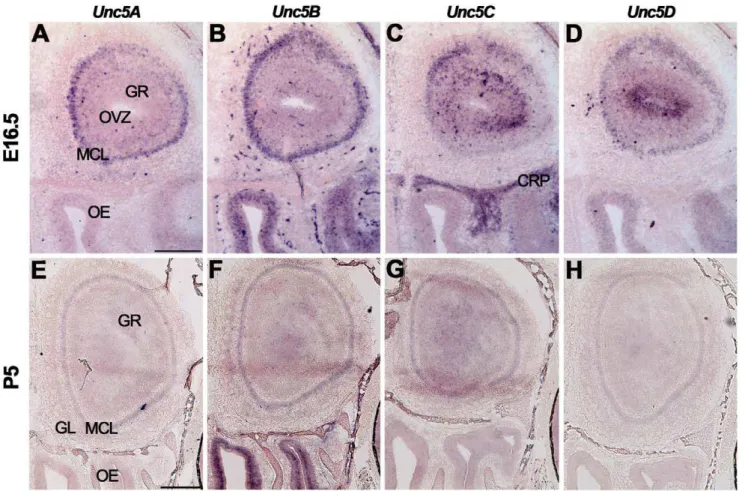
The expression ofUnc5A-Dwas studied usingin situ hybridiza-tion (Fig. 2, Table S2). At E16.5, expression of all fourUnc5family members was observed in the MCL of the olfactory bulb (Fig. 2A– D). Interestingly,Unc5C was only expressed by a small subset of dorsal mitral cells (Fig. 2C).Unc5B staining was observed in the OE and in blood vessels, in line with its proposed role in angiogenesis (Fig. 2B) [69].Unc5Cwas detected in the GR and the cribriform plate (Fig. 2C).Unc5D was strongly expressed in the olfactory ventricular zone and weakly in the OE and GR (Fig. 2D). At P5 and adult stages, expression levels ofUnc5A-Dwere reduced as compared to E16.5, but signals remained in the GL, MCL and GR (Fig. 2E–H, Table S2).
Cortex
The adult mammalian cerebral cortex consists of six layers comprised of morphologically and functionally distinct neurons. These layers are formed between E11 and E18, as cortical neurons undergo radial migration from the ventricular progenitor zone to their final position in the cortex [70]. The cortex is the origin of several major axon tracts in the forebrain, including the corticothalamic and corticospinal tracts. Cortical expression of
RGMa, RGMb and Neogenin has been reported and RGMa can inhibit the outgrowth of cortical axonsin vitro through Neogenin [13,15,18,19,22,67,71,72]. However, the precise role of RGMs and Neogenin during different stages of cortical development and maturationin vivois still incompletely understood.
At E16.5, expression of RGMa was restricted to the cortical plate (CP) and the ventricular zone (VZ) (Fig. 3A, A9, Table S3).
RGMbexpression was strongest in the pia, the upper part of the CP, and the subventricular zone (SVZ) (Fig. 3B, B9) [19].Neogenin
CP that gave rise to these projections. Interestingly, assessment of the cortical expression of Neogenin at E14.5 using an antibody directed against the C-terminal part of Neogenin revealed a different, more widespread pattern of expression including strong labeling of the ventricular zone [68,71,72]. It will therefore be interesting to determine whether this difference arises from the cleavage of Neogenin [61]. In line with previous observations, RGMa and Neogenin were also expressed on cells with the appearance of radial glia cells in the CP and in cells in the VZ (Fig. 3D9, F9) [50,72]. Strong binding of RGMa-AP was detected in the upper CP, in the striatum, and on axon bundles in the IZ and the IC (Fig. 1K, S3A, B). These observations are in line with the expression of Neogenin detected byin situ hybridization and immunohistochemistry in these brain areas (Fig. 3C, C9, F, F9, I). Sections incubated with isotype immunoglobulin controls did not show specific expression (Fig. 3J–L).
At P5,RGMawas expressed in layers 1–3, 5 and 6 of the cortex and expression of RGMb was confined to neurons of layer 5 displaying a medial high to lateral low gradient (Fig. 4A, A9, B, B9) [18].Neogeninwas expressed in layers 1–5 (Fig. 4C, C9, Table S3). Immunohistochemistry detected staining for RGMa in layers 1–3 and 5 (Fig. 4D). RGMb was virtually absent from the P5 cortex and Neogenin staining was most prominent in layers 1–3 (Fig. 4E, F). RGMa, RGMb and Neogenin were expressed on axons in the CC and the IC (data not shown). Strong immunostaining for RGMa and Neogenin was also detected in the corticospinal tract (CST), which is formed by cortical efferents from layer 5 cortical neurons (Fig. 4G–I). In the adult, expression of RGMa was detected in layers 5–6 and the VZ whileRGMbandNeogeninwere expressed throughout the cortex (Table S3). However, no specific signals for RGMa, RGMb or Neogenin were detected in the adult cortex by immunohistochemistry (data not shown).
Figure 1. RGM and Neogenin expression in the mouse olfactory system.In situhybridization on coronal mouse brain sections at E16.5 (A– C9) and P5 (L–N9). Panels A9–C9and L9–N9show higher magnifications of boxed areas in A–C and L–N, respectively. Immunohistochemistry (D–F, H–J) and RGMa-AP section binding (G, K) on E16.5 coronal mouse brain sections. Sections in D–F and H–J are counterstained in blue with fluorescent Nissl. (A–C9)In situhybridization shows differential expression patterns ofRGMa,RGMbandNeogeninin the olfactory bulb and olfactory epithelium (OE). In line with this, immunohistochemistry reveals that axons of olfactory sensory neurons in the OE stain strongly for RGMb and weakly for RGMa and Neogenin. Furthermore, RGMa, RGMb and Neogenin are expressed on olfactory bulb axon projections such as the lateral olfactory tract (LOT). a, apical; ACa, anterior commissure pars anterior; AOB, accessory olfactory bulb; b, basal; CP, cortical plate; CRP, cribriform plate; EPL, external plexiform layer; GL, glomerular layer; GR, granule cell layer; IPL, internal plexiform layer; IZ, intermediate zone; LV, lateral ventricle; MCL, mitral cell layer; ONL, olfactory nerve layer; OVZ, olfactory ventricular zone; S, septum; STR, striatum; VN, vomeronasal nerve. Scale bar A–C 200mm, A9–C9100mm, D–F
300mm, G 500mm, H–J 400mm, K 500mm, L–N 400mm and L9–N9200mm.
doi:10.1371/journal.pone.0055828.g001
Figure 2. Unc5 expression in the olfactory system.In situhybridization on coronal mouse brain sections at E16.5 (A–D) and P5 (E–H). AllUnc5s are differentially expressed in the olfactory bulb but the olfactory epithelium (OE) only expressesUnc5BandUnc5D. CRP, cribriform plate; GL, glomerular layer; GR, granule cell layer; MCL, mitral cell layer; OVZ, olfactory ventricular zone. Scale bar A–D 300mm and E–H 400mm.
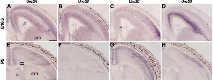
At E16.5, Unc5A-C were expressed in the CP, Unc5A and
Unc5C in the SP, andUnc5A-Din the SVZ and VZ (Fig. 5A–C, Table S3). Expression of Unc5D was especially strong in the SVZ (Fig. 5D). At P5 and in adulthood, Unc5A was expressed throughout the cortex, Unc5D in layers 1–4 and no signals for
Unc5B were detected [73] (Fig. 5E, F, H, Table S3).Unc5C was
expressed throughout the cortex at P5 but was restricted to layer 5 in the adult (Fig. 5G).
Hippocampus
The hippocampus is a multilayered structure with an essential role in learning and memory. It receives afferent projections from Figure 3. RGMa, RGMb and Neogenin display partially complementary patterns of expression in the developing cortex and on cortical projections.In situhybridization (A–C9) and immunohistochemistry (D–L) on coronal mouse brain sections at E16.5. Panels A9–C9and D9–F9 show higher magnifications of the boxed areas in A–C and D–F, respectively. Sections in D–L are counterstained in blue with fluorescent Nissl. (A–C9) In situhybridization reveals strong expression ofRGMaandNeogenin, and moderate expression ofRGMb, in the embryonic mouse cortex. Arrow in A indicates neurons of the lateral migratory stream. (D–I) RGMa and Neogenin protein are strongly expressed in the cortex and on various cortical axon projections. Strong staining for RGMb (E), and Neogenin (F), is detected in the corpus callosum (CC). The internal capsule (IC) stains strongly for RGMa (G). (J–L) Immunostaining with isotype-matched control antibodies did show significant staining. CP, cortical plate; FIM, fimbria; IZ, intermediate zone; LV, lateral ventricle; MZ, marginal zone; SP, subplate; STR, striatum; SVZ, subventricular zone; VZ, ventricular zone. Scale bar A–C 400mm, A9–C9
200mm, D–F 300mm, D9–F9150mm, G-I 300mm and J-M 300mm.
different regions in the brain. Axons from entorhinal cortex (CEn) neurons project via the perforant pathway to the molecular layer (ML) of the dentate gyrus (DG) and via the alvear pathway to the stratum lacunosum moleculare (SLM). Axons from the septum project to the stratum oriens (SO) and stratum radiatum (SR). Within the hippocampus, mossy fibers from DG granule cells synapse on cornu ammonis (CA)3 pyramidal neurons, while Schaffer collaterals from CA3 neurons project to the CA1 region [74].
At E16.5,RGMawas strongly expressed in the hippocampal VZ and specifically labeled neurons in the DG and CA layers (Fig. 6A, Table S4). Although weak to moderate RGMb expression was detected throughout the hippocampal formation, RGMb was prominently expressed in the pial layer lining the hippocampal fissure (Fig. 6B). Strong expression ofNeogeninwas observed in the DG, CA region and dentate migration stream. Lower signals were present in the SVZ and VZ (Fig. 6C) (for E18.5 patterns see [67]). Previous work has shown that pial RGMb expression directs the migration of Neogenin-positive granule cells in the dentate migratory stream [28,67]. Sections incubated with isotype immunoglobulin controls did not show specific expression (Fig. 6G–I). At E16.5,RGMa,RGMbandNeogeninwere expressed in the CEn and the septum (Table S4, S5). Immunohistochemistry detected RGMa in the hippocampal VZ, CA region, fimbria (FIM) and in the inner ML of the DG at E16.5 (Fig. 6D).In vitro
studies suggest that RGMa expression in the inner ML of the DG functions to restrict Neogenin-positive CEn axons to the outer ML [11]. Weak RGMb expression was detected in the CA region, DG and FIM. However, in line with the in situhybridization data, strong staining was detected in the pia (Fig. 6B, E). Neogenin was present in the outer ML of the DG, CA region, VZ and FIM
(Fig. 6F). In line with this pattern of expression, RGMa-AP bound to the DG, CA region and FIM (Fig. S3A, B).
Hippocampal expression ofRGMsandNeogeninpersisted at P5 (Fig. 6J–L, Table S4). In line with previous work, strongest expression ofRGMawas detected in CA1 neurons, whileRGMb
expression was most prominent in the CA2 and CA3 region (Fig. 6J, K) [11,18]. Neogenin was detected in CA2 and CA3 neurons and in a medial high to lateral low expression gradient in the CA1 region (Fig. 6L). Granule cells in the DG displayed weak expression ofRGMa and RGMb, and strong Neogenin expression (Fig. 6J–L). RGMa, RGMb and Neogenin were expressed in the polymorph layer (PO) of the DG (Fig. 6J–L). Immunohistochem-istry at P5 revealed expression of RGMa in the SO, SLM, FIM, ML and hippocampal commissure (Fig. 6M). Weak expression of RGMb was detected in the SLM, FIM and the outer ML of the DG (Fig. 6N). Neogenin was detected throughout the hippocam-pus, including in the FIM, hippocampal commissure, SLM, SO, SR and in the ML, granular layer and PO of the DG (Fig. 6O). Adult hippocampal expression of RGMa, RGMb and Neogenin
resembled that observed at P5. However,Neogeninexpression levels were decreased in the adult andRGMa,RGMbandNeogeninwere absent from the SR and ML (Table S4). Furthermore, it should be noted that another study failed to detectRGMaexpression in the adult hippocampus [19]. Immunohistochemistry in the adult hippocampus revealed weak expression of RGMa and RGMb in the SLM. Neogenin was prominently expressed in the PO and SR. Expression of RGMa, RGMb and Neogenin in the adult FIM was reduced as compared to P5 (data not shown).
Although Unc5A expression has been reported in hippocampal mossy fibers [75], the expression of Unc5 family members during hippocampal development is largely unknown. We detected Figure 4. Postnatal expression of RGMa, RGMb and Neogenin in the cortex and corticospinal tract.In situhybridization (A–C9) and immunohistochemistry (D–I) on coronal mouse brain sections at P5. Panels A9–C9show higher magnifications of boxed areas in A–C. Sections in D-I are counterstained in blue with fluorescent Nissl. (A–C9)In situhybridization detects strong expression ofRGMain cortical layers 1–3, 5 and 6.RGMbis mainly expressed in layer 5 in a medial to lateral gradient andNeogeninis expressed in layers 1–5. (D–F) RGMa protein is expressed in layers 1–3 and 5. Very weak staining is detected for RGMb and Neogenin is strongly expressed in cortical layers 1–3. (G–I) High levels of RGMa and Neogenin are detected in the corticospinal tract. AC, anterior commissure; CC, corpus callosum; LV, lateral ventricle; PD, pyramidal decussation; S, septum; STR, striatum. Scale bar A–C 600mm, A9–C9200mm, D–F 200mm and G–I 250mm.
doi:10.1371/journal.pone.0055828.g004
Figure 5. Unc5 expression in the cortex.In situhybridization on coronal mouse brain sections at E16.5 (A–D) and P5 (E–H). (A–D)Un5A-Care expressed in the cortical plate (CP), andUnc5AandUnc5C in the subplate (SP). AllUnc5s are expressed in the subventricular zone (SVZ) and ventricular zone (VZ). (E–H) At P5,Unc5A,Unc5CandUnc5Dare expressed in the cortex. CC, corpus callosum; LV, lateral ventricle; S, septum; STR, striatum. Scale bar A–D 300mm and E-H 600mm.
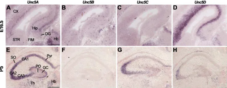
Unc5A-Dexpression in different regions of the E16.5 hippocampus with highest Unc5A-D expression in the CA region and DG (Fig. 7A–D, Table S4). Of the four Unc5 family members,Unc5A
displayed the most prominent expression in the developing hippocampus. At P5 and in the adult, Unc5A-D signals were detected in CA1-CA3 and the DG, though staining for Unc5B was not detected in the P5 hippocampus (Fig. 7E–H, Table S4).
Habenula
The habenula is part of the epithalamus and is subdivided into a medial (MHb) and lateral part (LHb). The MHb receives major inputs from the septum and the LHb receives afferents from the basal ganglia. The fasciculus retroflexus (FR) is the main output bundle of the Hb and carries LHb and MHb axons to the midbrain [76]. The expression and role of RGMs and Neogenin during the development of the habenula and its projections are unknown.
At E16.5,in situhybridization revealed strongRGMaexpression in the MHb and lower signals in the LHb. StrongRGMband weak
Neogenin expression was detected in the MHb and in the medial part of the LHb (Fig. 8A–F). In addition, strong expression for
RGMawas observed in the thalamus and forRGMbin the striatum (Fig. 8D, Table S5). Several of the synaptic targets of LHb and MHb axons in the FR expressed RGMa, RGMb and Neogenin, including the interpeduncular nucleus and the mesodiencephalic dopamine system (Table S5). Interestingly, the mesodiencephalic dopamine system not only receives habenular inputs but also projects axons to the LHb [77,78]. At E16.5,RGMawas expressed
in the ventral tegmental area (VTA) and substantia nigra (SN), which were identified bytyrosine hydroxylase(TH) labeling (Fig. 8M, N). Strong expression of RGMb was detected in the VTA and
Neogeninwas present in a subset of neurons in the VTA and SN (Fig. 8O, P). Immunohistochemistry revealed that RGMa expression was confined to the MHb while RGMb and Neogenin were expressed in the MHb and LHb (Fig. 8G–I). Furthermore, strong staining of the FR for RGMa and RGMb and only weak expression of Neogenin was detected (Fig. 8J–L). RGMb and Neogenin were also expressed in the stria medularis (SM) (Fig. 8H, I). The SM contains afferent projections to the Hb from different brain areas several of which display high levels of Neogenin and
RGMb (e.g. the septal nuclei and lateral hypothalamic region) (Table S5). RGMa-AP section binding detected moderate to weak binding to the FR and strong binding to the SM. Sections incubated with AP control did not exhibit specific staining (Fig. 8Q, R).
At P5 and in the adult, strongRGMaexpression was observed in the MHb andRGMbexpression was most prominent in the LHb and the lateral aspect of the MHb (Fig. 9A, B, G, H, Table S5) (see [18] for an equivalent pattern at P7). Only weak expression of
Neogeninwas detected (Fig. 9C, I). Expression ofRGMsandNeogenin
was also detected in the paraventricular thalamic nucleus, just ventral to the Hb (Fig. 9A–C, G–I). Immunohistochemistry revealed expression of RGMa and RGMb, and weak expression of Neogenin in the FR at P5 and in the adult (Fig. 9D–F, data not shown).
Figure 6. Subregion-specific expression of RGMs and Neogenin in the hippocampus. In situ hybridization (A–C, J–L) and immunohistochemistry (D–I, M–O) on coronal mouse brain sections at E16.5 (A–I) and P5 (J–O). Sections in D–I and M–O are counterstained in blue with fluorescent Nissl. (A–F) RGMa mRNA and protein are expressed in the ventricular zone (VZ), dentate gyrus (DG) and cornu ammonis (CA) region. Strong expression of RGMb mRNA and protein is detected in the pial surface lining the hippocampal fissure (HF). Neogenin transcripts and protein are widely expressed in the developing hippocampus (Hip). (G–I) Immunostaining with isotype matched controls. (J–L)In situhybridization at P5 shows strong but differential expression patterns ofRGMa,RGMbandNeogeninin the CA pyramidal cell layers (Pyr). In addition, strong expression ofNeogeninis detected in the granular layer (GC) of the DG. (M–O) Immunohistochemistry reveals expression of RGMa and weak expression of RGMb in the stratum lacunosum moleculare (SLM) and fimbria (FIM). Neogenin strongly labels different hippocampal layers. CX, cortex; Hb, habenula; HC, hippocampal commissure; PO, polymorph layer; SO, stratum oriens; SR, stratum radiatum; Th, thalamus. Scale bar A–C: 400mm, D–F: 300mm, G–I:
300mm, J–L: 500mm and M–O: 400mm.
doi:10.1371/journal.pone.0055828.g006
Figure 7. Unc5 expression in the hippocampus.In situhybridization on coronal mouse brain sections at E16.5 (A–D) and P5 (E–H). (A–D) Unc5A-Dare expressed in the E16.5 hippocampus (Hip) and dentate gyrus (DG). (E–H) At P5,Unc5AandUnc5Care expressed in cornu ammonis (CA) 1–3 and Unc5Dexpression is restricted to CA3. CX, cortex; FIM, fimbria; GC, granular layer; Hb, habenula; HC, hippocampal commissure; PO, polymorph layer; Pyr, pyramidal cell layers; SO, stratum oriens; SR, stratum radiatum; STR, striatum; Th, thalamus. Scale bar A–C: 400mm and D–F: 300mm.
In situ hybridization at E16.5 revealed strong expression of
Unc5A in the LHb and MHb (Fig. 10A, Table S5). Weak expression ofUnc5Bwas detected in the Hb, particularly staining blood vessels, and expression ofUn5CandUnc5Dwas confined to the LHb (Fig. 10B–D). At P5,Unc5Awas weakly expressed in the MHb and LHb and expression ofUnc5Dwas restricted to the LHb (Fig. 10E, H, Table S5). Expression ofUnc5BandUnc5Cwas not detected in the Hb (Fig. 10F, G). In adult, almost noUnc5staining
was observed in the Hb, except for very weak Unc5B labeling (Fig. 10I–L, Table S5).
Cerebellum
The cerebellum is located in the hindbrain and is associated with motor coordination and controlled movement. The adult cerebellum consists of three different layers: the molecular layer (ML), the Purkinje cell layer (PCL) and the granular cell layer (GCL). Interestingly, the neurons that occupy these layers derive Figure 8. Differential expression of RGMs and Neogenin in the habenula and its efferent and afferent projections.In situhybridization (A–F, M–P), immunohistochemistry (G–L) and RGMa-AP (Q) and AP (R) section binding on coronal (A–I, M–P) and sagital (J–L, Q–R) mouse brain sections at E16.5. Sections G–L are counterstained in blue with fluorescent Nissl. (A–L)In situ hybridization and immunostaining reveal strong expression of RGMa in the medial habenula (MHb) and strong expression of RGMb in the MHb and lateral habenula (LHb). Weak Neogenin expression is detected in the LHb and MHb. In line with this, strong RGMa and RGMb immunostaining is detected on the fasciculus retroflexus (FR), the major output bundle of the Hb. (M–P)In situhybridization fortyrosine hydroxylase(TH) stains dopaminergic neurons in the substantia nigra (SN) and ventral tegmental area (VTA).RGMaandNeogeninexpression is detected in the SN and VTA, whileRGMbis predominantly expressed in the VTA. (Q) RGMa-AP section binding shows strong staining of the stria medullaris (SM) and weak staining of the FR. (R) Section binding with AP control. CX, cortex; Hip, hippocampus; MB, midbrain; PC, posterior commissure; Th thalamus. Scale bars A–L: 400mm, M–P: 200mm and Q–R: 400mm.
doi:10.1371/journal.pone.0055828.g008
Figure 9. Postnatal RGM and Neogenin expression in the habenula and fasciculus retroflexus.In situhybridization (A–C, G–I) and immunohistochemistry (D–F) on coronal mouse brain sections at P5 (A–F) and in the adult (G–I). Sections D–F are counterstained in blue with fluorescent Nissl. (A–C, G–I) At P5 and in the adult,in situhybridization reveals strong expression ofRGMaandRGMbin the medial habenula (MHb). The lateral habenula (LHb) strongly expressesRGMb, while only weak expression of Neogenin is detected in Hb. (D–F) Immunohistochemistry detects strong expression of RGMa and RGMb and weak expression of Neogenin in the fasciculus retroflexus (FR). 3V, third ventricle; CX, cortex; SM, stria medullaris; PVT, paraventricular thalamic nucleus. Scale bars A–I: 200mm.
from two different progenitor zones. The cerebellar VZ gives rise to Purkinje cells (PCs), Bergmann glia (BG) and interneurons. Granule cells precursors (GCPs) are generated in the upper rhombic lip lining the fourth ventricle and migrate tangentially along the cerebellar surface to form the external granular layer (EGL). During the first two postnal weeks, cerebellar granule cells (CGCs) migrate from the EGL radially along BG in the ML to the internal granular cell layer (IGL) [79].
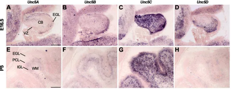
In situhybridization at E16.5 revealedRGMaexpression in the VZ of the cerebellum but not in the EGL (Fig. 11A, Table S6).
RGMbwas expressed in a subset of cells in the inner part of the EGL andNeogeninstaining was detected in the VZ and throughout the EGL (Fig. 11B, C) [18,19,23]. ThisRGMexpression profile is in line with previously reported expression patterns at E14.5 [18]. Interestingly, however, at E18.5 cerebellar RGMb signals are already much more restricted as compared to the expression observed at E16.5 (Fig. 11B) [19]. Furthermore, expression of
Neogeninin the EGL has been reported previously at E13 and is in line with RGMa-AP binding patterns (Fig. S3C), but could not be detected at E18.5 [23,67]. RGMa, RGMb and Neogenin were expressed in the PCL and deep cerebellar nuclei (DCN) (Fig. 11A– C). In line with this expression, immunostaining revealed expression of RGMa, RGMb and Neogenin in the DCN (Fig. 11D–F). RGMa was also expressed in cellular processes traversing the EGL (Fig. 11D). In line with thein situhybridization data, RGMb was confined to the inner part of the EGL (Fig. 11E). Neogenin expression was detected throughout the EGL and in a patch of GCPs in the EGL where the presumptive rhombic lip is located (Fig. 11F) [23].
At P5, RGMa was confined to the IGL (Fig. 11G, Table S6).
RGMbwas detected in the inner EGL, PCL and at lower levels in the IGL (Fig. 11H).Neogenin was strongly expressed in the outer part of the EGL, in the PCL and IGL (Fig. 11I). Immunohisto-chemistry showed staining for RGMa, RGMb and Neogenin in the EGL (Fig. 11J–L). Furthermore, RGMa, RGMb and Neogenin were expressed in the PCL and IGL including the white matter tracts in this structure. However, since no RGMa
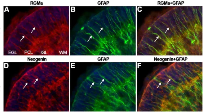
expression was detected in the PCL (Fig. 11G) this labeling may represent expression on migrating cerebellar granule neurons or BG. To determine whether RGMs and Neogenin are expressed on the radial processes of BG, co-immunostaining for glial fibrillary acidic protein (GFAP), a BG marker, and RGMa or Neogenin was performed. Both RGMa and Neogenin expression colocalized with the GFAP-positive BG fibers in the EGL (Fig. 12). In addition, cerebellar granule neurons along BG processes expressed Neogenin.
In the adult,in situhybridization revealed expression ofRGMa,
RGMbandNeogeninin the PCL and GCL (Fig. 11M–O, Table S6). It should be noted that other work does not detect RGMb in the adult cerebellum [19]. Immunohistochemistry revealed staining for RGMa, RGMb and Neogenin in the GCL, PCs and the ML. Staining in the ML may represent the dendritic processes of PCs (Fig. 11P–R).
Expression of Unc5A-D in the cerebellum has been reported in embryonic and postnatal stages and in the adult [64,80–86].
Unc5C knockout mice display severe defects in cerebellar development, including disturbed GCP migration and ectopically located PCs [80,83,84,87]. In line with these observations, Unc5A-Cbut notUnc5Dwere expressed in the EGL at E16.5 (Fig. 13A–D, Figure 10. Unc5 expression in the habenula.In situhybridization on coronal mouse brain sections at E16.5 (A–D), P5 (E–H) and adult (I–L). (A–D) At E16.5insitu hybridization shows expression ofUnc5A-Din the habenula (Hb).Unc5Bexpression labels blood vessels but not habenular neurons. (E–H) At P5,Unc5Ais weakly expressed in the Hb andUnc5Dexpression is restricted to the lateral habenula (LHb). (I–L) In the adult Hb, weakUnc5B expression is detected. 3V, third ventricle; CX, cortex; MHb, medial habenula; PVT paraventricular thalamic nucleus. Scale bar A–D: 300mm and E–L:
200mm.
Table S6). At P5,Unc5Awas restricted to the IGL, whileUnc5B
andUnc5Cwere detected in the EGL, PCL and IGL (Fig. 13E–G). Weak expression of Unc5D was only detected in the PCL (Fig. 13H). In adult Un5A-C were expressed in the PCL and IGL, while expression ofUnc5Dis only detected in the PCL (Table S6).
Discussion
Since their original identification in 2002, RGMs have been implicated in several different aspects of neural development. A large part of this work has focused on the important roles of RGMa and its receptor Neogenin during axon pathfinding in the chick retinotectal system [6,8–10,52]. However, experiments in
Xenopusembryos and on cultured rodent neuronal tissues have also highlighted more widespread roles for RGM-Neogenin signaling in axon guidance [11–17,20]. In addition, it has become clear that RGMs are pleiotropic and can regulate processes such as neurogenesis, differentiation, migration, and apoptosis [8,28,29,31,32,49–51,88]. Despite this progress, the precise role of RGMs and Neogenin in the development of many neuronal systems remains unknown, especially in the mouse. To begin to provide further insight into the possible roles of RGM-Neogenin signaling during mouse brain development, we performed a comparative analysis of the expression of RGMa, RGMb and
Neogenin transcript and protein at different developmental stages. Unc5s were included in this analysis as they are obligate co-receptors for the axon repulsive effects of RGMs [15].
Neurogenesis, Differentiation and Migration
Previous work has shown expression of RGMs and Neogenin in the proliferative zones of different brain structures [11,18,28,50,51,67,72,89]. Our data confirm and extend these findings and reveal expression of Neogenin, RGMs and Unc5s in the progenitor regions of the olfactory epithelium, olfactory bulb, cortex, hippocampus and cerebellum. For example, strong Neogenin expression was detected in the upper part of the cerebellar EGL, which contains progenitors for CGCs [90,91], and in the VZ of the olfactory bulb (Fig. 1C). Of the two RGMs expressed in the brain, RGMa was especially prominent in progenitor regions. On the other hand, expression of RGMb often appeared to mark regions containing differentiating cells imme-diately adjacent to the progenitor zones. For example, RGMb expression was detected in the SVZ of the cortex while RGMa was expressed in the adjacent VZ (Fig. 3A9, B9) [18,19]. Together these observations support widespread roles for RGMa and RGMb in neurogenesis and neuronal differentiation.
The moment that neuroblasts start to differentiate often coincides with their migration towards their target areas. The first indication that Neogenin regulates cell migration comes from Figure 11. Differential expression of RGMs and Neogenin in the cerebellum. In situ hybridization (A–C, G–I, M–O) and immunohistochemistry (D–F, J–L, P–R) on coronal mouse brain sections at E16.5 (A–F), P5 (G–L) and in the adult (M–R). (A–I, M–O) In situ hybridization and immunohistochemistry reveals strong and broad expression of RGMa, RGMb and Neogenin in the cerebellum (CB). (J–L) Immunostaining shows expression of RGMa and Neogenin in all cerebellar layers at P5. RGMb is expressed in the internal granular layer (IGL), Purkinje cell layer (PCL) and external granular layer (EGL). (P–R) In the adult, RGMa, RGMb and Neogenin protein are expressed in Purkinje cells (PCs) and axons in the granular cell layer (GCL), PCL and molecular layer (ML). Neogenin strongly labels PC dendrites in the ML. DCN, deep cerebellar nuclei; WM, white matter; VZ, ventricular zone. Scale bar A–C: 300mm, D–F: 250mm, G–I: 150mm, J–L: 150mm, M–O: 100mm and P–R: 50mm.
doi:10.1371/journal.pone.0055828.g011
Figure 12. RGMa and Neogenin are expressed on Bergmann glia.Immunohistochemistry for RGMa (A, C), glial fibrillary acidic protein (GFAP) (B–C, E–F) and Neogenin (D, F) on coronal mouse brain sections at P5 visualized by confocal microscopy. Sections are counterstained in blue with fluorescent Nissl. (A–F) RGMa and Neogenin immunostaining (in red) colocalizes with GFAP-positive staining (in green) on Bergmann glial fibers (arrows). Granule cells in the external granular layer (EGL) also express Neogenin. IGL, internal granular layer; PCL, Purkinje cell layer; WM, white matter.
the analysis of morpholino-induced Neogenin knockdown in zebrafish, which show defects in neural tube formation and somitogenesis [92]. It is clear, however, that RGM-Neogenin signaling also plays a crucial role in cell migration at later developmental stages. This is nicely illustrated by the ability of RGMb, expressed along the hippocampal fissure, to guide Neogenin-positive DG granule cells though the DG migratory stream towards the hippocampus [28]. Such a role for RGMs and Neogenin is likely to be more general. For example, we, and others, report Neogenin expression on radially migrating neurons in the cortex and young interneurons tangentially migrating from the ganglionic eminence (GE) [51,93]. Furthermore, Neogenin is expressed in migrating olfactory interneuron precursors in the rostral migratory stream [50,94]. The role of RGM-Neogenin signaling in these populations of migrating neurons is largely unknown. Repulsive signaling induced by RGMs may guide cortical interneurons during their migration from the GE to the cortex. A recent study shows that overexpression of Neogenin in the GE leads to a failure of interneurons to migrate out of the GE [93]. This migrational defect may be caused by an increased response of Neogenin-overexpressing cells to the attractive effects of Netrin-1, which is expressed in the GE [93]. Alternatively, however, increased Neogenin levels may enhance the responsive-ness of interneurons to repulsive cues around the GE. An interesting candidate is RGMb, which is expressed in the striatum around the time of interneuron migration (Fig. 3B). Overexpres-sion of Neogenin in interneurons may render these cells more sensitive to the repulsive effects of RGMb thereby confining them to the GE.
Another region where Neogenin and RGMs may regulate cell migration is the cerebellum. GCPs are generated in the rhombic lip and then migrate to the surface of the cerebellum to form the EGL. From this outer part of the EGL CGCs change their mode of migration from tangential to radial and migrate along radial glial projections towards the IGL. Strong Neogenin expression is detected in radially migrating CGCs in the outer EGL (Fig. 11I). In contrast,Neogeninis absent from CGCs in the inner EGL, where CGCs switch from tangential to radial migration. Interestingly,
strong expression of RGMb is detected in the inner EGL complementary to Neogenin expression in the outer EGL (Fig. 11H). It is therefore tempting to speculate that RGMb-Neogenin signaling is involved in the switch from tangential to radial migration. Of the Unc5 family members, Unc5C has been shown to have an important role in cerebellar development.Unc5C
is strongly expressed in the developing cerebellum [86] and mutations in theUnc5Cgene result in severe defects in cerebellar development, including a reduction of cerebellar size, abnormal cerebellar foliation and ectopic localization of PCs and CGCs [80,83,84,87]. Analysis of the Unc5C mutant mice reveals that Unc5C regulates migration of GCPs along the caudo-rostral and dorso-ventral axes. GCPs expressing mutant Unc5C invade the superior colliculus and brain stem [80,83]. A possible explanation for these migrational defects is the inability of migrating GCPs to respond to repellent guidance cues in the environment. In line with this hypothesis, RGMs are expressed in the superior colliculus and in the VZ of the cerebellum [18,19,52] and are therefore in the appropriate location to repel Neogenin- and Unc5C-express-ing migratUnc5C-express-ing GCPs and restrict their migration to ‘future cerebellar brain areas’. Further work is needed to examine whether or how interactions between RGMs, Neogenin and Unc5s regulate cerebellar development.
Axon Guidance and Axon Tract Development
RGMa- and RGMb-Neogenin signaling mediates neurite outgrowth inhibition of cultured chick retinal ganglion and mouse cortical, entorhinal cortical, cerebellar granule and dorsal root ganglia (DRG) neurons [6,9,11,12,14,15,17]. However, our understanding of the role of these neurite outgrowth inhibitory effects in regulating axon guidance events in vivo is far from complete. Our study together with previous work shows that RGMs and Neogenin are abundantly expressed throughout the developing mouse brain [18,19,67], indicating a potential role in regulating axon guidance events in different brain areas. Neogenin expression was detected in many different axonal tracts in the brain, including the LOT, cortical efferents, the ACa and CST, and axonal tracts in the hippocampus (fimbria) and cerebellum. Figure 13. Dynamic expression of Unc5 in the developing cerebellum.In situhybridization on coronal mouse brain sections at E16.5 (A–D) and P5 (E–H). (A–D) Expression ofUnc5A-Dis detected in the cerebellum (CB), although expression of Unc5Cis most prominent. Unc5A-Care expressed in the external granular layer (EGL). (E–H) At P5,Unc5BandUnc5Care expressed in the EGL, Purkinje cell layer (PCL) and internal granular layer (IGL). Expression ofUnc5Bin the EGL is restricted to the inner cell layers. Expression ofUnc5Ais only detected in the IGL and expression ofUnc5D is restricted to the PCL. VZ, ventricular zone; WM, white matter. Scale bar A–D: 300mm and E–H: 150mm.
Interestingly, many axon tracts also showed staining for RGMa and RGMb. The LOT, ACa, IC, CST, FR and axonal tracts in the hippocampus and cerebellum were strongly stained for RGMa. The LOT and FR also expressed RGMb and strong expression of RGMb was detected in OSN axons and the CC.
Axonal expression of RGMs may aid in organizing axon bundles into sub-bundles or in creating exclusion zones for axons. For example, clear differential expression of RGMs and Neogenin was found on axon bundles in the IC. In the IC, expression of RGMa is predominant in the core, while Neogenin expression is detected in axonal tracts in the outer regions of the IC (Fig. 3G, I). This suggests that axonally expressed RGMa may instruct Neogenin-positive axons to grow in the outer parts of the IC. In addition, axonal RGM expression may mediate the adhesion of axons into tight bundles. Evidence for a possible role for RGMb as an adhesive cue for DRG axons comes from a coculture assay of Neogenin- and RGMb-positive DRG neurons and HEK-293 cells transiently expressing RGMb. In this assay, DRG neurites make contacts with RGMb-expressing HEK293 cells [95]. Furthermore, RGMb enhances the adhesion of dissociated cultured DRG neurons to HEK293 cells transiently expressing RGMb. RGMa can also exert adhesive effects through Neogenin, for example during neural tube closure [30,52,88,92]. Further experiments are needed to confirm the role of these potential homophilic and heterophilic RGM interactions in axon tract developmentin vivo.
Neuronal RGM Receptor Complexes
The inhibitory effects of RGMs on neurite outgrowth depend on a multimeric receptor complex containing Neogenin and Unc5s. Although Unc5B has been shown to be required for these effects at the functional level, Neogenin can bind all Unc5 family members [15]. This suggests that Neogenin may interact with different Unc5s in different systems, cellular processes and/or at developmental stages. Our comparative analysis of Unc5A-D co-receptors during brain development revealed prominent and differential expression patterns at all developmental stages studied. At E16.5 multiple brain regions were identified that displayed expression of allUnc5s, including the olfactory bulb, hippocampus and hypothalamus (see Tables S2, S3, S4, S5, S6). However, even within these regions expression ofUnc5swas often highly specific and confined to specific substructures. For example in the VZ of the E16.5 olfactory bulb, prominent expression of Unc5D and Neogenin was detected, while expression ofUnc5A-Cwas absent (Fig. 1C, F, 2A–D, Table S2). This invites the speculation that a Neogenin/Unc5D receptor complex may play a role in neuronal cell proliferation and neurogenesis in the olfactory bulb. In the habenula, we detected very specific expression of Unc5A in the MHb, while the LHb expressedUnc5A,Unc5CandUnc5Dat E16.5 (Fig. 10A–D, Table S5). This indicates differential roles for Unc5s in the development of MHb and LHb neurons. Although binding of all Unc5 molecules to Neogenin has been shown, it is currently not known whether binding of different Unc5s results in different functional outcomes. Future work will focus on examining whether different Unc5 proteins serve as functional co-receptors for RGM receptors in different brain regions and during different develop-mental processes.
Conclusion
This study presents a comparative analysis of the expression of RGMa, RGMb, Neogenin and Unc5A-D in the mouse brain at three key developmental stages (E16.5, P5 and adult). The observed expression patterns support a widespread, and largely unexplored, role for RGMa/b and their Neogenin/Unc5 receptor complex in neuron proliferation, migration and axon guidance in
the mouse brain. Interestingly, RGMs may function both as exogenous cues that are detected by cells or neurites or as axon-derived factors involved in axon tract development. Analysis of Unc5 expression patterns suggests that the composition of the RGM receptor complex, i.e. which Unc5 family member it contains, may differ between different brain regions and/or cellular processes. In all, these data serve as a valuable framework for the further dissection of the role of RGMs during mouse neural development.
Supporting Information
Figure S1 No specific staining for sense probes. In situ
hybridization on coronal mouse brain sections at E16.5 (A–B), P5 (C–D) and in adulthood (E–F) using RGMa sense probes. No specific in situ hybridization signals were detected at any of the timepoints or in any of the brain regions examined. Sections hybridized with sense probes for RGMb, Neogenin, and Unc5A-D
displayed similar levels of background labeling (not shown). 3V, third ventricle; CP, cortical plate; CX, cortex; GCL, granular cell layer; Hb, habenula; Hip, hippocampus; IZ, intermediate zone; LHb, lateral habenula; MHb, medial habenula; ML, molecular layer; PVT, paraventricular thalamic nucleus STR, striatium; VZ, ventricular zone. Scale bar A: 400mm, B: 200mm, C: 700, D: 400mm, E: 200mm and F: 400mm.
(TIF)
Figure S2 Specific immunostaining for anti-RGMa, anti-RGMb and anti-Neogenin antibodies. COS-7 cells overexpressing RGMa (A, D), RGMb (B, E), GFP-Neogenin (G, J), GFP-DCC (H, K), pcDNA3.1 empty vector (C, F) or pEGFP-N1 (I, L). Cells are counterstained with DAPI in blue. Anti-RGMa and anti-RGMb antibodies specifically stain COS-7 cells overex-pressing RGMa (A–C) or RGMb (D–F), respectively. Anti-Neogenin antibody specifically stains COS-7 cells overexpressing GFP-Neogenin and does not stain COS-7 cells overexpressing GFP-DCC or pEGFP-N1 (G–L). Scale bar A-L: 200mm. (TIF)
Figure S3 RGMa-AP binding to E16.5 mouse brain slices.(A) RGMa-AP binding is detected in cells and neuronal projections in the cortical plate (CP) and intermediate zone (IZ) of the cortex. (B) The fimbria (FIM) of the hippocampus (Hip) and axonal projections in the internal capsule (IC) also bind RGMa-AP. In the hindbrain, the pontine nucleus (PN) and cerebellum (CB), in particular the external granular layer (EGL), are strongly stained for RGMa-AP. Scale bars A–C: 400mm. CA, cornus ammonis; DG, dentate gyrus; Hb, habenula; STR, striatum; Th, thalamus; VZ ventricular zone.
(TIF)
Table S1 Sense and antisense primer sequences for
RGMa, RGMb, Neo and Unc5A-D in situ hybridization probes.
(DOCX)
Table S2 Expression of RGMa, RGMb, Neogenin and
Unc5A-Din the primary olfactory system.
(DOCX)
Table S3 Expression of RGMa, RGMb, Neogenin and
Unc5A-Din the cortex.
(DOCX)
Table S4 Expression of RGMa, RGMb, Neogenin and
Unc5A-Din the hippocampus and entorhinal cortex.
Table S5 Expression of RGMa, RGMb, Neogenin and
Unc5A-Din the habenula, septum and thalamic area.
(DOCX)
Table S6 Expression of RGMa, RGMb, Neogenin and
Unc5A-Din the cerebellum.
(DOCX)
Acknowledgments
We are grateful to Silvia Arber, Valerie Castellani, Jean-Franc¸ois Cloutier, Roman Giger and Thomas Skutella for sharing cDNA constructs. We
thank Sharon Kolk, Roos Winter and Ewoud Schmidt and other members of the Pasterkamp lab for help and fruitful discussions.
Author Contributions
Conceived and designed the experiments: DMVDH RJP. Performed the experiments: DMVDH AJCGMH. Analyzed the data: DMVDH AJCGMH RJP. Wrote the paper: DMVDH RJP.
References
1. De Vries M, Cooper HM (2008) Emerging roles for neogenin and its ligands in CNS development. J Neurochem 106: 1483–1492.
2. Key B, Lah GJ (2012) Repulsive guidance molecule A (RGMa): A molecule for all seasons. Cell Adh Migr 6: 85–90. 20167.
3. Matsunaga E, Chedotal A (2004) Repulsive guidance molecule/neogenin: a novel ligand-receptor system playing multiple roles in neural development. Dev Growth Differ 46: 481–486.
4. Severyn CJ, Shinde U, Rotwein P (2009) Molecular biology, genetics and biochemistry of the repulsive guidance molecule family. Biochem J 422: 393– 403.
5. Yamashita T, Mueller BK, Hata K (2007) Neogenin and repulsive guidance molecule signaling in the central nervous system. Curr Opin Neurobiol 17: 29– 34.
6. Monnier PP, Sierra A, Macchi P, Deitinghoff L, Andersen JS, et al. (2002) RGM is a repulsive guidance molecule for retinal axons. Nature 419: 392–395. 7. Stahl B, Muller B, von Boxberg Y, Cox EC, Bonhoeffer F (1990) Biochemical
characterization of a putative axonal guidance molecule of the chick visual system. Neuron 5: 735–743.
8. Matsunaga E, Nakamura H, Chedotal A (2006) Repulsive guidance molecule plays multiple roles in neuronal differentiation and axon guidance. J Neurosci 26: 6082–6088.
9. Rajagopalan S, Deitinghoff L, Davis D, Conrad S, Skutella T, et al. (2004) Neogenin mediates the action of repulsive guidance molecule. Nat Cell Biol 6: 756–762.
10. Tassew NG, Chestopolava L, Beecroft R, Matsunaga E, Teng H, et al. (2008) Intraretinal RGMa is involved in retino-tectal mapping. Mol Cell Neurosci 37: 761–769.
11. Brinks H, Conrad S, Vogt J, Oldekamp J, Sierra A, et al. (2004) The repulsive guidance molecule RGMa is involved in the formation of afferent connections in the dentate gyrus. J Neurosci 24: 3862–3869.
12. Conrad S, Genth H, Hofmann F, Just I, Skutella T (2007) Neogenin-RGMa signaling at the growth cone is bone morphogenetic protein-independent and involves RhoA, ROCK, and PKC. J Biol Chem 282: 16423–16433. 13. Endo M, Yamashita T (2009) Inactivation of Ras by p120GAP via focal
adhesion kinase dephosphorylation mediates RGMa-induced growth cone collapse. J Neurosci 29: 6649–6662.
14. Hata K, Fujitani M, Yasuda Y, Doya H, Saito T, et al. (2006) RGMa inhibition promotes axonal growth and recovery after spinal cord injury. J Cell Biol 173: 47–58.
15. Hata K, Kaibuchi K, Inagaki S, Yamashita T (2009) Unc5B associates with LARG to mediate the action of repulsive guidance molecule. J Cell Biol 184: 737–750.
16. Lah GJ, Key B (2012) Dual roles of the chemorepellent axon guidance molecule RGMa in establishing pioneering axon tracts and neural fate decisions in embryonic vertebrate forebrain. Dev Neurobiol 72: 1458–1470.
17. Liu X, Hashimoto M, Horii H, Yamaguchi A, Naito K, et al. (2009) Repulsive guidance molecule b inhibits neurite growth and is increased after spinal cord injury. Biochem Biophys Res Commun 382: 795–800.
18. Oldekamp J, Kramer N, Alvarez-Bolado G, Skutella T (2004) Expression pattern of the repulsive guidance molecules RGM A, B and C during mouse development. Gene Expr Patterns 4: 283–288.
19. Schmidtmer J, Engelkamp D (2004) Isolation and expression pattern of three mouse homologues of chick Rgm. Gene Expr Patterns 4: 105–110. 20. Wilson NH, Key B (2006) Neogenin interacts with RGMa and netrin-1 to guide
axons within the embryonic vertebrate forebrain. Dev Biol 296: 485–498. 21. Metzger M, Conrad S, Skutella T, Just L (2007) RGMa inhibits neurite
outgrowth of neuronal progenitors from murine enteric nervous system via the neogenin receptor in vitro. J Neurochem 103: 2665–2678.
22. Yoshida J, Kubo T, Yamashita T (2008) Inhibition of branching and spine maturation by repulsive guidance molecule in cultured cortical neurons. Biochem Biophys Res Commun 372: 725–729.
23. Vielmetter J, Kayyem JF, Roman JM, Dreyer WJ (1994) Neogenin, an avian cell surface protein expressed during terminal neuronal differentiation, is closely related to the human tumor suppressor molecule deleted in colorectal cancer. J Cell Biol 127: 2009–2020.
24. Kang JS, Yi MJ, Zhang W, Feinleib JL, Cole F, et al. (2004) Netrins and neogenin promote myotube formation. J Cell Biol 167: 493–504.
25. Lejmi E, Leconte L, Pedron-Mazoyer S, Ropert S, Raoul W, et al. (2008) Netrin-4 inhibits angiogenesis via binding to neogenin and recruitment of Unc5B. Proc Natl Acad Sci U S A 105: 12491–12496.
26. Park KW, Crouse D, Lee M, Karnik SK, Sorensen LK, et al. (2004) The axonal attractant Netrin-1 is an angiogenic factor. Proc Natl Acad Sci U S A 101: 16210–16215.
27. Srinivasan K, Strickland P, Valdes A, Shin GC, Hinck L (2003) Netrin-1/ neogenin interaction stabilizes multipotent progenitor cap cells during mammary gland morphogenesis. Dev Cell 4: 371–382.
28. Conrad S, Stimpfle F, Montazeri S, Oldekamp J, Seid K, et al. (2010) RGMb controls aggregation and migration of Neogenin-positive cells in vitro and in vivo. Mol Cell Neurosci 43: 222–231.
29. Gessert S, Maurus D, Kuhl M (2008) Repulsive guidance molecule A (RGM A) and its receptor neogenin during neural and neural crest cell development of Xenopus laevis. Biol Cell 100: 659–673.
30. Kee N, Wilson N, De Vries M, Bradford D, Key B, et al. (2008) Neogenin and RGMa control neural tube closure and neuroepithelial morphology by regulating cell polarity. J Neurosci 28: 12643–12653.
31. Matsunaga E, Tauszig-Delamasure S, Monnier PP, Mueller BK, Strittmatter SM, et al. (2004) RGM and its receptor neogenin regulate neuronal survival. Nat Cell Biol 6: 749–755.
32. Shin GJ, Wilson NH (2008) Overexpression of repulsive guidance molecule (RGM) a induces cell death through Neogenin in early vertebrate development. J Mol Histol 39: 105–113.
33. Zhang AS, West AP, Jr., Wyman AE, Bjorkman PJ, Enns CA (2005) Interaction of hemojuvelin with neogenin results in iron accumulation in human embryonic kidney 293 cells. J Biol Chem 280: 33885–33894.
34. Babitt JL, Zhang Y, Samad TA, Xia Y, Tang J, et al. (2005) Repulsive guidance molecule (RGMa), a DRAGON homologue, is a bone morphogenetic protein co-receptor. J Biol Chem 280: 29820–29827.
35. Babitt JL, Huang FW, Wrighting DM, Xia Y, Sidis Y, et al. (2006) Bone morphogenetic protein signaling by hemojuvelin regulates hepcidin expression. Nat Genet 38: 531–539.
36. Corradini E, Babitt JL, Lin HY (2009) The RGM/DRAGON family of BMP co-receptors. Cytokine Growth Factor Rev 20: 389–398.
37. Hagihara M, Endo M, Hata K, Higuchi C, Takaoka K, et al. (2011) Neogenin, a receptor for bone morphogenetic proteins. J Biol Chem 286: 5157–5165. 38. Kuns-Hashimoto R, Kuninger D, Nili M, Rotwein P (2008) Selective binding of
RGMc/hemojuvelin, a key protein in systemic iron metabolism, to BMP-2 and neogenin. Am J Physiol Cell Physiol 294: C994–C1003.
39. Samad TA, Rebbapragada A, Bell E, Zhang Y, Sidis Y, et al. (2005) DRAGON, a bone morphogenetic protein co-receptor. J Biol Chem 280: 14122–14129. 40. Xia Y, Yu PB, Sidis Y, Beppu H, Bloch KD, et al. (2007) Repulsive guidance
molecule RGMa alters utilization of bone morphogenetic protein (BMP) type II receptors by BMP2 and BMP4. J Biol Chem 282: 18129–18140.
41. Xia Y, Babitt JL, Bouley R, Zhang Y, Da Silva N, et al. (2010) Dragon enhances BMP signaling and increases transepithelial resistance in kidney epithelial cells. J Am Soc Nephrol 21: 666–677.
42. Zhang AS, Yang F, Wang J, Tsukamoto H, Enns CA (2009) Hemojuvelin-neogenin interaction is required for bone morphogenic protein-4-induced hepcidin expression. J Biol Chem 284: 22580–22589.
43. Zhou Z, Xie J, Lee D, Liu Y, Jung J, et al. (2010) Neogenin regulation of BMP-induced canonical Smad signaling and endochondral bone formation. Dev Cell 19: 90–102.
44. Halbrooks PJ, Ding R, Wozney JM, Bain G (2007) Role of RGM coreceptors in bone morphogenetic protein signaling. J Mol Signal 2: 4.
45. Kanomata K, Kokabu S, Nojima J, Fukuda T, Katagiri T (2009) DRAGON, a GPI-anchored membrane protein, inhibits BMP signaling in C2C12 myoblasts. Genes Cells 14: 695–702.