Gas6 Promotes Oligodendrogenesis
and Myelination in the Adult
Central Nervous System and After
Lysolecithin-Induced Demyelination
Salman Goudarzi
1, Andrea Rivera
1, Arthur M. Butt
1, and Sassan Hafizi
1Abstract
A key aim of therapy for multiple sclerosis (MS) is to promote the regeneration of oligodendrocytes and remyelination in the central nervous system (CNS). The present study provides evidence that the vitamin K-dependent protein growth arrest specific 6 (Gas6) promotes such repair in in vitro cultures of mouse optic nerve and cerebellum. We first determined expression of Gas6 and TAM (Tyro3, Axl, Mer) receptors in the mouse CNS, with all three TAM receptors increasing in expression through postnatal development, reaching maximal levels in the adult. Treatment of cultured mouse optic nerves with Gas6 resulted in significant increases in oligodendrocyte numbers as well as expression of myelin basic protein (MBP). Gas6 stimulation also resulted in activation of STAT3 in optic nerves as well as downregulation of multiple genes involved in MS development, including matrix metalloproteinase-9 (MMP9), which may decrease the integrity of the blood–brain barrier and is found upregulated in MS lesions. The cytoprotective effects of Gas6 were examined inin vitromouse cerebellar slice cultures, where lysolecithin was used to induce demyelination. Cotreatment of cerebellar slices with Gas6 significantly attenuated demyelination as determined by MBP immunostaining, and Gas6 activated Tyro3 receptor through its phosphor-ylation. In conclusion, these results demonstrate that Gas6/TAM signaling stimulates the generation of oligodendrocytes and increased myelin production via Tyro3 receptor in the adult CNS, including repair after demyelinating injury. Furthermore, the effects of Gas6 on STAT3 signaling and matrixMMP9downregulation indicate potential glial cell repair and immunor-egulatory roles for Gas6, indicating that Gas6-TAM signaling could be a potential therapeutic target in MS and other neuropathologies.
Keywords
multiple sclerosis, myelin, oligodendrocyte, receptor tyrosine kinase, TAM receptors, Tyro3
Received March 24, 2016; Received revised July 13, 2016; Accepted for publication August 11, 2016
Introduction
Multiple sclerosis (MS) is an autoimmune disease of the central nervous system (CNS) that involves destruction of oligodendrocytes and of the myelin sheath they produce around axons. MS features an inflammatory response that causes gradual demyelination through formation of plaques or lesions in the CNS white matter, leading to disruption in nerve impulse transmission (Saxena et al., 2011). Remyelination occurs in MS by the recruitment of oligodendrocyte progenitor cells (OPCs) to the lesion site and their subsequent differentiation into myelinating oligodendrocytes, under the control of multiple extracel-lular and axon-derived factors (Wolswijk, 2002). However, remyelination by OPCs ultimately fails, due
in part to their differentiation being suppressed by inhibi-tory factors and the lack of a supportive cellular envir-onment (Franklin, 2002; Kuhlmann et al., 2008). Hence, there is a need for a greater understanding of the factors regulating OPC differentiation and the development of novel targets for promoting remyelination.
Growth arrest specific 6 (Gas6) is a vitamin K-depen-dent protein that is a ligand for the TAM (Tyro3, Axl,
1School of Pharmacy and Biomedical Sciences, Institute of Biomedical and
Biomolecular Science, University of Portsmouth, UK
Corresponding Author:
Sassan Hafizi, University of Portsmouth, St. Michael’s Building, White Swan Road, Portsmouth PO1 2UP, UK.
Email: sassan.hafizi@port.ac.uk
ASN Neuro
September-October 2016: 1–14
!The Author(s) 2016 DOI: 10.1177/1759091416668430 asn.sagepub.com
Creative Commons CC-BY: This article is distributed under the terms of the Creative Commons Attribution 3.0 License
Mer) family of receptor tyrosine kinases (RTKs). The Gas6–TAM interaction is involved in a number of cellu-lar processes including regulating cell survival, the immune system, inflammation, proliferation, migration, and removal of apoptotic cells and debris (Lu and Lemke, 2001; Hafizi and Dahlba¨ck, 2006a; Weinger et al., 2011), all of which are processes involved in MS. The TAMs appear to play a significant role in immune system regulation and macrophage activation, as TAM triple knockout mice feature a severe lymphoproliferative disorder accompanied by broad-spectrum autoimmunity (Lu and Lemke, 2001; Lemke and Lu, 2003). Gas6-TAM signaling also plays a role in oligodendrocyte survival (Shankar et al., 2003) and has been shown to enhance remyelination in a cuprizone-induced demyelination model (Binder et al., 2011). These observations together implicate Gas6 as a natural molecule with potential thera-peutic properties in MS, through its ability to promote oligodendrocyte survival and maturation, concomitant to its suppression of the innate immune response. In the present study, we characterize the expression of TAM receptors in the mouse CNS and demonstrate that Gas6 promotes oligodendrogenesis directly via Tyro3 receptor, and consequently myelination after lysolecithin-induced demyelination in vitro.
Materials and Methods
Animals
C57/BL6 mice aged postnatal day 7 (P7), P14, and adults were used for developmental studies, and 2-month-old adults were used for ex vivo experiments utilizing optic nerves and organotypic cerebellar slices cultures. Also, a transgenic mouse line was used in which green fluorescent protein (GFP) was expressed under control of theSOX10
gene promoter (Stolt et al., 2006; Azim and Butt, 2011). All experiments involving animals were approved by University of Portsmouth Ethics Committee and also by the Home Office Animals Scientific Procedures Act (1986). Animals were killed humanely by cervical disloca-tion, and brains were removed rapidly and placed in ice-cold saline or fixative prior to experiments.
Real-Time Quantitative PCR
Total RNA was isolated from human oligodendrocyte precursor cells (HOPC; 1600; ScienCell, CA), human astrocyte-cerebellar (HA-c; 1810; ScienCell), cultured optic nerves, and cortex and cerebellum of mice at differ-ent ages (P7, P14, and adult) using GeneJET RNA puri-fication kit (Thermo Scientific, Loughborough, UK). The RNA was reverse transcribed into first-strand cDNA (NanoScript RT kit, Primerdesign, Southampton, UK) prior to use in real-time quantitative polymerase chain
reaction (qPCR) analysis. Samples from human cell lines and from cortex and cerebellum of different ages were used to quantify gene expression, using specific pri-mers and probes (Integrated DNA Technologies, IDT; Leuven, Belgium; Supplementary Table 1) and a qPCR master mix (FastStart Essential DNA Probes Master; Roche, Burgess Hill, UK). Also, cDNA from cultured optic nerves was analyzed for expression of 84 MS-related genes in a qPCR mini-array format (RT2
ProfilerTM PCR Array Mouse Multiple Sclerosis;
PAMM-125 Z; Qiagen, Hilden, Germany). The human
and mouse samples were normalized to GAPDH and
Cdc40as respective housekeeping genes for each species. TheCdc40 gene was selected based on our own prelim-inary study of 12 reference genes (geNorm; Primerdesign Ltd), which revealedCdc40to be among the most stable of mouse reference genes (not shown). The qPCR data from various mouse CNS tissues were analyzed based on the relative standard curve method as described earlier (Vouri et al., 2015). The qPCR amplification data from human samples and from optic nerves used in qPCR mini-array were analyzed based on the 2Ct method, whereCt is CtTarget gene– Cthousekeeping geneandCt is CtGas6 – CtMock, and 2Ct shows the fold up- or
downregulation, where values>1 are upregulated and<1 are downregulated (Livak and Schmittgen, 2001). The qPCR mouse mini-array contained 84 different genes involved in different aspects of MS, including myelin-ation, T-cell activation and signaling, adaptive immunity, inflammation, and apoptosis (listed in Supplementary Table 2).
Western Blot
Total protein extracts were obtained from fresh brain regions from mice at P7, P14, and adult ages, as well as from cultured optic nerves, using lysis buffer composed of 50 mM Tris-HCl, 150 mM NaCl, 1% Triton X-100, 0.5% NP-40, 1 mM EDTA, 10 mM Na4P2O7, pH 8.0. The extracts were loaded in equal total protein amounts on a 10% SDS-polyacrylamide gel, and proteins were sepa-rated by electrophoresis as described earlier (Goudarzi et al., 2013). Separated proteins were then transferred to a polyvinylidene fluoride membrane (Immobilon-P; Millipore, Watford, UK). Membranes were first blocked in 3% nonfat dry milk (for normal antibodies) or 3% bovine serum albumin (for phospho-specific antibodies) in 25 mM Tris, 150 mM NaCl, 0.05% Tween-20, pH 8.0, for 1 h at room temperature (RT), after which they were incubated at 4
Signaling Technology, Leiden, The Netherlands), anti-phospho-Tyro3 (‘‘anti-phospho-MER/SKY’’; 1:1000, Phospho-Tyr749/681; Abbexa, Cambridge, UK), and
anti-myelin basic protein (MBP, 1:500, MAB386;
Millipore, Darmstadt, Germany). Postincubation wash-ing of the membrane (25 mM Tris-HCl, 150 mM NaCl, 0.05% Tween-20, pH 8.0) was followed by 1 h incubation with a horseradish peroxidase-conjugated secondary
anti-body (1:5000; Promega, Madison, USA; Dako,
Denmark) at RT. Chemiluminescence detection reagent (Luminata Forte Western HRP Substrate, Millipore) was used to generate the signal, and bands were visualized using a CCD-based digital gel imaging system (Bio-Rad ChemiDocTM MP Imaging System, Hemel Hempstead, UK). The intensity of each band was quantified by densi-tometry using ImageJ software (Schneider et al., 2012), and query protein band intensities were normalized against those of GAPDH or actin protein in each sample.
Optic Nerve Culture
Mouse optic nerves were isolated and established in cul-ture for several days as described earlier (Azim and Butt, 2011). Briefly, 2-month-old transgenic Sox10-GFP or wild-type mice were killed by cervical dislocation, and optic nerves were removed while still attached to the eye-ball and immediately placed in ice-cold artificial cerebro-spinal fluid. The tissue was placed onto semiporous membrane inserts (0.4mm; Millipore) and covered with 2 ml culture medium containing 50% Opti-MEMTM, 25% horse serum, 25% Hanks Balanced Salt Solution (Gibco Invitrogen, Paisley, UK), supplemented with 25 mM D-glucose (Sigma), and antibiotics (Penicillin G sodium 10,000 U/ml, streptomycin sulfate 1,000mg/ml; Gibco Invitrogen) diluted to 1:500, in a six-well culture plate. For experiments, recombinant Gas6 protein (diluted in cell culture medium containing Dulbecco’s Modified Eagle Medium, supplemented with 10% fetal calf serum, 100 U/ml penicillin, 100mg/ml streptomycin, and 2 mM L-glutamine; Lonza, Slough, UK) was added directly to the culture medium at 400 ng/ml final concen-tration. In some experiments, Gas6 or Axl inhibitors were added, including the Gas6 antagonists soluble Axl/Fc as ligand quencher (R&D Systems, Minneapolis, MN) and warfarin-Gas6 (produced in-house) (both previously used by us and not toxic; Stenhoff et al., 2004), and the Axl-specific small molecule inhibitor BGB324 (Stratech Scientific, Newmarket, UK), which was used at a concen-tration of 1mM as previously reported by us as effective in culture (Vouri et al., 2015). The cultures were incubated for 3 days, or for 3 h in signaling activation experiments. Cell culture medium alone was used as mock treatment, the same as the medium containing recombinant Gas6, as described earlier (Vouri et al., 2015). The culture medium and treatments were replenished on the second day of
incubation. After 3 days, optic nerves were detached from the eye and the tissue processed for either Western blotting or qPCR, and samples from transgenic Sox10-GFP mice were fixed (4% paraformaldehyde, 15% picric acid) for 1 h at RT, prior to whole mounting on slides in mounting medium (Vectashield; Vector Labs, Peterborough, UK) and confocal microscopic examination of Sox10þcells. All experiments were repeated with cul-tures prepared from different animals at different times.
Organotypic Cerebellar Slice Culture
Sagittal sections of 300mm thickness were made from brains of C56/BL6 mice at ages P8 to P12, using a tissue chopper (Campden Instruments, Loughborough, UK). The sections were placed onto semiporous mem-brane inserts (0.4mm; Millipore) and covered with 1 ml culture medium composed of 50% Minimum Essential Medium with Glutamax-1, 18% Earle’s Balanced Salt Solution (EBSS), 5% EBSSþD-glucose, 1% penicillin-streptomycin, and 10% horse serum (Gibco Invitrogen; De Simoni and Yu, 2006). The slices were maintained on 0.4mm semiporous membrane inserts in a six-well plate at 37
C in a humidified atmosphere with 5% CO2, for a total of 7 days. After 3 days in culture, the slices were either treated with Gas6 for 3 h to investigate signaling mechanisms or in separate experiments to induce demye-lination; on Day 3 of incubation, the medium was replaced with fresh media containing 1 mg/ml lysopho-sphatidylcholine (LPC, lysolecithin; Sigma) or methanol as vehicle. To investigate the effect of Gas6 on demyelin-ation, 400 ng/ml of recombinant Gas6 in conditioned cul-ture medium and mock medium as control were added to the same wells as the LPC treatments. All treatments with LPC were incubated further for 24 h, following which the medium was removed and the slices were incubated for another 3 days in fresh medium in the presence of freshly added Gas6 or mock treatments. Symmetrical cerebellar sections were used for direct comparisons between mock and Gas6 treatments. MBP immunofluorescent staining was then performed on the sections and analyzed by con-focal microscopy. For quantification of myelination, the numbers of complete myelinated axons of length greater than 50mm were counted for each treatment usingImageJ software. The values were converted to percentages and normalized against the values for vehicle-treated sections.
Immunofluorescence Confocal Microscopy
intervals. The values of cell counts represent the mean (SEM) percentage (%) of cells per field of view.
Cerebellar sections were fixed in 4% paraformalde-hyde overnight, then blocked for 1 h in 20% normal horse serum diluted in TBS-TX (41.9 mM, Tris HCl; 8 mM Tris base; 154 mM NaCl; and 4.6 mM Triton-X). The sections were then incubated overnight with primary antibodies diluted in TBS-TX at the following dilutions:
anti-MBP (1:300, MAB386; Millipore), anti-APC
(5ug/ml, Ab-7; Millipore), anti-NeuN (1:250, MAB377; Millipore), and anti-glial fibrillary acidic protein (GFAP, 1:400, AB5541; Millipore). The next day, after washing with TBS-TX, the slices were incubated with the second-ary antibodies for 2 h.
Statistical Analysis
All statistical analyses were performed using the software IBM SPSS Statistics 20 or Prism 6 (GraphPad Inc, La Jolla, CA). Animals were randomly assigned to treatment groups. All results are expressed as meanSEM, with each experiment performed a minimum 3 times unless otherwise stated in the figure legend, using multiple rep-licates per treatments. Differences between multiple treat-ments in optic nerve cultures and organotypic cerebellar slice culture were compared using one-way analysis of variance followed by Bonferroni post hoc test, and com-parison between two groups was carried out using unpaired Student t test. A p value of less than .05 was considered to be statistically significant.
Results
Expression of Gas6 and TAM Receptors
in the Mouse CNS and in Human Glial Cells
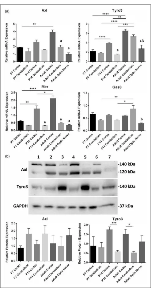
TAM receptors are expressed in a wide variety of tissues and organs throughout the body and, while it is indicated that Tyro3 might be the main receptor in the CNS (Pierce and Keating, 2014), the expression profile of TAMs and Gas6 in the CNS remains unclear. To obtain a comprehen-sive picture of the whole TAM RTK subfamily and Gas6 in the mouse CNS, qPCR was performed on mRNA from different regions of the CNS and at different ages of post-natal development. TAM and Gas6 mRNA expression were detected in cortex and cerebellum of P7, P14, and adult mice and in adult optic nerve (Figure 1(a)). Furthermore, mRNA expression of all three TAMs increased significantly with age in the cortex but not in the cerebellum. This qPCR expression data are valid only for comparisons between samples per gene and not between samples for different genes. This is because the primer or probe sets for different genes will not have the same amp-lification dynamics and so the arbitrary relative expression values are valid only for a sample set for each gene.
In addition, Western blots showed clear expression of both Axl and Tyro3 proteins in all mouse brain regions and at all ages (Figure 1(b)), as well as in the adult optic nerve. However, Tyro3 protein expression was signifi-cantly higher in cortex versus cerebellum in P14 and adult, while the opposite was true of Axl expression (Figure 1(b)). The two bands seen in Axl Western blots are120 kDa and140 kDa forms of Axl due to differ-ent levels of glycosylation, reflecting differdiffer-ent levels of maturation of the RTK.
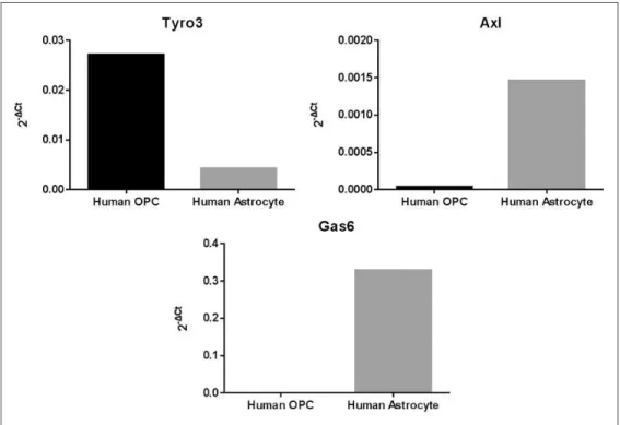
The mRNA expression of Gas6, Tyro3, and Axl was also analyzed by qPCR in cultured HOPCs and HA-c, using the 2Ct method where Ct is Cttarget gene
Ctinternal controland 2
Ct
is the relative gene expression (Schmittgen and Livak, 2008). Tyro3 was expressed clearly in HOPC, whereas Axl was absent in this type of glial cells. In stark contrast, Gas6 was highly expressed in HA-c, while it was absent in HOPC (Figure 2).
Gas6 Promotes Oligodendrogenesis
in the Mouse Optic Nerve
To assess the effect of Gas6 on oligodendrogenesis, we used the optic nerve from adult Sox10-EGFP reporter mice, in which the reporter is expressed by myelinating oligodendrocytes, which make up the bulk of glia in the optic nerve, together with adult OPCs, which make up 3% to 5% of cells in the adult optic nerve (Butt et al., 2004). Optic nerves were isolated with retina intact and maintained in organotypic culture for 3 days, in control medium or medium containing Gas6. Samples also included Gas6 together with warfarin, which blocks the vitamin K-dependentg-carboxylation of the protein
post-translation (Korshunov, 2012) and therefore produces Gas6 that is uncarboxylated and inactive, or the recom-binant extracellular domain of Axl (Axl/Fc), which can act as a ligand antagonist by quenching the ligand before it binds to the receptor. After 3 days, optic nerves were fixed and whole mounted for confocal analysis and cell counts. The results show that Gas6 significantly increased the number of Sox10-GFP positive cells, and this effect was blocked by both Axl/Fc and warfarin (Figure 3;
p<.001). These results were obtained from a number of experiments performed on different days, using separate cultures from different animals.
Gas6 Enhances Myelination Following
Lycolecithin-Induced Demyelination
in Organotypic Cerebellar Slice Cultures
Figure 1. Expression of TAM receptors and Gas6 in different mouse brain regions and at different postnatal ages. (a) qPCR expression analysis of mRNA for Gas6 and TAM receptors. Values represent meanSEMof relative gene expression;n¼4 for all samples except
also features spontaneous repair (Birgbauer et al., 2004; Zhang et al., 2011). Cerebellar slices were obtained from mice aged between P8 and P12 and cultured for 3 days to allow myelin formation to occur prior to adding LPC for 24 h, in normal medium or medium containing Gas6, fol-lowed by a further 3 days culture following LPC removal. Myelin loss was observed in slices treated with LPC, whereas demyelination was significantly decreased by approximately twofold in slice cultures coincubated with Gas6 (Figure 4; p<.05).
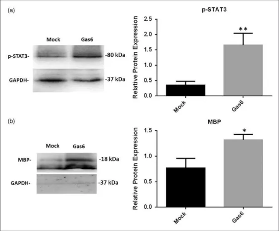
Gas6 Activates STAT3 Signaling
and Myelination in Optic Nerves
The JAK/STAT signaling pathway is a primary target of Gas6 signaling (Yanagita et al., 2001) and has been shown to promote oligodendrocyte regeneration and
remyelination (Hesp et al., 2015), as well as being a pro-mising therapeutic target in multiple animal models of MS (Liu et al., 2014). Therefore, we used Western blot to determine STAT3 activation through detecting levels of phosphorylated STAT3 (p-STAT3; Figure 5(a)) as well as levels of MBP as a measure of differentiation or mye-lination (Figure 5(b)). The protein extracts from cultured adult optic nerves were analyzed, and Western blot band intensities were quantified by densitometric analysis, nor-malizing against GAPDH protein (Figure 5). The results demonstrate that compared with controls, Gas6 stimula-tion (400 ng/ml) resulted in a significant increases in p-STAT3 protein (Figure 5(a); p<.01) as well as MBP, which was increased approximately twofold (Figure 5(b);p<.05). The results demonstrate that Gas6 activates STAT3 signaling and stimulates the molecular process of differentiation or myelination in the adult optic nerve.
Figure 1.Continued
for optic nerve (n¼3) and P14 cerebellum (n¼2). All samples were normalized againstCdc40as internal control; *p<.05, **p<.01, ***p<.001, ****p<.0001 for comparisons as indicated. Columns containing letteraare significant compared with cortex of the same age, and columns containing letterbare significant compared with cerebellum of the same age. (b) Representative Western blot of Axl, Tyro3, and GAPDH (loading control) proteins. Lanes correspond to the following: (1) P7 cortex, (2) P7 cerebellum, (3) P14 cortex, (4) P14 cerebellum, (5) adult cortex, (6) adult cerebellum, and (7) adult optic nerve. The histograms show the quantification of Axl and Tyro3 protein expression by densitometric analysis. Values represent meanSEM(n¼4 blots); *p<.05, #p¼.056 between samples as indicated by lines. Columns containing letteraare significant compared with cortex of the same age, and columns containing letterbare significant compared with cerebellum of the same age.
Figure 2. Expression of Tyro3, Axl, and Gas6 mRNA in human glial cell cultures. Extracted mRNA from human astrocytes and human oligodendrocyte precursors were analyzed by qPCR using specific primers for human Tyro3, Axl, and Gas6 genes. Results were analyzed based on 2Ct
Gas6 Stimulates STAT3 Phosphorylation
in Mature Oligodendrocytes and Astrocytes
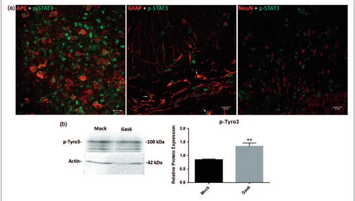
Having observed by Western blot that Gas6 stimulates STAT3 phosphorylation in cultured optic nerves, we then investigated the specific cell types in which this STAT3 activation occurred, through confocal microscopic ana-lysis of cerebellar slice cultures. After 3 h culture incuba-tion in the presence of Gas6, fixed cerebellar secincuba-tions were double stained with p-STAT3 antibody together with antibodies for mature oligodendrocyte, astrocyte, and neuronal markers. The double immunostaining of Gas6-treated tissues showed p-STAT3 to be present pre-dominantly in mature oligodendrocytes and also in some
astrocytes but not in neurons (Figure 6(a) and
Supplementary Figure S3). Our data indicate that treat-ment of cerebellar slices with Gas6 for only 3 h is enough for activating STAT3, and thus the effect of Gas6 on increased level of STAT3 phosphorylation is not due to
increased level of STAT3 protein, which is a longer term process.
small molecule inhibitor of Axl and BGB324 (Barcena et al., 2015; Vouri et al., 2015) and observed that blocking Axl did not affect the Gas6 effect on mye-lination, further indicating that the myelination effect occurs specifically via Tyro3 receptor (Supplementary Figure S1).
Gas6 Alters MS-Related Gene Expression in
Cultured Optic Nerves in a Promyelinating Direction
The effect of Gas6 on expression of 84 different MS-related genes was examined in the adult optic nerve,
using the Mouse Multiple Sclerosis PCR array
(Qiagen). Fifteen genes were identified as being signifi-cantly altered by Gas6 treatment compared with controls (Table 1), seven being upregulatedtwofold and eight
downregulatedtwofold. Among those, some of
the altered genes were selected for further individual analysis by qPCR, including the genes for matrix
metalloproteinase-9 (MMP9), which is able to mediate blood–brain barrier (BBB) injury (Ravindran et al., 2011), the astrocyte marker GFAP (Scheller and Kirchhoff, 2009), and Epha1, which is an RTK (Owshalimpur and Kelley, 1999). The individual follow-up qPCR analyses confirmed that MMP9, Gfap, and
Epha1 genes were all significantly downregulated in the optic nerve following Gas6 treatment (Figure 7).
Discussion
that their expression increases during myelination and is highest in the adult cortex. In addition, we show that Gas6 promotes the generation of oligodendrocytes and myelin in the white matter of the adult mouse optic nerve and also attenuates myelin loss in the lysolecithin model of demyelination in cerebellar slice cultures. Furthermore, analysis of the molecular mechanisms of action of Gas6 demonstrates that it activates the Tyro3 receptor, intracellular STAT3 signaling, and the pro-gramming of a host of MS-related genes in a prorepair and myelinating direction. These results therefore indi-cate that Gas6 or TAM signaling is a potential target for stimulating repair mechanisms that are relevant to demyelination and MS.
All three TAM receptors were observed to be expressed throughout the brain tissue, although Tyro3 was expressed in more regions and at higher levels com-pared with Axl and Mer. This supports previous indica-tions that Tyro3 is the primary TAM receptor mediating the actions of Gas6 in the CNS (Prieto et al., 2000). In addition, our data show that Tyro3 expression increases
during development in the CNS. These observations are in line with previous time-course expression data, show-ing Tyro3 protein to be very low at embryonic stages while its expression begins to increase shortly after birth, dramatically increasing between postnatal days P3 and P15, reaching its peak level at P24, and remaining at that level thereafter in the adult (Prieto et al., 2000). Although most myelination occurs early in life, myelin-ation carries on at least into late adolescence and, in some regions of the CNS, may increase throughout much of adult life (Emery, 2010). Therefore, the concomitant tem-poral profiles of myelination and Tyro3 expression in the developing mouse CNS suggest that Tyro3 plays a role in oligodendrocyte development or maturation and myelin-ation. In the Axl blot, a double band was observed at both 120 kDa and 140 kDa, which are the well-known alternately glycosylated versions of Axl (the 140 kDa pro-tein being most mature; O’Bryan et al., 1995). Our results showed clear expression of Axl protein in all brain regions and ages analyzed, with no significant differences in levels of expression among them.
We also conducted qPCR analysis for Tyro3, Axl, and Gas6 genes in separate pure cultures of human OPCs and astrocytes. Tyro3 was highly expressed in oligodendro-cytes, in which Axl expression was absent. This key find-ing therefore suggests that Gas6 acts directly on OPCs or oligodendrocytes via the Tyro3 receptor to affect their fate and function. In contrast, Tyro3 expression was lower in astrocytes compared with oligodendrocytes but instead astrocytes showed a high level of Gas6 expres-sion. This therefore suggests an indirect and supportive role for astrocytes in regulating the fate and function of oligodendrocytes, by providing the ligand through para-crine signaling. However, this interpretation of relative TAM expression comes with a certain degree of caution as the human isolated glial cell cultures were not only from different brain regions but were also derived from different brains and at different ages. Nevertheless, within a specific cell type, our data clearly show that Tyro3 is present in oligodendrocytes, while Axl is virtually absent in these cells. Furthermore, according to Figure 1(a), Mer mRNA was also observed in the CNS, although we did not characterize its expression pattern in glial cells, but according to previous reports, it is mainly expressed in microglia within the CNS and is involved in phagocytic Figure 6. Gas6 activates STAT3 in mature oligodendrocytes and astrocytes and activates Tyro3 receptor after 3 h stimulation in culture. (a) Cerebellar slices were treated with mock or Gas6 medium for 3 h, and fixed sections were double stained for p-STAT3 and APC (mature oligodendrocyte marker), p-STAT3 and GFAP (astrocyte marker), p-STAT3 and NeuN (neuronal marker; scale bar¼20mm). The stainings reveal that p-STAT3 is present in oligodendrocytes and astrocytes but not in neurons. The arrows point to cells that are both GFAPþand p-STAT3þ. (b) Representative Western blot of p-Tyro3 protein in optic nerve lysate. The graph shows the densitometric quantification of protein level relative to actin in the same sample. Values represent meanSEM(n¼5 blots); **p<.01 versus control, Studentttest. Gas6 significantly increased the activation of Tyro3.
Table 1. Summary of Genes in Cultured Mouse Optic Nerves Altered by Gas6 Stimulation by2-Fold.
Gene Symbol
Ct
Fold upregulation or downregulation
Gas6 Mock Ct 2Ct
Ccl3 3.06 4.18 1.12 2.173469725
Ccl7 0.45 1.62 1.17 2.250116969
Cxcl11 8.86 10.31 1.45 2.732080514
Fasl 11.71 13.39 1.68 3.20427951
H2-Eb1 12.12 14.07 1.95 3.863745316
Il13 11.82 13.29 1.47 2.770218936
Il1b 8.65 9.69 1.04 2.056227653
Ednra 8.08 6.65 1.43 0.371131
Epha1 9.36 8.06 1.3 0.406126198
GFAP 1.06 0.38 1.44 0.368567304
Mag 2.09 0.79 1.3 0.406126198
MMP9 6.43 5.41 1.02 0.493116352
Nr2f1 7.27 5.47 1.8 0.287174589
Tubb4 3.04 2.01 1.03 0.489710149
activity of these cells (Fourgeaud et al., 2016). Axl has also been reported to be present in microglia, and no Tyro3 was observed in these cells (Fourgeaud et al., 2016). Also, it was shown that, in addition to Mer, Axl is also essential for the phagocytic activity and nonin-flammatory properties of microglia cells, including removal of cell debris (Fourgeaud et al., 2016).
Moreover, as we have only analyzed glial cells in this study, it is important to note that expression of Gas6 expression has been reported in different neuronal cells including Purkinje neurons, motor neurons of spinal cord, as well as large dorsal root ganglia neurons (Li et al., 1996; Prieto et al., 1999). Therefore, it is also pos-sible that neurons may play a similar supportive role in regulating oligodendrocyte development and function, as astrocytes do.
As well as in the brain, qPCR analysis of adult mouse optic nerve demonstrated that Gas6 or TAM signaling components are also present in this tissue, with relative levels of expression indicated in the following order Tyro3>Axl>Mer. Also, Gas6 stimulation of optic nerve from Sox10-EGFP mice further confirmed the pres-ence and activity of TAM receptors in CNS white matter, through which they promote the generation of oligo-dendrocyte lineage cells. The Sox10-EGFP reporter iden-tifies both oligodendrocytes and OPCs, since Sox10 marks all stages of oligodendrocyte development (Stolt et al., 2006); but in the adult, OPCs are continuously generating new oligodendrocytes and are the likely tar-gets of Gas6 in our study (Gonzalez-Pereza and Alvarez-Buyllab, 2011). In addition to oligodendrogenesis, Gas6 stimulated increases the levels of MBP protein in the
adult optic nerve, which indicates that Gas6 also pro-motes myelination in mature oligodendrocytes (Mi et al., 2011). Therefore, these observations together dem-onstrate that Gas6 boosts both the number of OPCs as well as mature myelinating oligodendrocytes. In this regard, it is worthwhile considering protein S, a protein homologous to Gas6 that is also a TAM receptor ligand. However, its expression appears restricted in the CNS, with low levels of protein S mRNA observed in locus coeruleus and high levels of mRNA in the choroid plexus (Prieto et al., 1999). Furthermore, the expression of protein S appears to be primarily neuronal, having been detected in the cortex, hippocampal pyramidal neu-rons, and granule neurons of the dentate gyrus of the rabbit brain (He et al., 1995; Prieto et al., 2007).
Gas6 stimulation also led to a decreased demyelination in a cerebellar slice culture model of demyelination by lysolecithin (Birgbauer et al., 2004), which is one of the most commonly used toxins to induce focal demyelin-ation in different parts of the brain (van der Star et al., 2012). Our results show that treatment with Gas6 signifi-cantly attenuated the extent of demyelination that con-tinues for days after exposure to lysolecithin (Birgbauer et al., 2004). This is consistent with studies in the cupri-zone model of demyelination, which showed systemic administration of Gas6 to enhance remyelination (Tsiperson et al., 2010), while Gas6 ablation decreased the number of myelinating oligodendrocytes and delayed recovery (Binder et al., 2011). Together, these studies demonstrate that Gas6 or TAM signaling plays a major role in oligodendrocyte generation and myelination in the adult CNS. However, our experiment was focused on the protective effect of Gas6 during demyelinating injury, whereas it is also important to investigate the acceleratory effect of Gas6 on remyelination after damage. Thus far, we did not observe an enhanced remyelination when Gas6 was added 3 days after LPC damage (Supplementary Figure S2). However, further detailed time-course studies are warranted to explore the time window during which Gas6 can be effective as an enhancer of remyelination post damage. Furthermore, although Gas6 increases MBP and blocks demyelination in culture, it is also worthwhile to measure the axon diameter or g-ratio by electron microscopy to determine specifically the location of myelination as being around axons, and hence contributing to increased axon thick-ness. Also, although this study was focused on the non-microglial response to Gas6, it is nevertheless possible, considering the phagocytosis-mediating role of Mer (Chen et al., 1997), that Gas6 could also activate Mer in microglia in our culture model; this requires further investigation.
In addition to the ligand Gas6, the role of Axl receptor has also investigated by other research groups in demye-lination models. In a study using the cuprizone model of Figure 7. Effect of Gas6 on expression of selected MS-related
genes in cultured mouse optic nerves. qRT-PCR was performed on extracts from mock and Gas6-treated optic nerve cultures, using specific primer or probe sets, and normalizing expression against theGAPDHgene. Treatment of optic nerves with Gas6 resulted in downregulation ofEpha1,GFAP, andMMP9genes. Values represent meanSEM(n¼4 for qPCR experiments for all genes except for
demyelination, Axl knockout mice showed no significant differences to wild-type mice in axon diameter, myelin thickness, and g-ratio of myelin-containing axons (Hoehn et al., 2008). This therefore suggests that Axl is not the main receptor involved in remyelination or that loss of signaling through all three TAM receptors is more harmful than loss of signaling through a single TAM receptor. This is consistent with our observations, which point toward the prominence of Tyro3 in oligo-dendrocyte biology and myelination, reinforced by its unique mRNA expression in oligodendrocytes.
As our expression analysis indicated Tyro3 as the most prominent TAM receptor in the CNS, at least as concerns oligodendrocytes, we therefore hypothesized that the Gas6 effects on OPC proliferation, oligodendrocyte maturation, and myelination are likely occurring via a direct activation of Tyro3 in these cell populations. We therefore utilized our optic nerve culture model to also investigate the rela-tively acute activation of Tyro3 in response to Gas6 stimu-lation. Thus, Gas6 stimulated a higher activation state of Tyro3 after 3 h in the optic nerve, as observed by increased p-Tyro3 protein levels. This observation is supported by a recent study in the peripheral nervous system that showed Tyro3 to be an important modulator of myelination by Schwann cells (Miyamoto et al., 2015).
We also observed that the transcriptional regulator protein STAT3 was mobilized by Gas6 stimulation using both of ourin vitroculture models. Gas6 increased levels of phosphorylated STAT3 protein in the optic nerve, and using the slices for microscopy, we were able to localize the pSTAT3 to oligodendrocytes as well as to a lesser extent astrocytes. Gas6 has previously been reported to act via STAT3 to promote proliferation of mesangial cells (Yanagita et al., 2001). This is consistent with our current observations of Gas6-induced activation of STAT3 and proliferation of OPCs or oligodendrocyte in the optic nerve. STAT3 controls a large number of genes that are involved in apoptosis, cell migration, cell cycle regulation, and angiogenesis as well as modulation of immune suppressive factors (Ferguson et al., 2015). Once STAT3 becomes activated via phosphorylation, it translocates to the nucleus and binds to specific DNA sequences to activate the expression of target genes that regulate proliferation and differentiation (Qi and Yang, 2014). There is also evidence that shows STAT3 activa-tion is potentially important during neuroinflammaactiva-tion by protecting myelin development (Nobuta et al., 2012). Although neurons also express Tyro3 (Schulz et al., 1995), we found Gas6 to induce STAT3 activation pre-dominantly in OPCs or oligodendrocytes, as would be expected for differentiating or proliferating cells (Fukada et al., 1996). Therefore, STAT3 appears to be a key intracellular mediator of Gas6 signaling in oligo-dendrocytes, most likely via Tyro3, to affect their devel-opment and the subsequent myelination of axons.
From a mini-array screen, we also observed Gas6 to induce significant changes in expression of multiple genes relevant to glial biology and MS in the cultured optic nerve, including downregulation of MMP9, Epha1, and
GFAPgenes. GFAP is an astrocyte-specific protein and is used as a marker of reactive astrogliosis (Beckerman et al., 2015), suggesting that Gas6 may modulate astro-glial responses in the optic nerve. MMP9 is a proteolytic enzyme that degrades type IV collagen, laminin, and fibronectin, all of which are major components of the basement membrane of the BBB (Zheng et al., 2014). During a relapse course of MS, active B cells increase the expression and secretion of MMP9 protein, ultimately resulting in digestion of myelin as well as neurological disability (Aung et al., 2015). Furthermore, it has been shown that MMP9 can be secreted by OPCs that have proliferated at sites of demyelination in the CNS, which results in opening of the BBB and the subsequent second-ary cascades of cerebrovascular injury and demyelination (Seo et al., 2013). Moreover, disruption of the BBB has been considered as one of the initial steps in the develop-ment of disease in MS patients (Alvarez et al., 2011). Also, the role of MMP9, at least produced by active B lymphocytes, in the disruption of the BBB and degrad-ation of MBP protein is well recognized (Aung et al., 2015). Thus, our observation of Gas6 causing a twofold downregulation of the MMP9 gene suggests that Gas6 may help maintain BBB integrity and hence inhibit immune cell infiltration into the CNS during the course of MS; this is a focus of investigation in subsequent studies.
In conclusion, this study shows that components of Gas6 or TAM signaling are present throughout the adult CNS, and that, specifically, Tyro3 is present on OPCs or oligodendrocytes and mediates their develop-ment and function in response to the ligand Gas6, while astrocytes are the main source of the Gas6. Gas6 pro-motes the development and maturation of oligodendro-cytes from OPCs and myelination in the adult CNS, as well as stimulating remyelination after toxic injury by lysolecithin. In addition, we show that the effects of Gas6 involve regulation of a set of genes that, through their coordinated upregulation or suppression, push the glial cell developmental pathway in a promyelinating dir-ection. These results support a prominent role for Gas6 in promoting CNS repair after demyelination, and thus an attractive consideration for novel therapeutic approaches for MS.
Summary
toxin-induced demyelination. Gas6 acted via Tyro3 receptor to activate intracellular STAT3 signaling in oligodendrocytes.
Declaration of Conflicting Interests
The authors declared no potential conflicts of interest with respect to the research, authorship, and/or publication of this article.
Funding
The author(s) disclosed receipt of the following financial support for the research, authorship, and/or publication of this article: SG was supported by a PhD studentship from the Multiple Sclerosis Society UK (grant number 953/11).
References
Alvarez, J. I., Cayrol, R., & Prat, A. (2011). Disruption of central nervous system barriers in multiple sclerosis. Biochimica et Biophysica Acta,1812, 252–264.
Aung, L. L., Mouradian, M. M., Dhib-Jalbut, S., & Balashov, K. E. (2015). MMP-9 expression is increased in B lymphocytes during multiple sclerosis exacerbation and is regulated by microRNA-320a.Journal of Neuroimmunology,278, 185–189. Azim, K., & Butt, A. M. (2011). GSK3beta negatively regulates oligodendrocyte differentiation and myelination in vivo. Glia, 59, 540–553.
Barcena, C., Stefanovic, M., Tutusaus, A., Joannas, L., Menendez, A., Garcia-Ruiz, C.,. . .Morales, A. (2015). Gas6/Axl pathway is activated in chronic liver disease and its targeting reduces fibrosis via hepatic stellate cell inactivation. Journal of Hepatology,63, 670–678.
Beckerman, S. R., Jimenez, J. E., Shi, Y., Al-Ali, H., Bixby, J. L., & Lemmon, V. P. (2015). Phenotypic assays to identify agents that induce reactive gliosis: A counter-screen to prioritize compounds for preclinical animal studies. Assay and Drug Development Technologies,13, 377–388.
Binder, M. D., Xiao, J., Kemper, D., Ma, G. Z., Murray, S. S., & Kilpatrick, T. J. (2011). Gas6 increases myelination by oligo-dendrocytes and its deficiency delays recovery following cupri-zone-induced demyelination.PLoS One,6, e17727.
Birgbauer, E., Rao, T. S., & Webb, M. (2004). Lysolecithin induces demyelination in vitro in a cerebellar slice culture system. Journal of Neuroscience Research,78, 157–166.
Butt, A. M., Pugh, M., Hubbard, P., & James, G. (2004). Functions of optic nerve glia: Axoglial signalling in physiology and path-ology.Eye (Lond),18, 1110–1121.
Chen, J., Carey, K., & Godowski, P. J. (1997). Identification of Gas6 as a ligand for Mer, a neural cell adhesion molecule related receptor tyrosine kinase implicated in cellular transform-ation.Oncogene,14, 2033–2039.
De Simoni, A., & Yu, L. M. (2006). Preparation of organotypic hippocampal slice cultures: Interface method.Nature Protocols, 1, 1439–1445.
Emery, B. (2010). Regulation of oligodendrocyte differentiation and myelination.Science,330, 779–782.
Ferguson, S. D., Srinivasan, V. M., & Heimberger, A. B. (2015). The role of STAT3 in tumor-mediated immune suppression. Journal of Neuro-oncology,123, 385–394.
Fourgeaud, L., Traves, P. G., Tufail, Y., Leal-Bailey, H., Lew, E. D., Burrola, P. G.,. . .Lemke, G. (2016). TAM receptors
regulate multiple features of microglial physiology. Nature, 532, 240–244.
Franklin, R. J. M. (2002). Why does remyelination fail in multiple sclerosis?Nature Reviews,3, 9.
Fukada, T., Hibi, M., Yamanaka, Y., Takahashi-Tezuka, M., Fujitani, Y., Yamaguchi, T.,. . .Hirano, T. (1996). Two signals are necessary for cell proliferation induced by a cytokine recep-tor gp130: Involvement of STAT3 in anti-apoptosis.Immunity, 5, 449–460.
Gonzalez-Pereza, O., & Alvarez-Buyllab, A. (2011). Oligodendrogenesis in the subventricular zone and the role of epidermal growth factor.Brain Research Reviews,67, 9. Goudarzi, S., Smith, L. J., Schutz, S., & Hafizi, S. (2013).
Interaction of DISC1 with the PTB domain of Tensin2. Cellular and Molecular Life Sciences: CMLS,70, 1663–1672. Hafizi, S., & Dahlba¨ck, B. (2006a). Gas6 and protein S. Vitamin
K-dependent ligands for the Axl receptor tyrosine kinase subfam-ily.FEBS Journal,273, 5231–5244.
Hafizi, S., & Dahlba¨ck, B. (2006b). Signalling and functional diver-sity within the Axl subfamily of receptor tyrosine kinases. Cytokine Growth Factor Reviews,17, 295–304.
He, X., Shen, L., Bjartell, A., & Dahlback, B. (1995). The gene encoding vitamin K-dependent anticoagulant protein S is expressed in multiple rabbit organs as demonstrated by northern blotting, in situ hybridization, and immunohistochemistry.The Journal of Histochemistry and Cytochemistry: Official Journal of the Histochemistry Society,43, 85–96.
Hesp, Z. C., Goldstein, E. A., Miranda, C. J., Kaspar, B. K., & McTigue, D. M. (2015). Chronic oligodendrogenesis and remyelination after spinal cord injury in mice and rats. The Journal of Neuroscience: The Official Journal of the Society for Neuroscience,35, 1274–1290.
Hoehn, H. J., Kress, Y., Sohn, A., Brosnan, C. F., Bourdon, S., & Shafit-Zagardo, B. (2008). Axl-/- mice have delayed recovery and prolonged axonal damage following cuprizone toxicity. Brain Research,1240, 1–11.
Korshunov, V. A. (2012). Axl-dependent signalling: A clinical update.Clinical Science,122, 361–368.
Kuhlmann, T., Miron, V., Cui, Q., Wegner, C., Antel, J., & Bruck, W. (2008). Differentiation block of oligodendroglial progenitor cells as a cause for remyelination failure in chronic multiple sclerosis.Brain: A Journal of Neurology,131, 1749–1758. Lemke, G., & Lu, Q. (2003). Macrophage regulation by Tyro 3
family receptors.Current Opinion in Immunology,15, 31–36. Li, R., Chen, J., Hammonds, G., Phillips, H., Armanini, M., Wood,
P.,. . .Mather, J. P. (1996). Identification of Gas6 as a growth factor for human Schwann cells.The Journal of Neuroscience: The Official Journal of the Society for Neuroscience, 16, 2012–2019.
Liu, Y., Holdbrooks, A. T., De Sarno, P., Rowse, A. L., Yanagisawa, L. L., McFarland, B. C.,. . .Qin, H. (2014). Therapeutic efficacy of suppressing the Jak/STAT pathway in multiple models of experimental autoimmune encephalomyelitis. Journal of Immunology,192, 59–72.
Livak, K. J., & Schmittgen, T. D. (2001). Analysis of relative gene expression data using real-time quantitative PCR and the 2(-Delta Delta C(T)) Method.Methods,25, 402–408.
Mi, S., Lee, X., Hu, Y., Ji, B., Shao, Z., Yang, W.,. . .Pepinsky, R. B. (2011). Death receptor 6 negatively regulates oligodendro-cyte survival, maturation and myelination.Nature Medicine,17, 816–821.
Miyamoto, Y., Torii, T., Takada, S., Ohno, N., Saitoh, Y., Nakamura, K.,. . .Yamauchi, J. (2015). Involvement of the Tyro3 receptor and its intracellular partner Fyn signaling in Schwann cell myelination. Molecular Biology of the Cell,26, 3489–3503.
Nobuta, H., Ghiani, C. A., Paez, P. M., Spreuer, V., Dong, H., Korsak, R. A.,. . .Waschek, J. A. (2012). STAT3-mediated astrogliosis protects myelin development in neonatal brain injury.Annals of Neurology,72, 750–765.
O’Bryan, J. P., Fridell, Y. W., Koski, R., Varnum, B., & Liu, E. T. (1995). The transforming receptor tyrosine kinase, Axl, is post-translationally regulated by proteolytic cleavage.The Journal of Biological Chemistry,270, 551–557.
Owshalimpur, D., & Kelley, M. J. (1999). Genomic structure of the EPHA1 receptor tyrosine kinase gene.Molecular and Cellular Probes,13, 169–173.
Pierce, A. M., & Keating, A. K. (2014). TAM receptor tyrosine kinases: Expression, disease and oncogenesis in the central ner-vous system.Brain Research,1542, 206–220.
Prieto, A. L., O’Dell, S., Varnum, B., & Lai, C. (2007). Localization and signaling of the receptor protein tyrosine kinase Tyro3 in cortical and hippocampal neurons. Neuroscience,150, 319–334.
Prieto, A. L., Weber, J. L., & Lai, C. (2000). Expression of the receptor protein-tyrosine kinases Tyro-3, Axl, and mer in the developing rat central nervous system. The Journal of Comparative Neurology,425, 295–314.
Prieto, A. L., Weber, J. L., Tracy, S., Heeb, M. J., & Lai, C. (1999). Gas6, a ligand for the receptor protein-tyrosine kinase Tyro-3, is widely expressed in the central nervous system1. Brain Research,816, 646–661.
Qi, Q. R., & Yang, Z. M. (2014). Regulation and function of signal transducer and activator of transcription 3. World Journal of Biological Chemistry,5, 231–239.
Ravindran, J., Agrawal, M., Gupta, N., & Rao, P. V. (2011). Alteration of blood brain barrier permeability by T-2 toxin: Role of MMP-9 and inflammatory cytokines.Toxicology,280, 44–52.
Saxena, A., Martin-Blondel, G., Mars, L. T., & RSL. (2011). Role of CD8 T cell subsets in the pathogenesis of multiple sclerosis. FEBS Letters,585, 6.
Scheller, A., & Kirchhoff, F. (2009). Astrocyte: Identification Methods. In: L. R. Squire (ed.) Encyclopedia of neuroscience Oxford, England: Academic Press, pp. 573–577.
Schmittgen, T. D., & Livak, K. J. (2008). Analyzing real-time PCR data by the comparative C(T) method. Nature Protocols, 3, 1101–1108.
Schneider, C. A., Rasband, W. S., & Eliceiri, K. W. (2012). NIH Image to ImageJ: 25 years of image analysis.Nature Methods, 9, 671–675.
Schulz, N. T., Paulhiac, C. I., Lee, L., & Zhou, R. (1995). Isolation and expression analysis of tyro3, a murine growth factor recep-tor tyrosine kinase preferentially expressed in adult brain. Molecular Brain Research,28, 273–280.
Seo, J. H., Miyamoto, N., Hayakawa, K., Pham, L. D., Maki, T., Ayata, C.,. . .Arai, K. (2013). Oligodendrocyte precursors induce early blood-brain barrier opening after white matter injury.The Journal of Clinical Investigation,123, 782–786. Shankar, S. L., O’Guin, K., Cammer, M., McMorris, F. A., Stitt, T.
N., Basch, R. S.,. . .Shafit-Zagardo, B. (2003). The growth arrest-specific gene product Gas6 promotes the survival of human oligodendrocytes via a phosphatidylinositol 3-kinase-dependent pathway. The Journal of Neuroscience, 23, 4208–4218.
Stenhoff, J., Dahlback, B., & Hafizi, S. (2004). Vitamin K-depen-dent Gas6 activates ERK kinase and stimulates growth of cardiac fibroblasts. Biochemical and Biophysical Research Communications,319, 871–878.
Stolt, C. C., Schlierf, A., Lommes, P., Hillgartner, S., Werner, T., Kosian, T.,. . .Wegner, M. (2006). SoxD proteins influence multiple stages of oligodendrocyte development and modulate SoxE protein function.Developmental Cell,11, 697–709. Tsiperson, V., Li, X., Schwartz, G. J., Raine, C. S., &
Shafit-Zagardo, B. (2010). GAS6 enhances repair following cupri-zone-induced demyelination.PLoS One,5, e15748.
van der Star, B. J., Vogel, D. Y., Kipp, M., Puentes, F., Baker, D., Amor, S. (2012). In vitro and in vivo models of multiple scler-osis. CNS & Neurological Disorders Drug Targets, 11, 570–588.
Vouri, M., An, Q., Birt, M., Pilkington, G. J., & Hafizi, S. (2015). Small molecule inhibition of Axl receptor tyrosine kinase potently suppresses multiple malignant properties of glioma cells.Oncotarget,6, 16183–16197.
Weinger, J. G., Brosnan, C. F., Loudig, O., Goldberg, M. F., Macian, F., Arnett, H. A.,. . .Shafit-Zagardo, B. (2011). Loss of the receptor tyrosine kinase Axl leads to enhanced inflam-mation in the CNS and delayed removal of myelin debris during experimental autoimmune encephalomyelitis. Journal of Neuroinflammation,8, 49.
Wolswijk, G. (2002). Oligodendrocyte precursor cells in the demyelinated multiple sclerosis spinal cord.Brain: A Journal of Neurology,125, 338–349.
Yanagita, M., Arai, H., Nakano, T., Ohashi, K., Mizuno, K., Fukatsu, A.,. . .Kita, T. (2001). Gas6 induces mesangial cell proliferation via latent transcription factor STAT3. The Journal of Biological Chemistry,276, 42364–42369.
Zhang, H., Jarjour, A. A., Boyd, A., & Williams, A. (2011). Central nervous system remyelination in culture—A tool for multiple sclerosis research.Experimental Neurology,230, 138–148. Zheng, M., Wei, J., Tang, Y., Yang, C., Wei, Y., Yin, X.,. . .Liu, Q.