A Comparative Study of Variables
Influencing Ischemic Injury in the Longa and
Koizumi Methods of Intraluminal Filament
Middle Cerebral Artery Occlusion in Mice
Gary P. Morris1,2, Amanda L. Wright1☯, Richard P. Tan1,3☯, Amadeus Gladbach4, Lars M. Ittner4,5, Bryce Vissel1,2,6*
1Neurodegenerative Disorders, Garvan Institute of Medical Research, Sydney, Australia,2Faculty of Medicine, University of New South Wales, Sydney, Australia,3Heart Research Institute, 2042 New South Wales, Sydney, Australia,4Dementia Research Unit, Department of Anatomy, School of Medical Sciences, Faculty of Medicine, University of New South Wales, Sydney, Australia,5Neuroscience Research Australia, Sydney, Australia,6Faculty of Science, University of Technology Sydney, Sydney, Australia
☯These authors contributed equally to this work.
*brycevissel@gmail.com
Abstract
The intraluminal filament model of middle cerebral artery occlusion (MCAO) in mice and rats has been plagued by inconsistency, owing in part to the multitude of variables requiring control. In this study we investigated the impact of several major variables on survival rate, lesion volume, neurological scores, cerebral blood flow (CBF) and body weight including fil-ament width, time after reperfusion, occlusion time and the choice of surgical method. Using the Koizumi method, we found ischemic injury can be detected as early as 30 min after reperfusion, to a degree that is not statistically different from 24 h post-perfusion, using 2,3,5-Triphenyltetrazolium chloride (TTC) staining. We also found a distinct increase in total lesion volume with increasing occlusion time, with 30–45 min a critical time for the develop-ment of large, reproducible lesions. Furthermore, although we found no significant differ-ence in total lesion volume generated by the Koizumi and Longa methods of MCAO, nor were survival rates appreciably different between the two at 4 h after reperfusion, the Longa method produces significantly greater reperfusion. Finally, we found no statistical evidence to support the exclusion of data from animals experiencing a CBF reduction of<70% in the MCA territory following MCAO, using laser-Doppler flowmetry. Instead we suggest the main usefulness of laser-Doppler flowmetry is for guiding filament placement and the identifica-tion of subarachnoid haemorrhages and premature reperfusion. In summary, this study pro-vides detailed evaluation of the Koizumi method of intraluminal filament MCAO in mice and a direct comparison to the Longa method.
OPEN ACCESS
Citation:Morris GP, Wright AL, Tan RP, Gladbach A, Ittner LM, Vissel B (2016) A Comparative Study of Variables Influencing Ischemic Injury in the Longa and Koizumi Methods of Intraluminal Filament Middle Cerebral Artery Occlusion in Mice. PLoS ONE 11(2): e0148503. doi:10.1371/journal.pone.0148503
Editor:Lucio Annunziato, University of Naples Federico II, ITALY
Received:August 16, 2015
Accepted:January 19, 2016
Published:February 12, 2016
Copyright:© 2016 Morris et al. This is an open access article distributed under the terms of the Creative Commons Attribution License, which permits unrestricted use, distribution, and reproduction in any medium, provided the original author and source are credited.
Data Availability Statement:All relevant data are within the paper and its Supporting Information files.
Introduction
The most common human focal cerebral ischemia occurs due to thrombotic or embolic occlu-sion of the middle cerebral artery (MCA). Rodent models have therefore predominantly aimed to mimic MCA occlusion (MCAO), however no single rodent model fully recapitulates the fea-tures of human MCAO and each model has various advantages and disadvantages [1,2]. The intraluminal filament method developed by Koizumi et al., [3] and later modified by Longa et al., [4] has become a widespread model of choice for mimicking MCAO in rodents, due to the minimally invasive technique involved, and ability to allow reperfusion post-occlusion [5]. The later point is particularly important considering early restoration of blood supply is a major determinant of the severity of ischemic injury [6], reflected in the success of thrombo-lytic therapy following acute ischemic stroke in some individuals [7].
However, despite having been utilised for almost three decades, and many attempts to reduce variability [8,9], the intraluminal filament method still displays large inconsistency in the volume of ischemic lesions generated, both within studies and from lab to lab. For example, following similar protocols, different groups have obtained mean ischemic lesion volumes ranging from as much as 11.1–55.6% of the ipsilateral cortex, for a 30 min intraluminal thread occlusion in mice [2]. Dirnagl highlighted standard deviations in infarct volumes are as much as 40% of the mean in many studies [10]. These figures are indicative of the difficulty in gener-ating robust and reproducible data from pre-clinical stroke studies.
Although simple in theory, the intraluminal filament method of MCAO requires practice to master the surgical skills necessary for success, and becomes more complex surgically when moving from a rat to a relatively smaller mouse. Furthermore, the complexity increases when considering the many variables shown to impact resulting ischemic lesion volume. These include the physical properties of the chosen embolus, such as the diameter and material [11,12], occlusion time [13], recovery time [14], temperature during and post-surgery [15], choice of anaesthetic [16], minor differences in surgical technique [8], experience of the sur-geon [17], choice of animal (rat vs. mouse) [2], strain of animal [18], animal to animal variation in cerebrovasculature (such as the patency of the posterior communicating artery (PComA)) [19], sex [20] and age [21], among many others.
In both the Koizumi and Longa methods of intraluminal filament MCAO, one of the fore-most causes of inconsistency derives from the choice of embolus (intraluminal filament), which can vary widely in thickness, length and in the material used to create it. A recent popu-lar choice has been silicon coated silk sutures, which offer greater consistency in preparation compared to traditionally used homemade versions, such as heat-blunted sutures [11]. Consid-ering the relatively recent introduction of these filaments, few comprehensive studies have been undertaken to determine the efficacy of silicone coated sutures to produce consistent lesions, or how often they may induce common side effects of the intraluminal filament sur-gery, such as subarachnoid haemorrhage (SAH), or high rates of premature death after reperfu-sion, which have in the past plagued the reliability of the method.
Secondly, despite the widespread use of both the Koizumi and Longa methods in rats and mice, the Longa method appears to have proliferated more widely throughout the literature. The use of mice is becoming more commonplace owing to the availability of many different genetically modi-fied strains, and therefore it is important to compare these two methods to determine when and why each should be utilised in the mouse. There are two key differences between the methods; 1. The Longa method allows reperfusion via both central carotid arteries (CCA), as opposed to reper-fusion via only one CCA in Koizumi’s method and 2. The Longa method traditionally employs fil-aments that only have a bulbous tip produced by heat-blunting sutures, whereas the Koizumi method initially utilised filaments coated with silicone for 5 mm at a consistent width [22].
preparation of the manuscript. No research costs or authors’salaries were funded by a tobacco company.
In this study we have therefore compared three types of silicone coated filaments, varying in thickness and coating length, time after reperfusion, occlusion time and the efficacy of the Koi-zumi vs. the Longa method against various outcome measures including ipsilateral lesion vol-ume, body weight loss, survival rates and neurological scores.
Finally, it has become commonplace to employ a tool to measure regional cerebral blood flow, such as laser-Doppler flowmetry, or synchrotron radiation angiography, to assist with confirmation of successful MCAO [23]. Data obtained from these recordings is often utilised to indicate SAH and premature reperfusion during occlusion, as well as being used to remove data on the basis of predetermined cut-off points for relative changes in CBF following MCAO. A commonly cited figure for exclusion of data is a<70% reduction in CBF following MCAO by an intraluminal filament. If measuring blood flow continuously throughout occlusion, or reapplying at a later time, these tools can also allow a measurement of‘reperfusion’following withdrawal of an embolus. In this study we examine the value of laser-Doppler measurement in determining SAH and premature reperfusion, and determine whether there is any valid sta-tistical support for exclusion of data based on the‘<70%’alteration in CBF following MCAO in mice.
Materials and Methods
Animals
Male C57BL/6JAusb mice aged 8–13 weeks were obtained from Australian BioResources in New South Wales, Australia. Prior to surgery mice were housed in groups of 4–5 per cage and post-surgery mice were single housed, withad libitumaccess to food and water. They were maintained on a 12 h light-dark cycle, with lights being switched on at 7.00 am. All surgeries were performed during light hours. All animal experiments were performed with the approval of the Garvan Institute and St. Vincent's Hospital Animal Ethics Committee, in accordance with National Health and Medical Research Council animal experimentation guidelines and the Australian Code of Practice for the Care and Use of Animals for Scientific Purposes (8th edition, 2013).
Ischemic model
Pre-surgery. All surgical tools were sterilized for 1 min in a 250°C Dry Glass Bead
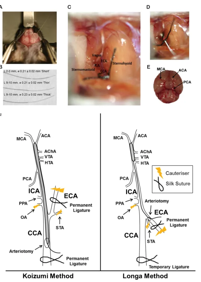
Steril-izer (Cole-Parmer, #EW-10779-00). The surgical table (a styrofoam platform) and associated equipment were sanitised using 70% (w/v) ethanol. Mice were anaesthetised with ketamine (8.7 mg/ml; Mavlab, Slacks Creek, QLD,http://www.mavlab.com.au) and xylazine (2 mg/ml; Troy Laboratories Pty Ltd, Smithfield, Australia,http://www.troylab.com.au). Both the head and neck of each mouse was shaved then cleaned with an alcohol swab. Mice were placed in a prone position and, using a scalpel blade, a 2 cm midline incision was made on the skin from the nose to between the eyes. Mice were secured, still prone, into a stereotaxic apparatus. The skin was then pulled laterally to expose the skull (Fig 1A). Eye ointment was applied liberally to both eyes.
Measurement of local CBF. CBF was monitored before, during and after surgery using a
was adhered perpendicular to the right temporal skull at Antero-Posterior (AP) ~-1.0 and Medio-Lateral (ML) ~3.0 from bregma, to measure blood flow in the territory supplied by the right MCA [24] (Fig 1A). Once Loctite Gel firmed the mice, with the laser-Doppler now attached, were carefully released from the stereotaxic apparatus and gently placed supine upon the surgical table, with an area cut from the styrofoam platform to allow room for the attached laser-Doppler probe.
MCAO surgery. Mouse body temperature was monitored throughout surgery, occlusion
and reperfusion with the use of a rectal thermometer (PhysioSuite1, Kent Scientific Corpora-tion) and maintained between 36.0°C and 38.0°C via the use of a flat reptile heat pad
(#HMAT5CN, Reptapets). Refer toS1 Videofor a brief video of the surgery. Shaved areas under the chin were sterilised with an alcohol swab and 1 cm midline incisions were made between the manubrium and the jaw, under a stereo dissecting microscope (Leica M50, Leica Microsystems). Underlying submandibular glands were bluntly divided, and retractors (#17000–02, Fine Science Tools) were applied to expose the surgical field beneath the glands. The thin omohyoid muscle sometimes covered the central carotid artery (CCA), and in these cases it was divided to expose the right central CCA. Once identified, the right CCA was sepa-rated from the vagus nerve, which lies lateral to it, taking great care not to damage or puncture either structure with surgical tools (Dumont #5/45 #1125–31 and Dumont #JF-5TC Forceps #00632–11, Fine Science Tools). The bifurcation of the CCA into the right external carotid artery (ECA) and right internal carotid artery (ICA) was identified, and the ECA and ICA were isolated from surrounding nerves and fascia (Fig 1C). Damage was minimised to both the tra-chea and the sternocleidomastoid muscle. Several small nerve fibers are adjacent to the CCA, ECA and ICA, including the main branches of the vagus nerve and hypoglossal nerves and great care was also taken not to cauteriser, pinch, or cut these throughout surgery. The superior thyroid artery (STA) and the Occipital Artery (OA) were both isolated and cauterised
(#18010–00, Fine Science Tools,S1 Video).
Next, the ECA was permanently ligated as distal as possible from the bifurcation of the CCA, with silk sutures (Dynek, USP 6/0, #S604), then cauterised. In C57BL/6JAusb a fat pad is often adhered to, and overlays the ICA. This was isolated and either cauterised or clipped away with the use of reverse action scissors (#15000–08, Fine Science Tools), taking care not to dam-age the ICA. At this point some operators suggest removal of the pterygopalatine artery, which bifurcates from the ICA [8], however due to surgical difficulty reaching it in mice and possible adverse side effects of attempting to do so [19], this was not attempted. From here, the Koizumi and Longa methods diverge.
Koizumi method. The CCA was ligated permanently 5 mm from the bifurcation to the
ECA and ICA resulting in a reduction in CBF, detected by laser-Doppler. A second loose collar suture was tied around the CCA, 2 mm from the bifurcation. A microvascular clamp (#00325–
00, #12018–12, Fine Science Tools) was applied to the ICA. An arteriotomy was performed between the two sutures around the CCA using reverse action scissors. Either one of 3 silicone coated filaments (#60SPREPK5, Doccol (Fig 1B)), were introduced into the arteriotomy and advanced into the right ICA, until they reached the microvascular clamp. The microvascular clamp was then removed to allow insertion of the filament towards the MCA (Fig 1F). Correct placement of the filament at the MCA was confirmed by a sudden drop in CBF, detected by External Carotid Artery (ECA), Internal Carotid Artery (ICA), Central Carotid Artery (CCA). (D) Underside of mouse brain, identifying the Middle Cerebral Artery (MCA), Anterior Carotid Artery (ACA) and Posterior Cerebral Artery (PCA). (F) Koizumi’s method (left) and Longa’s method (right) of the intraluminal filament MCAO (not to scale). Anterior choroidal artery (AChA), HTA, hypothalamic artery (HTA), pterygopalatine artery (PPA), occipital artery (OA), superior thyroid artery (STA), ventral thalamic artery (VTA).
laser-Doppler. The filament was advanced 0.5 mm past the first appearance of the drop to ensure it sat over the origin of the MCA. The loose collar suture was tightened around the inserted fila-ment to prevent movefila-ment during the occlusion period. Mice were left supine throughout the occlusion period, to prevent the risk of filament movement, and rectal temperature and CBF were continuously monitored. Following the desired occlusion period (0 min, 15 min, 30 min, 45 min or 60 min), the filament was withdrawn as far as the bifurcation, and the microvascular clamp reapplied to the ICA. The filament was then fully withdrawn and the loose collar suture was tightened around the CCA, to prevent backflow through the arteriotomy. The microvascular clamp was then removed. Sham surgeries were achieved by inserting the filament to the MCA, then immediately withdrawing. Following withdrawal of the filament, CBF was continuously recorded for up to 5 min, to confirm reperfusion. Surgical time was 15–25 min.
Longa method. The CCA was ligated temporarily 5 mm from its bifurcation to the ECA
and ICA resulting in a drop in CBF, detected by laser-Doppler. A second loose collar suture was tied around the ECA as close to the ICA/ECA bifurcation as possible. A microvascular clamp was applied to the ICA. An arteriotomy was performed between the cauterised stump of the ECA, and the loose collar suture. Either one of the 3 silicone coated filaments (Fig 1B) were introduced into the arteriotomy and advanced up the right ICA, toward the microvascular clamp. The loose collar suture was tightened around the filament and the microvascular clamp was then removed to allow insertion of the filament towards the MCA. As with the Koizumi method, correct placement of the filament at the MCA was confirmed by a sudden drop in CBF, detected by laser-Doppler and the filament was advanced 0.5mm past the first appearance of the drop to ensure it sat over the origin of the MCA. The loose collar suture was then tight-ened around the inserted filament to prevent movement during the occlusion period. Once the filament was withdrawn following occlusion, the loose collar suture was completely closed around the ECA to prevent backflow through the arteriotomy. The temporary suture around the CCA was then removed, allowing reperfusion through the right CCA. Again, CBF was recorded for 5 min to confirm reperfusion. Surgical time was 20–30 min.
Post-surgery. Following surgery the laser-Doppler probe was removed and both neck and
head incision sites were sutured. A 0.5 mL saline injection was given subcutaneously to each mouse as fluid replacement immediately post-surgery. Mice were returned to home cages to recover, single-housed and with one half of the cage placed on a heating pad, allowing mice to choose their environment during recovery. Recovery times were 30 min, 4 h, 12 h or 24 h. To assist recovery mice were provided with recovery gel (#72-01-1062, ClearH2O) and chow
soft-ened in water.
Alterations in CBF following CCAO and MCAO were expressed as a percentage of baseline CBF, prior to CCAO, reflecting the alteration in CBF in the MCA territory from the initial arbi-trary baseline value prior to any manipulation from the CCAO or MCAO. The change in CBF over occlusion was measured as a fold change between the arbitrary values of CBF from imme-diately post-MCAO to immeimme-diately pre-reperfusion. Reperfusion was calculated in two ways; 1. Reperfusion was calculated via a method traditionally used, by determining percentage increase in CBF following reperfusion, relative to the baseline immediately prior to MCAO and 2. Reperfusion was calculated via a modified method, due to the tendency of the arbitrary val-ues of CBF to gradually reduce over occlusion periods, by expressing it as a fold increase in CBF following MCA reperfusion, normalised against the original fold decrease following MCAO and compared to traditionally calculated values. Thus, via this method a reperfusion of 1 represents complete reperfusion back to the level of CBF prior to MCAO. I.e. (seeFig 2A and 2Bfor explanation of letters a-g);
CCAOðLonga and KoizumiÞ ¼b=a100
MCAOðLonga and KoizumiÞ ¼d=a100
MCAO ReperfusionðKoizumi; ‘traditional’calculationÞ ¼ ðf eÞ=c dÞ
MCAO ReperfusionðKoizumi;‘modified’calculationÞ ¼ ðf=eÞ=ðd=cÞ
CCAO Reperfusion following MCAO ReperfusionðLonga;via‘traditional’calculationÞ ¼ ðg eÞ=c dÞ
CCAO Reperfusion following MCAO ReperfusionðLonga; via‘modified’calculationÞ ¼ ðg=eÞ=ðd=cÞ
Fold change in PUðCBFÞover the occlusion periodðLonga and KoizumiÞ: d=e
Neurological Severity Scoring
All neurological severity scoring (NSS) was carried out at 4 h after reperfusion, for animals sac-rificed at 4 h, 12 h after reperfusion for animals sacsac-rificed at 12 h, or 24 h after reperfusion for animals sacrificed at 24 h. Animals sacrificed after 30 min reperfusion were not neurologically scored as they were still under anaesthetic and motor movements were not yet restored. Mice were scored as described previously [26] according to a 6 point scale, by an individual blinded to experimental group; 0, normal; 1, mild turning behaviour with or without inconsistent curl-ing when picked up by tail,<50% attempts to curl to the contralateral side; 2, mild consistent curling,>50% attempts to curl to contralateral side; 3, strong and immediate consistent curl-ing, mouse holds curled position for more than 1–2 s, the nose of the mouse almost reaches tail; 4, severe curling progressing into barrelling, loss of walking or righting reflex; 5, comatose or moribund.
Volumetric assessment of ischaemic injury
Of the ten coronal levels cut, only the first eight were quantified, as they contained territories at risk of ischemic injury following MCAO. Brain regions were denoted based on the Compara-tive Cytoarchitectonic Atlas of the C57BL/6 and 29/Sv Mouse Brains [27] (S1 Fig). An investi-gator blinded to experimental groups conducted planimetric measurements using ImageJ. Quantification of lesions for each section was made via the indirect Swanson formula, to account for edema, as edema can affect the accuracy of lesion estimation. Volumes were calcu-lated for both the top and bottom of each section. The top and bottom lesion volumes were Fig 2. Representative laser-Doppler flowmetry during Koizumi or Longa intraluminal filament MCAO(A) Representative laser-Doppler flowmetry during Koizumi’s method of intraluminal filament middle cerebral artery occlusion (MCAO). (B) Representative laser-Doppler flowmetry during Longa’s method of intraluminal filament MCAO. Perfusion Units (PU) are arbitrary units of cerebral blood flow. a. Baseline. b. Immediately post CCAO. c. Immediately pre-MCAO. d. Immediately post-MCAO. e. Immediately pre-MCA reperfusion. f. 5 min post-MCA reperfusion. g. CCA reperfusion (Longa method only).
then averaged to provide an overall lesion volume assessment for each section. The estimated total ipsilateral cortex volume for each brain was then derived from summation of the lesion volumes in each of the each sections, before being expressed as a percentage of the total cortex volume in the control hemispheres ± the standard error of the mean (SEM), using the Swanson formula [28]. The Swanson formula relies upon the left and right brain volumes being identical. To ensure this, the left and right lesion volumes were also analysed in all sham animals. SeeS1 Figfor details of area A and B.
%infarction¼100 ðvolume left hemisphereðArea‘A’Þ volume non infarcted right hemisphereðArea‘B’Þ=volume left hemisphereðArea‘A’Þ
Statistical analysis
Statistical analysis was performed using GraphPad Prism Version 6.0 (GraphPad Software, Inc), SPSS (Graduate pack) (SPSS Inc., Chicago, IL,http://www.spss.com), or STATA (STA-TACorp 2015). For Neurological severity, scores were square root transformed to produce a normal distribution. Outliers were detected in ipsilateral lesion data via Grubbs’test and removed accordingly. Differences between means were assessed, as appropriate, by, one-, two-or three-way ANOVA, followed by Bonferronipost-hocanalysis. For analysis of weight loss, three-way ANCOVA was performed with starting weight as a covariate. Correlations were assessed by simple linear regression.
Results
Filament width and time point of collection post-reperfusion do not alter
the volume of ischemic lesions
Previous studies have shown that the volume of ischemic lesion grows between 0–24 h, peaking at 24 h, when assessed by TTC staining following the intraluminal filament model of MCAO in mice [14]. However, contrary results have suggested the volume of ischemic lesions following a permanent MCAO in rats is not significantly different when assessed 30 min after reperfusion, compared with 24 h [29]. We therefore tested whether the collection time after reperfusion impacts the volume of resulting ischemic lesions in the acute phase of recovery (0–24 h). Mice underwent a 60 min intraluminal filament MCAO using the Koizumi method, or sham sur-gery. 60 min occlusions were initially chosen based on preliminary results indicating high mor-tality following longer periods of occlusion (S1 Table) and was in line with commonly used occlusion periods in other studies in mice [2]. Following occlusion, mice were euthanised and analysed via TTC staining at various time points after reperfusion (30 min, 4 h, 12 h and 24 h).
Previous studies have also suggested the tip width of intraluminal filaments used for MCAO can alter resulting volumes of ischemic lesions [30]. Recently, two different tip widths for occluding filaments have been utilised in mice, 0.21mm [31–33] and 0.23mm [8], although the type of filaments used in both rats and mice have not only varied in size and composition, but also tensile strength, elasticity, material and length of coating [34]. We therefore also per-formed the intraluminal filament MCAOs using standardised‘thick’(0.23 mm) and‘thin’
(0.21 mm) silicone coated monofilaments, produced by the Doccol corporation, coated with silicone for 9–10 mm (Fig 1B).
starting weight between any groups undergoing occlusion (S8,S9andS10Tables). A 60 min occlusion produced a significant lesion compared with sham mice across all times after reper-fusion (Fig 3A and 3B,p<0.05). The total percentage of ischemic lesions in the ipsilateral hemisphere was not significantly different at any time point assessed after reperfusion, when comparing animals undergoing occlusion with thin or thick filaments, indicating 0.21 and 0.23 mm diameter filaments are both capable of inducing large, reproducible ischemic lesions (Fig 3A and 3B). However, there was a significantly larger lesion volume in animals assessed at 30 min after reperfusion (44.6% ± 3.4%) than animals assessed at 12 h after reperfusion
Fig 3. Post-reperfusion time-course of ischemic lesion volume detected by TTC following 60 min intraluminal filament MCAO using the Koizumi method(A) Representative TTC stained brain sections indicating areas of healthy tissue (red) and ischemic injury (white) for each group. (B) Total volume of ischemic lesion in the ipsilateral hemisphere, expressed as a percentage of the total contralateral hemisphere volume, 30 min, 4 h, 12 h and 24 h after reperfusion following sham surgeries or 60 min MCAOs, with thin or thick silicone coated filaments, via the Koizumi method. (C) No alteration in neurological severity scores between animals assessed at 4 h, 12 h and 24 h after reperfusion, following a 60 min occlusion. (D) Body weight loss post-MCAO significantly increased from 24 h as compared to 4 h and 12 h after reperfusion. Each value represents the mean±the standard error of the mean (SEM).*p<0.05. N = 3–4 for sham surgeries and n = 5–8 for animals undergoing occlusion.
(23.8% ± 5.3%), after undergoing 60 min occlusion with thin filaments (Fig 3B,p<0.05). Lesion volume was largest by percentage of the total ipsilateral hemisphere in coronal level 3 (corresponding to Bregma 0.0), compared with the other 7 coronal levels, in the thin 4 h, thin 12 h, thin 24 h and thick 4 h groups. Lesion volume was largest in coronal level 4 (Bregma -1.0) in the thick 30 min, thick 12 h and thick 24 h groups, while the thin 30 min group had the larg-est area of lesion in coronal level 5 (Bregma -2.0) (S2A Fig). This indicates the core infarct was most often between Bregma 0.0 and -1.0, regardless of time after reperfusion.
To determine the effect of filament width and time after reperfusion, following MCAO, on ischemia in various brain structures, we assessed the total number of animals with lesions in areas of the brain commonly injured during intraluminal MCAO in rodents [2]. Ischemic lesions were observed in the striatum and cortex in all animals undergoing 60 min occlusion with either a thin or thick filament and at every time point after reperfusion (S7 Table). 8/14 and 6/15 (thin and thick filament occlusions combined) animals assessed at 30 min and 4 h after reperfusion, respectively, had ischemic lesions in the dorsal hippocampus and 8/14 and 8/15 had evidence of ischemic lesions in the ventral hippocampus at 30 min and 4 h after reperfusion respectively. 7/ 14 and 6/13 had lesions in the thalamus at 30 min and 4 h after reperfusion respectively. How-ever, fewer animals had injury in these areas at 12 h and 24 h after reperfusion (2/11 in dorsal and ventral hippocampus, and 3/11 and 2/11 in the thalamus for 12 h and 24 h respectively). On the contrary, more animals assessed at 12 h and 24 h after reperfusion had ischemic lesions in the amygdala in comparison to the 30 min and 4 h groups (7/11 and 11/11 vs. 4/14 and 1/15), sug-gesting injury in this area may appear later in the evolution of the ischemic infarct.
Following intraluminal MCAO, animals present with neurological deficits that can be assessed on a simple 6 point scale [26]. In our study there were no significant differences in neurological scores between animals occluded with thin and thick filaments, or between any animals assessed at different time points after reperfusion (Fig 3C). No sham animals showed any sign of lesion or neurological deficit. Weight loss was significantly different between ani-mals euthanised at 24 h after reperfusion, and aniani-mals euthanised at 4 h or 12 h after reperfu-sion (Fig 3D,p<0.05), but there was no difference in weight loss between thin and thick groups. There were also no significant differences in relative levels of CBF following CCAO, MCAO or reperfusion (based on the modified calculation) in any groups, indicating a standard consistency in surgical technique across all groups (Table 1). There was also no significant dif-ference in reperfusion based on the traditional calculation between any groups undergoing occlusion. However reperfusion was significantly higher in sham groups compared to occlu-sion groups (F(15,59)= 9.090,p<0.05), based on the traditional method, indicating that if the
drop in CBF over occlusion (also evident inTable 1) is not controlled for in the calculation, as it is in the modified method, reperfusion appears significantly decreased.
Mortality is known to be high during the acute phase of recovery (0–24 h) from intralum-inal MCAO in mice [35]. Consistent with this, our data revealed the percentage of animals sur-viving decreased between 0–24 h. There was 87.5% and 100% survival at 30 min, 75% and 70% survival at 4 h, 60% and 83.3% survival at 12 h and only 37.5% and 26.3% survival at 24 h after reperfusion, for animals occluded with thin and thick filaments, respectively (S4 Table). Con-sidering the high survival rate in all animals undergoing sham surgeries in this study (47/49 (96%)), death was considered to result from ischemic injury, rather than surgical complications (S4,S5andS6Tables).
MCAO work are used to compare to the volume of injury following MCAO in humans, it is also important to determine which occlusion time in rodents can reproduce similar size lesions to those seen in human MCAO. Mice therefore underwent varying levels of MCAO (0 min, 15 min, 30 min, 45 min and 60 min) with‘thick’intraluminal filaments, were euthanised and ischemic lesion volumes were analysed via TTC staining at 4 h and 24 h after reperfusion.
A two-way ANOVA revealed no significant interaction of post-reperfusion and occlusion time (F(4,37)= 2.277,p>0.05), therefore the main effects were assessed. We revealed no effect
of time after reperfusion on lesion size (F(1,37)= 3.630,p>0.05), however there was a
signifi-cant difference in occlusion time (F(4,37)= 3.630,p<0.05), indicating the time of occlusion
alters lesion size outcome. Post-hoc analysis with Bonferroni corrections indicated significant differences between the total percentage of ischemic lesion volume in the ipsilateral hemisphere following 45 min occlusion after 4 h and 24 h reperfusion (24.6% ± 2.5% and 39.1% ± 1.25%, respectively), compared with sham (0 min), 15 min and 30 min occlusions after 4 h and 24 h Table 1. Relative alterations in CBF following CCAO, MCAO, during occlusion and following reperfusion, for thin and thick filaments at 30 min, 4 h, 12 h, and 24 h after reperfusion.
Recovery Time (h)
Filament n CCA MCAO CBF Reduction over occlusion (Fold Change)
Reperfusion (modified calculation)
Reperfusion (traditional calculation)
0.5 Sham Thin
3 30.41±2.9 76.49 ±4.47
1 0.949±0.19 92.19±3.81*
Sham Thick 3 29.13 ±10.28 77.82 ±2.33
1 1.01±0.01 101.75±2.01*
Thin 7 29.61
±2.41
78.42±1.8 3.87±1.15 1.58±0.57 38.35±6.62
Thick 7 33.36
±5.35
75.48 ±3.72
2.73±0.87 1.1±0.25 42.69±5.94
4 Sham Thin 3 44.48 ±10.88 81.82 ±5.03
1 1.07±0.02 108.16±2.40*
Sham Thick
3 39.8±1.49 81.82 ±5.03
1 1.04±0.04 105.50±6.33*
Thin 8 26.16
±4.74
82±1.66 1.72±0.24 0.87±0.13 55.41±14.45
Thick 7 22.78
±3.67
74.43±3.4 1.56±0.17 0.64±0.09 31.53±7.31
12 Sham Thin
3 32±2.38 77.82 ±1.16
1 1.07±1.063 109.28±0.14*
Sham Thick 3 41.97 ±3.36 86.85 ±2.04
1 1.013±0.01 113.27±5.62*
Thin 6 23.23
±0.57
79.09±3.7 1.81±0.31 0.9±0.17 48.25±10.42
Thick 5 25.93±6.5 68.12 ±3.04
2.04±0.35 0.8±0.09 35.85±3.99
24 Sham Thin
3 34.14±2.2 81.179 ±3.09
1 1.03±0.02 104.50±2.58*
Sham Thick 3 45.28 ±7.92 77.92 ±4.34
1 1.02±0.02 101.68±2.59*
Thin 6 28.54±3.9 72.56 ±6.94
1.58±0.23 0.81±0.15 49.68±13.94
Thick 5 32.23
±3.39
80.61 ±3.63
2.4±0.45 0.90±0.06 40.68±6.83
Each value represents the mean±the standard error of the mean (SEM)
*p<0.05.
reperfusion (0.7% ± 0.8% and -0.2% ± 0.5%, 0.9% ± 1.2% and 2.3% ± 2.7%, 7.7% ± 4.4% and 21.4% ± 0.79%, respectively,Fig 4A and 4B,p<0.05) 7.7% ± 4.4%,Fig 4A and 4B,p<0.05). The same was true for 60 min occlusions at 4 h and 24 h (39.7% ± 5.4% and 36.8% ± 2.0%, respectively) compared to 0, 15 and 30 min occlusions (p<0.05). However there was no signif-icant difference in total ischemic lesion volume after 4 h or 24 h when comparing 45 and 60 min occlusions (Fig 4B). Lesion volume was largest by percentage of the total ipsilateral hemi-sphere in coronal level 3 (corresponding to Bregma 0.0), compared with the other 7 coronal levels in the 15 min occlusion, 24 h after reperfusion, 30 min occlusion, 4 h and 24 h after reperfusion and the 45 min, 4 h after reperfusion groups (S2B Fig). Coronal level 4 (Bregma -1.0) had the largest percentage lesion for the remaining groups. The core infarct was therefore Fig 4. Ischemic lesion volume detected by TTC following 15 min, 30 min, 45 min or 60 min intraluminal filament MCAO using the Koizumi method (A) Representative TTC stained brain sections indicating areas of healthy tissue (red) and ischemic injury (white) for each group. (B) Total volume of ischemic lesions in the ipsilateral hemisphere, expressed as a percentage of the total contralateral hemisphere volume, measured at 4 and 24 h after reperfusion, following 0 min, 15 min, 30 min, 45 min or 60 min of MCAO with a thick silicone coated filament, via the Koizumi method. (C) Significant increases in neurological severity scores following 45 or 60 min MCAO, compared to 15 min MCAO. (D) Body weight loss significantly increased from 4 h to 24 h after reperfusion in every group. Each value represents the mean±the standard error of the mean (SEM).*p<0.05. N = 3 for sham surgeries and n = 4–7 for animals undergoing occlusion.
consistently between Bregma 0.0 and -1.0, independent of occlusion length, or time after reperfusion.
As above, we assessed the effect of occlusion on ischemic lesions in various brain structures commonly affected by MCAO in rodents. Following 15 min occlusion, only 3/5 and 0/5 had ischemic lesions in the cortex, and 2/4 and 1/4 mice had ischemic lesions in the striatum, 4 h and 24 h after reperfusion, respectively. After 30 min occlusion and 4 h after reperfusion 3/5 and 2/5 mice had ischemic lesions in the cortex and striatum, respectively. Ischemic lesions were observed in the striatum and cortex in 100% of other animals, regardless of occlusion or time after reperfusion (S7 Table). There was no ischemic injury noticeable in any other brain region in mice undergoing 15 min and 30 min occlusions, regardless of time after reperfusion. The dorsal and ventral hippocampus were injured in 1/5 and 4/5 mice subjected to 45 min of occlusion, 4 h and 24 h after reperfusion respectively whereas the dorsal and ventral hippocam-pus were injured in 3/5 and 1/5 mice undergoing 60 min occlusion 4 h and 24 h after reperfu-sion, respectively. Only mice undergoing 45 min or 60 min of occlusion showed any evidence of ischemic lesions in the thalamus, although thalamic injury was more prevalent following a 60 min stroke, after 4 h reperfusion (3/5 animals) than all other groups. Also, only 45 min occlusion with 24 h reperfusion (5/5 mice), and 60 min occlusion with 4 h (4/5 mice) or 24 h (4/5 mice) reperfusion, produced any injury in the amygdala. This indicates that longer occlu-sion times increase the likelihood of ischemic injury in the thalamus, hippocampus and amyg-dala, although lesions in the amygdala are more noticeable after 24 h of reperfusion, rather than 4 h, supporting the trend observed in the post-reperfusion time-course above. However, in contradiction to the results obtained 24 h after a 60 min occlusion, hippocampal lesions were still visible after 24 h reperfusion in the majority of animals undergoing a 45 min stroke (4/5), suggesting the prevalence of ischemic injury in the hippocampus may not always be reduced after 24 h reperfusion.
The neurological scores following the varying occlusion periods largely followed a similar pattern to the lesion data, with significant differences between both 45 min and 60 min occlu-sions when compared with 15 min occluocclu-sions (Fig 4C,p<0.05). No sham animals showed signs of lesion or neurological deficit. Weight loss was significantly different between animals euthanised at 24 h after reperfusion, and those euthanised at 4 h after reperfusion for every occlusion time (Fig 4D,p<0.05). There were no significant differences in relative levels of CBF following CCAO, MCAO or reperfusion (based on the modified calculation) in any groups, once again indicating a standard consistency in surgical technique across all groups (Table 2). There was also no significant difference in reperfusion based on the traditional calcu-lation between any groups undergoing occlusion, although there was a trend toward lower reperfusion the longer the occlusion time (F(9,36)= 3.81,p= 0.0803).
There was a significant difference in the decrease of CBF over time in the 45 min occlusion group, with a 4 h reperfusion, compared to both 15 min groups (Table 2, F(9,36)= 3.542 p<
0.01), with a trend towards a significant increase in the 45 min occlusion group, with a 24 h reperfusion, mirroring the trend seen with reperfusion calculated via the traditional method. Once again, this indicates the drop in CBF over occlusion time appears to heavily influence the level of reperfusion based on the traditional calculation. There was also a trend toward increased CBF reduction over time in the 30 min and 60 min occlusion groups compared to the 0 min (sham) and 15 min occlusions, indicating the longer the occlusion the more likely a reduction in CBF.
occlusion, 4 h and 24 h after reperfusion and 81.8% and 38.5% after 60 min occlusion, 4 h and 24 h after reperfusion, respectively (S5 Table).
The Longa method of intraluminal filament MCAO produces more
complete reperfusion than the Koizumi method without altering ischemic
infarct size at 4 h reperfusion
As mentioned earlier, there are two key differences between the original intraluminal filament methods of Koizumi and Longa; 1. The entry point of the intraluminal filament, which is via the CCA in Koizumi, but the ECA in Longa, allowing reperfusion via both CCAs in Longa’s method and 2. The type of intraluminal filament employed in the Koizumi method was initially a consistent width 5 mm silicone coated filament, differing to that of the Longa method which utilised a heat blunted suture with an enlarged width only at the tip [36]. Despite the use of both methods throughout the literature, in both rat and mouse studies, it is surprising that very few studies exist comparing the two methods side-by-side.
In this study we therefore compared the Longa and Koizumi methods of intraluminal MCAO, and aimed to determine if coating length of the intraluminal filament, or the difference in entry point of the filament (ECA vs. CCA) were responsible for any difference in resulting lesion volumes or levels of reperfusion. Animals underwent 60 min MCAO via either the Koi-zumi, or the Longa method, using either‘thick’(silicone coated for 9–10 mm) filaments, simi-lar to those originally used by Koizumi et al., [3] or‘short’(silicone coated for 2–3 mm) filaments, coated for a shorter length than those used by Koizumi et al., but more similar to the bulbous-head, heat-blunted filaments used by Longa et al., [4]. Mice were compared at 4 h after reperfusion, as survival was high at this point, reducing the possibility that any potential Table 2. Relative alterations in CBF following CCAO, MCAO, during occlusion and following reperfusion for 15 min, 30 min, 45 min and 60 min occlusions measured at 4 h or 24 h after reperfusion.
Occlusion Time (min)
Recovery Time (h)
n CCA MCAO CBF Reduction over occlusion (Fold Change)
Reperfusion (modified calculation)
Reperfusion (traditional calculation)
0 4 3 45.2
±2.24
86.67 ±2.04
1 1.11±0.02 113.1±2.68
24 3 28.27±6 79.31 ±1.76
1 1.04±0.03 105.1±4.50
15 4 5 32.56
±3.31
79.66 ±2.2
0.88±0.09 0.69±0.08 68.4±11.11
24 4 29.68 ±1.93
79.11 ±2.47
0.94±0.07 0.78±0.11 79.5±18.55
30 4 5 25.48
±1.93
78.42 ±3.23
1.73±0.22 1.37±0.40 87.2±23.18
24 4 29.68 ±4.77
82.86 ±2.48
1.26±0.07 1.03±0.15 82.7±12.45
45 4 5 23.52
±5.93
74.54 ±1.91
2.8±0.71* 1.68±0.48 66.7±15.67
24 5 30.35 ±3.61
74.23 ±2.43
2.11±0.4 1.09±0.22 56.7±18.77
60 4 7 22.05
±3.77
79.76 ±3.82
2.34±0.43 0.85±0.08 40.5±6.3
24 5 25.60 ±2.86
69.14 ±7.16
1.16±0.13 1.1±0.24 88.3±19.7
Each value represents the mean±the standard error of the mean (SEM).
*p<0.05
differences in survival would alter resulting lesion comparisons. Furthermore, our earlier results with the Koizumi method suggested no difference in lesion volume between 4 h and 24 h when assessed by TTC following a 60 min occlusion.
There was a similar level of survival following both Koizumi and Longa methods, with both filament types (62.5–83.3%,S6 Table). The total percentage of ischemic lesion volume in the ipsilateral hemisphere was not significantly different between the Longa or Koizumi method, or when using thick or short coated filaments in either model (Fig 5A and 5B). Lesion volumes were largest in coronal levels 3 (Bregma 0.0) for the thick Longa group and coronal level 4 (Bregma -1.0) for all other groups, indicating the core infarct was once again between Bregma 0.0 and -1.0 (S2C Fig).
We compared the various brain regions injured following occlusion in the Koizumi and Longa methods. Ischemic lesions were observed in the striatum and cortex in all animals (S7 Table). There was little difference in the proportion of animals with evidence of injury in the ventral and dorsal hippocampus, thalamus and amygdala across the groups, indicating neither the type of surgery, nor the type of filament altered the areas of injury after 4 h reperfusion.
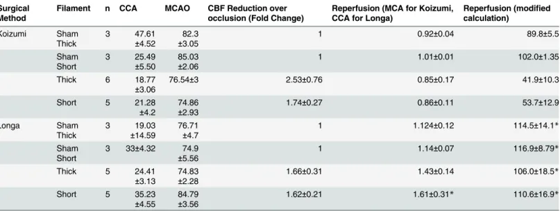
There were no significant differences in relative levels of CBF following CCAO, MCAO or reperfusion in any Koizumi groups, once again indicating consistency (Table 3). There was however significantly increased reperfusion (via the modified calculation) in animals undergo-ing the Longa surgeries with short filaments, followundergo-ing the release of the temporary suture occluding the CCA, when compared with reperfusion following the Koizumi method with short filaments (Table 3,F(7,25)= 2.639,p<0.05), which leaves the right CCA permanently
blocked (Table 3). Significantly increased reperfusion in the Longa vs. the Koizumi method was also verified by the traditional calculation of reperfusion, with a significant increase detected when comparing any of the Longa groups to the Koizumi thick filament group (F(7,25)= 3.81,p<0.01).
Relative changes in CBF following CCAO, MCAO and reperfusion do
not correlate to ischemic infarct volume
In intraluminal filament MCAO studies there is a tendency to remove animals with laser-Doppler calculated MCAO reductions lower than‘70%’relative to baseline CBF following MCAO [37–39]. However, to the best of our knowledge there is no study in mice indicating animals with relative alterations in CBF of<70%, calculated using laser-Doppler derived mea-surements, have different size ischemic lesions to animals with relative alterations in CBF of
>70% in the MCAO territory. Determining if this 70% cut-off point is statistically relevant is important as many studies may be unnecessarily removing data on the basis of an arbitrary cut-off point. We therefore analysed whether there was any statistical evidence to support the removal of data on the basis of relative CCAO, MCAO and reperfusion values calculated from laser-Doppler readouts, by correlating these values to resulting lesion volumes in each mouse, via linear regression. The laser-Doppler data was pooled where appropriate, in all animals undergoing the same conditions (i.e. Koizumi surgeries with thick filaments, 60 min occlusion and 4 h recovery, and Koizumi surgeries with thick filaments, 60 min occlusion and 24 h ery), but where conditions differed, such as the variety of different occlusion periods and recov-ery times, which can influence the resulting lesions volumes, data was correlated separately.
experienced a reduction in CBF of>70% from baseline following intraluminal filament MCAO (138/153 (87.9%)), there were a small proportion of animals with MCAO drops falling between 60–70% (15/153 (9.6%)) and an even smaller proportion with values falling between 40–60% (4/153 (2.5%)).
Fig 5. Ischemic injury detected after 4 h reperfusion by TTC following 60 min intraluminal filament MCAO via the Koizumi or Longa method(A) Total volume of ischemic lesion in the ipsilateral hemisphere, expressed as a percentage of the total contralateral hemisphere volume, measured at 4 h after reperfusion, following 60 min MCAO via the Koizumi or Longa method with a thick or short silicone coated filament. (B) Representative TTC stained brain sections indicating areas of healthy tissue (red) and ischemic injury (white) for each group. (C) Body weight loss following either Koizumi or Longa surgeries measured at 4 h after reperfusion. Each value represents the mean±the standard error of the mean (SEM). N = 3 for sham surgeries and n = 5–6 for animals undergoing occlusion.
There was no correlation between the size of CCAO and resulting lesion volume in each group assessed (Table 4). For all groups there was also no significant correlation between the levels of MCAO and reperfusion (calculated via both methods) compared to resulting lesion Table 3. Relative alterations in CBF following CCAO, MCAO, during occlusion and following reperfusion for Koizumi vs. Longa at 4 h after reperfusion.
Surgical Method
Filament n CCA MCAO CBF Reduction over occlusion (Fold Change)
Reperfusion (MCA for Koizumi, CCA for Longa)
Reperfusion (modified calculation) Koizumi Sham Thick 3 47.61 ±4.52 82.3 ±3.05
1 0.92±0.04 89.8±5.5
Sham Short 3 25.49 ±5.50 85.03 ±2.06
1 1.01±0.01 102.0±1.35
Thick 6 18.77
±3.06
76.54±3 2.53±0.76 0.85±0.17 41.9±10.3
Short 5 21.28
±4.2
74.86 ±2.93
1.74±0.27 0.86±0.11 53.7±12.9
Longa Sham Thick 3 19.03 ±14.59 76.71 ±4.7
1 1.124±0.12 114.5±14.1*
Sham Short
3 33±4.32 74.9 ±5.56
1 1.14±0.07 116.9±8.79*
Thick 5 24.41
±3.13
74.83 ±2.28
1.66±0.31 1.43±0.14 106.0±18.5*
Short 5 35.23
±4.55
84.79 ±3.56
1.62±0.21 1.61±0.31* 110.6±16.9*
Each value represents the mean±the standard error of the mean (SEM).
*p<0.05
doi:10.1371/journal.pone.0148503.t003
Table 4. Correlations of relative alterations in CBF following CCAO, MCAO, reperfusion, neurological score, weight loss, age and starting weight to ischemic infarct volume.
Occlusion Time (min)
Recovery Time (h)
n Filament Surgical Method
CCAO MCAO Reperfusion (modified calculation) Reperfusion (traditional calculation) Neurological Score Weight Loss (% of starting weight) Starting Body Weight Starting Age
60 0.5 7 Thin Koizumi 0.1268 0.7003 -0.3991 -0.6205 N/A N/A 0.2233 0.09996
60 4 8 Thin Koizumi 0.09871 -0.375 -0.4095 -0.4299 0.1839 0.3474 0.2558 -0.07882
60 12 6 Thin Koizumi 0.2873 0.5682 0.09023 0.05019 -0.7407 0.7662 0.4453 0.01676
60 24 6 Thin Koizumi 0.2076 0.6478 -0.3418 -0.4294 0.4349 -0.4070 -0.1856 -0.1225
60 0.5 7 Thick Koizumi -0.222 -0.04689 -0.49 -0.518 N/A N/A 0.04971 0.2553
60 4 20 Thick Koizumi 0.2835 0.3207 0.3383 0.1962 0.1111 0.06063 0.4602* 0.4344
60 12 5 Thick Koizumi -0.0462 0.3353 -0.06473 0.7795 0.8748 -0.7795 0.1223 0.3851
60 24 10 Thick Koizumi 0.4724 -0.07115 0.0807 0.07662 0.3416 -0.1274 0.07993 0.2628
15 4 5 Thick Koizumi 0.554 -0.4102 -0.3433 -0.7058 0.1576 -0.03727 -0.1738 0.2323
30 4 5 Thick Koizumi -0.4309 0.8331 -0.608 -0.7601 0.7643 -0.7601 -0.1565 -0.8328
45 4 5 Thick Koizumi -0.3467 0.3023 -0.03974 -0.7842 0.3102 -0.1438 -0.193 0.7155
15 24 4 Thick Koizumi -0.1864 0.2054 -0.7422 -0.8587 -0.7998 0.2103 -0.947 0.3452
30 24 4 Thick Koizumi 0.773 0.7218 0.0176 -0.321 -0.2203 0.01763 -0.4964 -0.249
45 24 5 Thick Koizumi -0.1106 0.7955 -0.5429 -0.5452 -0.3235 -0.3235 -0.6357 -0.7553
60 24 5 Short Koizumi -0.5642 -0.8274 0.3913 0.3697 N/A 0.2984 0.4118 -0.1326
60 4 5 Short Longa -0.6358 0.1284 -0.8624 -0.4658 -0.8624 -0.4658 -0.5046 -0.8457
60 4 5 Thick Longa 0.2314 -0.2747 -0.4232 0.2166 N/A 0.5530 -0.28 -0.4075
Values represent r values of correlations.
*p<0.05
volume. This indicates that the relative alteration in CBF following MCAO, calculated using laser-Doppler, does not determine the lesion volume. These results therefore provide support for the argument it is unnecessary to remove data on the basis of MCAO drops calculated from laser-Doppler readouts and indicates that the level of reperfusion does not provide any statisti-cal indication of resulting lesion volume.
Previous studies of intraluminal filament MCAO have also indicated that the percentage body weight loss post-MCAO [40] can correlate to lesion volume. Starting age and starting body weight may also influence lesion volume, based on the premise that different weight mice have different size arteries, and therefore occluding filaments may be either too big or too small depending on the initial size of a mouse. To determine the influence of these factors in our model we also correlated them final lesion volume. There was no correlation between the neu-rological score, percentage body weight loss, or starting age and lesion volume in any of the groups assessed. In 16 out of the 17 experimental groups there was also no correlation between starting body weight and lesion volume. However heavier animals had a statistically significant increase in lesion volume following a 60 min occlusion via the koizumi method with thick fila-ments, assessed 4 h post-reperfusion (r = 0.4602,p<0.05).
Although laser-Doppler provides only arbitrary readouts in CBF value during surgery, occlusion and reperfusion, its main usefulness not only lies in the identification of when the occluding filament is in the right location, but also in the identification of rare events such as possible SAHs and premature reperfusion during occlusion, which can assist with the correct removal of data as a result. As a guide for identification we have provided some examples of these events occurring in our own data sets (Fig 6). SAH was identified as previously described [25,41], via a distinct drop in CBF following removal of occluding filament, or a failure or reperfusion. In our study these cases were rare, with only 7 out of the 158 animals surviving to their collection time points requiring removal from the final data set due to possible SAHs occurring and only 1 out of the 158 removed due to possible premature reperfusion (Fig 6F). As a proof of principle that SAH can influence the lesion volumes, in the case of the animal represented inFig 6C, a large lesion was apparent as a result of a possible SAH, despite this ani-mal not undergoing any occlusion as they were in a designated sham group.
However, we also found that the laser-Doppler can provide false-positives of SAH. As repre-sented inFig 6D, there were two cases of MCAO occlusion failing to produce a distinct drop in CBF, but filament withdrawal causing a large drop in CBF, indicating SAH. Both these cases occurred in animals undergoing a SHAM surgery. In the case of the animal represented inFig 6D, which was also undergoing a sham surgery, there was visible evidence of a blood clot hav-ing formed near the MCA, validathav-ing the Doppler result, but no ischemic lesion was visible, indicating the perforation of the vessel wall causing the blood clot may have repaired itself suf-ficiently to prevent any cellular injury. However, considering the case of the animal inFig 6C, we immediately removed any data showing possible SAH in case lesion volume was inadver-tently affected.
Discussion
Surgical Experience Improves Surgical Outcome in the Intraluminal
Filament Model of MCAO
Surgical skill, when performing intraluminal filament models of MCAO in rodents, is often cited as a major possible cause of inconsistency in resulting lesion volumes [17,42]. To reduce the impact of surgical experience on variability in this study, the surgeon (GM) was trained extensively over>100 surgeries, with guidance from two experienced practitioners (RT and AG), prior to gathering data. When mapping the progress of this training period, it was clear the ability to perform a successful surgery increased over time. Mortality caused by surgical error (S2 Table) reduced from 60.6% in an initial group of practice surgeries, to only 4.8% in a later group (S1 Table). We also attributed high mortality rates in our early study to the long
Fig 6. Representative laser-Doppler flowmetry traces of SAH and premature reperfusion(A) A typical representative laser-Doppler flowmetry during Koizumi’s method of middle cerebral artery occlusion (MCAO), during a 60 min occlusion. Insert: representative resulting ischemic lesion analysed by TTC staining. (B) A typical representative laser-Doppler flowmetry during Longa’s method of MCAO, during a 60 min occlusion. (C) Representative laser-Doppler flowmetry of subarachnoid haemorrhage (SAH) during the Koizumi method of MCAO, with resulting ischemic injury. (D) Representative laser-Doppler flowmetry of false-positive SAH. Inserts: image of the same animal, with a brain bleed resulting from vessel perforation, yet leading to no discernible ischemic injury. (E) Representative laser-Doppler flowmetry showing arbitrary units of CBF falling gradually over the course of a 60 min MCAO occlusion time, conducted via the Koizumi method. (F) Representative laser-Doppler flowmetry indicating premature reperfusion, due filament movement out of place, followed by immediate recovery of MCAO by repositioning the filament. Perfusion Units (PU) are arbitrary units of cerebral blood flow.
(90 min) occlusion time we were initially using, shifting to a 60 min occlusion period as the longest utilised in this study. Mortality of animals 24 h post-surgery markedly decreased with practice (>80% down to<52%,S1 Table), and was similar to levels seen in other studies, which have been as high as 60–85% in mice [35,43,44]. Considering mice are more susceptible to occlusion time than rats, where a 90 min occlusion is common, this is not surprising [2]. Another important factor we noted was that the insertion distance of filaments, based on laser-Doppler guided placement, was highly consistent at 9.0 ± 0.5 mm from the bifurcation of the CCA, in line with other studies using similar age and weight C57BL/6 mice [45]. However, it is common to see an insertion distance of 11 mm [46–48] or insertion until‘a mild resistance is felt’[49], which we are concerned raises the possibility of inadvertent perforation of the ACA, increasing lesion volume and inconsistency in the model, a view shared by others [32].
As a result of undertaking the rigorous training progress, the number of mice required in this study was profoundly decreased, considering the markedly increased survival rate follow-ing extensive trainfollow-ing (S1 Table). Of the 274 animals entered into the main study, only 8 out of 274 (2.9%), were deceased due to error during surgery, and only 16 out of 158 (10.1%) that sur-vived to their designated times after reperfusion were removed from final analysis due to either surgical anomalies such as the movement of the filament during occlusion, possible SAH (Fig 6), or because they were outliers. This compares favourably to other studies, where as many as 30% of animals have been excluded from final analysis [50]. A particularly encouraging statistic was the 95.9% (47/49) survival rate in animals undergoing sham surgeries, indicating the vast majority of deaths post-surgery are likely due to the impact of the occlusion and resulting lesion, rather than surgery related injury.
A major factor contributing to the difficulty of surgery, particularly in mice that have a nar-row surgical window compared to rats, is the anatomical differences between mice, even amongst the same strain. As two quantifiable examples, the occipital artery (OA) and superior thyroid artery (STA), each isolated and cauterised in our surgical procedure, can originate from several different structures. As reported by Chen et al., [8], the OA was originally described as bifurcating from the proximal segment of the ECA in mice. However, similar to their observations, we found the OA instead originated from the ICA, the proximal segment of the ECA and the bifurcation of the CCA 66.5%, 10% and 23.5% of the time, respectively (S3 Table). The STA on the other hand had a more consistent appearance, originating from the ICA, the proximal segment of the ECA and the bifurcation of the CCA 6.8%, 91.3% and 1.9% of the time. The impact of these differing origins on surgery is to increase the difficulty of their removal. For instance, an OA arising more distal to the bifurcation is likely in closer proximity to the vagus nerve, increasing the risk of inadvertent damage to this structure.
provided any difference in post-stroke measures following occlusion. We found no significant difference in lesion volume when comparing these two filaments at any time point after reper-fusion, nor were there any differences in resulting CCAO, MCAO or reperreper-fusion, indicating both diameter filaments occluded the MCA to the same reproducible degree. Survival post-sur-gery was also similar for both groups. Therefore, both 0.21 mm and 0.23 mm thick silicone coated filaments are suitable for use in the intraluminal filament method of MCAO in mice, within the body weight and age ranges used in this study.
No changes in total ischemic lesion volumes were observed following
varying times after reperfusion using TTC staining
Somewhat surprisingly, no significant differences in total lesion volume were observed when we measured the levels of ischemic lesions just 30 min and 4 h after reperfusion, compared to 24 h after reperfusion. This result is in contrast to a previous study in mice, where lesion was shown to evolve from 1.5 h to 24 h in mice [14], and also from 0 to 24 h using anin vitroTTC staining technique in rats [52].
However, Hatfield et al., showed that TTC staining using a perfusion technique could clearly identified lesions at 30 min after reperfusion that are not significantly different to those at 24 h in rats [29]. More recently, and also using anin vivoTTC-staining perfusion technique in rats, infarct areas were shown to be large immediately after reperfusion, but significantly decreased after 16h reperfusion, before significantly increasing once more by 24 h [52]. This is similar to our results with thin filaments, whereby we have a significantly smaller ischemic area at 12 h after reperfusion than at 30 min, which then increases again by 24 h. As Benedek et al., suggested, this could represent recovery of penumbra at 12 h [52], an observation also partially supported by our results showing fewer mice with a lack of staining in the thalamus and hippocampus by 12 h compared with earlier time points. Reversible injury following longer reperfusion has also been detected using TTC staining, suggesting some viable cells with perturbed mitochondrial activity, early after MCAO, may recover following longer periods of reperfusion [53].
Another more simple explanation for ischemic injury failing to increase over time in our study may be due to a rapid decline in survival post-12 h. It is entirely possible animals with larger lesions were amongst those less likely to survive, hence a lack of clear evidence for lesion and neurological deficit evolution at later time points after reperfusion.
In any case, our results, and those by others, question the reliability of the TTC stain to accurately determine the evolution of infarct during early time points after reperfusion, espe-cially considering the stain may produce a false-positive detection of injury due to transient alterations in the ability of mitochondrial enzymes to metabolise TTC [52–55]. There is also a suggestion TTC may be unreliable at time after reperfusion longer than 24 h, considering later infiltration of healthy marcophages into infarcted tissue could increase the level of TTC stain-ing [56].
Despite these qualms, a debate could be made that early, reversible cell death detectable by TTC staining, is a useful marker of regions likely to undergo degeneration [14], and there has been a number of studies that have validated TTC staining as a reliable tool for infarct assess-ment in comparison to other common methods such as Fluoro-Jade B, NeuN and cresyl violet staining, and light microscopic evaluation [14,54,57,58].
Intraluminal filament MCAO with long coated filaments consistently
causes ischemic injury in regions outside of the MCA territory
arising in sections corresponding to Bregma 0.0 to -1.0. However, with increasing occlusion time, we noticed an interesting trend in the hippocampus, where the dorsal and ventral hippo-campus often lacked staining, suggesting ischemic injury, at earlier time points (8/18 and 9/18 respectively for all animals with 60 min MCAO via the Koizumi method, with thick filaments, analysed 4 h after reperfusion), yet tended to have little obvious hippocampal injury at 24 h (1/ 10 and 1/10 respectively for all animals with 60 min MCAO via the Koizumi method, with the thick filaments, after 24 h after reperfusion).
A similar trend was seen in the thalamus with 30 min (2/7), 4 h (7/18), 12 h (1/5) and 24 h (1/10) animals showing ischemic lesions in this territory (following a 60 min MCAO via the Koizumi method with the thick filaments), suggesting there may be reversible injury in these areas, when analysed by TTC staining. However, the proportion of animals with ischemic injury in the amygdala increased with increasing time after reperfusion, with 30 min (2/7), 4 h (8/18), 12 h (5/5) and 24 h (9/10) animals showing lesions in this territory (following 60 min MCAOs via the Koizumi method with the thick filaments). No ischemic lesions were seen in the any of these areas after shorter durations occlusions (15 and 30 min), indicating the extent of ischemic injury was dependent on duration of occlusion, as confirmed in other studies [19]. Despite the fact the hippocampus, thalamus and amygdala are not supplied by the MCA, it is not uncommon for these regions to show evidence of ischemic injury following MCAO [19,44,45,59], for several reasons. First and foremost, C57BL/6 mice may be more susceptible to variation in intraluminal filament MCAO compared to other strains owing to a common lack of patency in posterior communicating arteries (PComA) [31,44]. Up to 30% of C57BL/6 may be deficient in both PComAs, with perhaps only 10% having both [19]. The consequence of missing PComAs is that territory supplied by the posterior communicating artery (PCA) will also experience a lack of blood flow when an intraluminal filament is present, as a lack of a PComA will prevent collateral blood supply to this area during occlusion [19,44]. Considering the PCA supplies blood to the hippocampus and thalamus, these areas may therefore experi-ence ischemia during occlusion. Even with shorter coated filaments it is impossible to circum-vent the issue of poorly developed or absent PComAs, as in this case blood supply to the ipsilateral ACA, and therefore PCA, will still be blocked.
Second, blood supply may also be inadvertently blocked to the anterior choroidal artery (AChA), ventral thalamic artery (VTA) and hypothalamic artery (HTA,Fig 1). This is espe-cially likely with longer coated filaments, as the AChA, VTA and HTA branch from and are supplied by the internal ICA (Fig 1) [23,60]. Despite using longer coated filaments, we only saw evidence of hypothalamic lesions in one mouse, across all groups, which is less than has been reported previously [19]. As mentioned above however, we did see lesions in the thala-mus, which may not only be due absent PComAs, but perhaps also by blockage of the VTA. Interestingly, the likelihood of injury in the thalamus did appear to decrease when shorter coated filaments were used.
Finally, regions outside of the MCA territory may be susceptible to collateral damage from the effects of a variety of pathophysiological mechanisms of ischemic injury including excito-toxicity, spreading depression, reactive oxygen species, inflammation and apoptosis [36].
accurately measured [61]. Defining these distances may aid in determining the optimal length of filament coating to prevent ischemic lesions occurring in areas provided by these arteries.
Ischemic injury increases sharply between 30 and 45 min of occlusion
Studies using the intraluminal filament model of MCAO generally utilise one distinct occlusion period which can range from brief (15 min) to longer (180 min, or permanent). Despite this, lesion volumes have varied greatly from lab to lab, with some producing much larger lesions volumes than others, even with the same occlusion period [2]. In our study few ischemic lesions were noticeable following minor (15 min) periods of occlusion when analysed by TTC at 4 h, and only inconsistent, small lesions were visible following 24 h reperfusion after 15 min occlusion in the striatum and cortex. However, with increasing occlusion periods, the increase in lesion volume was remarkably consistent, until 45 min, wherein lesion volume appeared to peak, with no statistically significant difference between the overall lesion volume generated following a 45 min or 60 min occlusion. This does not necessarily mean 45 min is the‘ thresh-old’level of occlusion in mice, as 2 h occlusion has in the past been shown to significantly increase lesion volume over a 1 h occlusion [47], however it is a fair indication there is a consid-erable jump in lesion volume between 30 min and 45 min occlusion, suggesting>30 min of occlusion dramatically increases ischemic injury in the mouse.Others have found a steep difference in lesion volume between only 15 and 30 min of occlu-sion, when performing a similar time course, although these results were obtained with differ-ent filamdiffer-ent properties, differ-entry via the ECA rather than the CCA, and older mice than in our model [19]. Remarkably in the rat, lesion volume may not peak until after 2–3 h [15], or per-haps even 3–4 h of occlusion [63]. Together these results indicate a much greater susceptibility of mice to occlusion time than rats.
The Longa and Koizumi methods of intraluminal filament MCAO do not
produce differences in lesion volume in mice at 4 h reperfusion
To the best of our knowledge, only two studies exist directly comparing the Longa and Koizumi methods of intraluminal filament MCAO in rodents, despite widespread use of both tech-niques. The first, from Laing et al., in rats, illustrated that the method of Longa was shown to be unreliable in reproducibly inducing infarct, compared to the Koizumi method [22]. This was thought likely due to the properties of the filament employed in Longa’s method, which were more likely to induce vessel perforation during insertion [22]. A more recent study in mice compared the two methods, but employed a consistent 180μm silicone-tipped filament when performing both methods [64]. Contrary to the study of Laing, this study concluded that the Koizumi method produced a higher mortality rate after 24 h reperfusion, but the lesion vol-umes did not differ at 24 h.
likely to impact resulting lesion volumes at both 24 h and 7 d, as animals with larger lesions may not survive to these time points. Further research is required to compare and contrast the usefulness of these two models under different scenarios, utilising different filaments, different occlusion periods, different analysis times after reperfusion, and different techniques for cell death assessment.
Relative CCAO, MCAO and reperfusion laser-Doppler measurements
do not correlate with lesion volume, but can identify rare cases of SAH
and premature reperfusion
Laser-Doppler measurement has long been utilised as a tool to assist with intraluminal filament surgeries as it offers a simple, continuous, instantaneous, inexpensive and non-invasive way to assess relative CBF alterations in specific brain regions. Although absolute values obtained from laser-Doppler instruments are arbitrary, relative changes in CBF correlate well with other methods of CBF detection such as autoradiography [65]. When laser-Doppler data is reported, it is often accompanied by a statement that animals failing to register a 70% drop from baseline CBF, following intraluminal filament occlusion of the MCAO, are excluded from final datasets [66,67]. In some cases this cut-off has been 60% [17,68–70], 80% [71] or as high as 85% [14], but 70% has been a popular choice, in both mouse and rat studies [37–39].
The 70% figure perhaps derives from studies aiming to determine‘thresholds’for CBF, below which tissue will become irreversibly injured during MCAO [72,73]. For instance, using the [14C]iodoantipyrine (IAP) autoradiographic technique, changes in local CBF were mapped to the probably of infarct in a rat model of intraluminal filament MCAO [74,75]. It was deter-mined that decreases in local blood flow of 0–20% gave a 96% probability of infarction, leading to irreversibly injured core areas of infarct. Even minor differences in this level substantially altered the probability of infarction, with penumbral regions (surrounding the core) only experiencing decreases in CBF to 20–40% of control.
However, the applicability of these thresholds to intraluminal filament MCAO in the mouse, using laser-Doppler to derive relative changes in CBF, is unclear. Early papers using laser-Doppler flowmetry in the intraluminal filament technique of MCAO did not appear to exclude animals failing to meet thresholds [25,41,45,76], nor do some more recent papers [77]. Furthermore, to the best of our knowledge there is no statistical comparison between laser-Doppler measurements of MCAO and resulting lesion volume in mice that suggests differences in the percentage drop in CBF following MCAO leads to alterations in lesion volume. There-fore there is no statistical support for the exclusion of animals with a relative change in CBF of
<70% in the MCA territory following MCAO, calculated using laser-Doppler measurements. There is one relevant study in rats suggesting perfusion deficits, measured by laser-Doppler in the territory of the MCA-ACA leptomeningeal branches, but not the MCA territory, may be a useful predictor of resulting infarct volume and neurological deficit [78]. A lack of these corre-lations in mice may be the cause of the current varied approaches to animal exclusion on the basis of laser-Doppler measurements.