Vibrio cholerae
Response Regulator VxrB
Controls Colonization and Regulates the Type
VI Secretion System
Andrew T. Cheng, Karen M. Ottemann, Fitnat H. Yildiz*
Department of Microbiology and Environmental Toxicology, University of California, Santa Cruz, California, United States of America
Abstract
Two-component signal transduction systems (TCS) are used by bacteria to sense and re-spond to their environment. TCS are typically composed of a sensor histidine kinase (HK) and a response regulator (RR). TheVibrio choleraegenome encodes 52 RR, but the role of these RRs inV.choleraepathogenesis is largely unknown. To identify RRs that controlV. choleraecolonization, in-frame deletions of each RR were generated and the resulting mu-tants analyzed using an infant mouse intestine colonization assay. We found that 12 of the 52 RR were involved in intestinal colonization. Mutants lacking one previously uncharacter-ized RR, VCA0566 (renamed VxrB), displayed a significant colonization defect. Further ex-periments showed that VxrB phosphorylation state on the predicted conserved aspartate contributes to intestine colonization. The VxrB regulon was determined using whole ge-nome expression analysis. It consists of several genes, including those genes that create the type VI secretion system (T6SS). We determined that VxrB is required for T6SS expres-sion using several in vitro assays and bacterial killing assays, and furthermore that the T6SS is required for intestinal colonization.vxrBis encoded in a four gene operon and the othervxroperon members also modulate intestinal colonization. Lastly, thoughΔvxrB ex-hibited a defect in single-strain intestinal colonization, theΔvxrBstrain did not show anyin vitrogrowth defect. Overall, our work revealed that a small set of RRs is required for intesti-nal colonization and one of these regulators, VxrB affects colonization at least in part through its regulation of T6SS genes.
Author Summary
Pathogenic bacteria experience varying conditions during infection of human hosts and often use two-component signal transduction systems (TCSs) to monitor their environ-ment. TCS consists of a histidine kinase (HK), which senses environmental signals, and a corresponding response regulator (RR), which mediates a cellular response. The genome of the human pathogenV.choleraecontains a multitude of genes encoding HKs and RRs proteins. In the present study, we systematically analyzed the role of eachV.choleraeRR
a11111
OPEN ACCESS
Citation:Cheng AT, Ottemann KM, Yildiz FH (2015)
Vibrio choleraeResponse Regulator VxrB Controls Colonization and Regulates the Type VI Secretion System. PLoS Pathog 11(5): e1004933. doi:10.1371/ journal.ppat.1004933
Editor:Andreas J Baumler, University of California, Davis, UNITED STATES
Received:April 29, 2015
Accepted:May 4, 2015
Published:May 22, 2015
Copyright:© 2015 Cheng et al. This is an open access article distributed under the terms of the Creative Commons Attribution License, which permits unrestricted use, distribution, and reproduction in any medium, provided the original author and source are credited.
Data Availability Statement:All relevant data are within the paper and its Supporting Information files.
Funding:This study is funded by National Institutes of Health, National Institute of Allergy and Infectious Diseases (AI055987 and AI102584) (http://www.niaid. nih.govto FHY. The funders had no role in study design, data collection and analysis, decision to publish, or preparation of the manuscript.
for its role in pathogenesis. We identified a previously uncharacterized RR, VxrB, as a new virulence factor. We demonstrated that VxrB controls expression of the type VI secretion system (T6SS), a virulence nanomachine that directly translocates effectors into bacterial or host cells, thereby facilitating colonization by competing with sister cells and intestinal microbiota. This study represents the first systematic analysis of the role of all RRs inV.
choleraepathogenesis and provides a foundation for understanding the signal transduc-tion pathways controllingV.choleraepathogenesis.
Introduction
Vibrio choleraecauses the diarrheal disease cholera that affects 3 to 5 million people worldwide every year, resulting in 100,000–120,000 deaths annually [1].V.choleraeproduces a number of virulence factors which facilitate colonization of the intestine and subsequent disease. Major virulence factors are cholera toxin (CT), which is responsible for production of profuse watery diarrhea, and a type IV pilus called the toxcoregulated pilus (TCP), which is required for in-testinal colonization [2].V.choleraevirulence factors are well known to be under extensive transcriptional control. CT and TCP production are controlled by the transcriptional activator ToxT [3,4]. Expression oftoxT, in turn, is controlled by a virulence regulatory cascade involv-ing the membrane-bound transcriptional activators ToxRS and TcpPH. These two regulators activatetoxTtranscription directly [5–7]. TcpPH expression is activated by the transcriptional activators AphA and AphB [8,9]. The quorum sensing (QS) regulatory system is also linked to the virulence gene regulatory cascade through HapR, the master QS regulator, which represses
aphAexpression [10].
Recently, the type VI secretion system (T6SS) has been identified as a new virulence factor inV.cholerae[11,12]. T6SSs deliver effector proteins into both eukaryotic and bacterial cells in a contact-dependent manner [12,13].V.choleraehas one T6SS system with multiple T6SS effectors: VrgG1 and VrgG3 (valine-glycine repeat protein G), which have actin cross-linking activity and peptidoglycan-degrading activity, respectively [14–17]; TseL, which has lipase ac-tivity [15,18]; and VasX, which perturbs the cytoplasmic membrane of target cells [15,19]. Ac-tivity of these effectors is antagonized by corresponding immunity proteins: TsiV3, TsiV1, and TsiV2, respectively, to prevent killing by strains bearing these proteins [15,16,20,21].
The T6SS can be divided into functional sections consisting of the core structural compo-nents, the T6SS effector and immunity proteins, and transcriptional regulators. The base of the T6SS apparatus spans the cell envelope, and is a tube within a tube. The inner tube is composed of polymers of the hemolysin coregulated protein (Hcp). The outer tube, also called the con-tractile sheath, is formed by polymers of VipA and VipB [14,22]. The Hcp inner tube is capped with a spike complex of trimeric VgrG proteins. The effectors are delivered by contraction of the VipA/VipB sheath, which in turn results in ejection of the inner tube along with VgrG and the effectors towards the target cell [12].
The genes encoding the T6SS components are organized into one large cluster
(VCA0105-VCA0124) and two auxiliary clusters (VCA0017-VCA0022 and VC1415-VC1421) [11,23]. A key positive transcriptional regulator of theV.choleraeT6SS is VasH (VCA0117), which is related to enhancer binding proteins that activate transcription in aσ54 (RpoN) de-pendent manner [24,25]. VasH acts on the T6SS auxiliary clusters andvgrG3of the large clus-ter, but does not affect expression of the structural genes encoded in the large T6SS gene cluster [24,26]. Additionally, Hcp production is positively regulated by the master quorum sensing regulator HapR and the global regulator cyclic AMP (cAMP) receptor protein CRP, and
negatively regulated by QS regulator LuxO and by global regulator TsrA, a protein homologous to heat-stable nucleoid-structuring (H-NS) [27,28]. These studies have thus shown that nu-merous global regulators control T6SS expression, as well as one specific regulator (VasH).
V.choleraeT6SS studies have mainly focused on theV.choleraeO37 serogroup V52 strain because it assembles a T6SS apparatus constitutively [11]. In this strain, the T6SS is required for cytotoxicity towardsDictyostelium discoideumand J774 macrophages, and induces inflam-matory diarrhea in the mouse model [29]. InV.choleraeO1 strain C6706, the T6SS is not con-stitutively produced and conditions that promote T6SS production are unknown. However, production of T6SS can be achieved in other O1 strains by inactivating mutations in genes en-coding the LuxO and TsrA negative regulators. In O1 strains, the T6SS translocates T6SS effec-tors into macrophages, and increases fecal diarrhea and intestinal inflammation in infant rabbits [27]. It was also shown that theV.choleraeO1 C6706 strain T6SS mediates antagonistic interbacterial interactions during intestinal colonization. A strain unable to produce the TsiV3 immunity protein, which provides immunity against the effector VgrG3, exhibited an intestinal colonization defect only when co-infected with strains harboring an intact T6SS locus and VrgG3 [30]. Although T6SS is regulated and expressed differently betweenV.choleraestrains, production of this system in multiple strains promotes virulence against both eukaryotic and bacterial cells, suggesting the function is largely conserved but the regulation varies.
Pathogenic bacteria experience varying conditions during infection of human hosts and often use two-component signal transduction systems (TCSs) to monitor their environments [31]. TCSs play important roles in the regulation of virulence factors, metabolic adaptation to host environments, and response to numerous environmental stresses including pH, osmolar-ity, oxygen availabilosmolar-ity, bile salts, and antimicrobial peptides [32]. TCS rely on a phosphore-lay-based signal transduction system. The prototypical TCS consists of a membrane-bound histidine kinase (HK), which senses environmental signals, and a corresponding response reg-ulator (RR), which mediates a cellular response. Response regreg-ulators are typically multi-do-main proteins harboring a conserved receiver domulti-do-main (REC) and C-terminal output domulti-do-main such as DNA-binding, diguanylate cyclase, or methyltransferase [33–35]. Upon environmen-tal stimulation, the HK caenvironmen-talyzes an ATP-dependent autophosphorylation reaction on a con-served histidine residue. The phosphoryl group is transferred from the HK to a concon-served aspartate residue on the RR, eliciting a conformation change and subsequent cellular response [32,34,35].
TheV.choleraegenome reference genome of O1 EL Tor N16961 strain is predicted to en-code 43 HK and 49 RR (http://www.ncbi.nlm.nih.gov/Complete_Genomes/RRcensus.htmland
regulators and concomitant CT and TCP production; lipid A modification enzymes; or motility and chemotaxis. 39/52, however, were not yet analyzed at the time of this study.
To systematically evaluate the role ofV.choleraeTCSs in intestinal colonization, we generat-ed in-frame deletion mutants of each RR gene and analyzgenerat-ed thein vivocolonization phenotypes of the resulting mutants. We found 12 RR were required for wild-type intestinal colonization. One RR in particular had a very strong defect, encoded by genomic locus VCA0566. We deter-mined that VCA0566 (now termedVibriotype six secretion regulator,vxrB) controls expression of several genes including the T6SS genes. We used multiple methods to substantiate that VxrB is required for expression of the T6SSin vitroandin vivo. Lastly, we report that the T6SS con-tributes to colonization of theV.choleraeO1 strain used in this study.
Results
Multiple RRs impact intestinal colonization
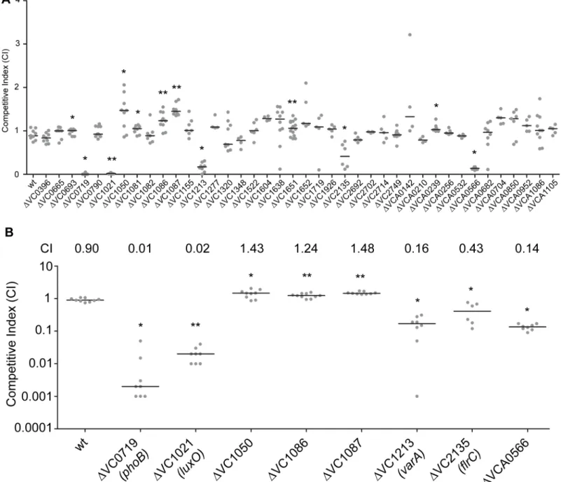
We have a limited understanding of theV.choleraeTCSs and their role in colonization and ad-aptation to host environments. To evaluate the importance of the 52 TCS RRs in colonization, we generated in-frame deletion mutants of the 40 RRs. For this analysis, we excluded 12 RR that were either predicted to be involved in chemotaxis (11 CheY, CheV, and CheB proteins) or that we were unable to mutate (VC2368, ArcA) [43,45]. We then analyzed the ability of 40 RR deletion mutants to colonize the small intestine in anin vivocompetition assay wherein vivofitness of a mutant strain is compared to that of wild type strain using the infant mouse in-fection model (Fig 1A) [46]. While the vast majority of mutants—28—were not different from wild type, we identified 12 RR mutants that had a statistically significant colonization differ-ence as compared to wild type (Fig 1A). We focused on 8 mutants with a statistically significant colonization difference and exhibited at least 1.2-fold difference in CI (Fig 1B). Consistent with previous studies, we identified thatΔVC0719 (phoB),ΔVC1021(luxO),ΔVC1213 (varA),and
ΔVC2135(flrC)were defective in colonization [36,37,40,44]. The competitive indices (CI) for
ΔphoB,ΔluxO,ΔvarA, andΔflrCwere 0.01, 0.02, 0.16, and 0.43, respectively (Fig 1B). Additionally, we identified a set of genes whose absence slightly but statistically signifi-cantly enhanced colonization (at least 1.2 fold higher CI), suggesting that inhibition of their expression and activity may be needed for wild-type colonization. These mutants were
ΔVC1050,ΔVC1086, andΔVC1087, which exhibited subtle and enhanced colonization phe-notypes with CIs of 1.43, 1.24, and 1.48, respectively (Fig 1B). VC1050 is classified as an Hnr-type RR, [47] but its function is yet to be determined. VC1086 and VC1087 are part of a pre-dicted eight gene operon encompassing VC1080-VC1087. Both VC1086 and VC1087 have domains that suggest they function in cyclic guanylate (c-di-GMP) regulation. Specifically, VC1086 contains an EAL domain with conserved residues required for enzymatic function, while VC1087 harbors an HD-GYP domain, but this domain lacks the conserved residues re-quired for enzymatic activity.
We also identified one RR that was defective for colonization that had not been previously characterized. This mutant,ΔVCA0566, had a colonization defect with a CI of 0.14 (Fig1A
and1B). Because this uncharacterized RR was important for colonization, we focused the rest of our studies on this protein.
Δ
VCA0566/VxrB impacts colonization
VCA0566 is the second gene of a predicted five gene operon and had been previously annotat-ed as a RR of the OmpR family. The encodannotat-ed protein, which we namannotat-ed VxrB for reasons de-scribed below, is 245 amino acids in length with an N-terminal REC domain and a C-terminal winged helix-turn-helix DNA-binding domain (Fig 2B). Previously characterized members of
the OmpR family inV.choleraeinclude PhoB, CarR, and ArcA [40–42]. Amino acid sequence alignment of theV.choleraeRRs in the OmpR family and the previously characterizedE.coli
OmpR [48] was used to identify the aspartate residue that is predicted to be phosphorylated in the REC domain (Fig 2A). Since the phosphorylation state of a RR is likely to determine its ac-tivity, we mutated the aspartate residue in the REC domain of VxrB to mimic constitutively
Fig 1. Identification of RRs impacting colonization in the infant mouse infection model.(A) Ability of 40ΔRR mutants inV.choleraestrain A1552 to colonize the infant mouse intestine was analyzed using a competition assay with the isogenic wild-type strain. (B) Same data presented in Fig 1A, expanded to highlight the mutants showing a statistically significant difference in colonization p<0.05 and a minimum 1.2-fold change in colonization ability. Competitive
index (CI) is defined as the output ratio of mutant to wild-type bacteria divided by the input ratio of mutant to wild-type bacteria. Each symbol represents the CI in an individual mouse; horizontal bars indicate the median. Statistical analysis was carried out using Wilcoxon Signed Rank Test, comparing the CI of each strain to the CI of wtlacZ+/ wt lacZ-(shown as wt) (*, p<0.05;**, p<0.01).
active (D78E) and inactive (D78A) versions, as used in other work [48], and replaced the wild-type gene in the chromosome with these altered genes. These mutants were competed against wild type in the infant mouse colonization assay to determine if the phosphorylation state of VxrB is important for colonization. In accordance with our initial colonization screen,ΔvxrB
displayed a CI of 0.15 (Fig 2B). Somewhat surprisingly, the CI forvxrB::D78A (inactive form) was 0.53, indicating a modest defect in colonization. This result indicates that the“inactive” form of VxrB does not phenocopy theΔvxrBmutant, suggesting that VxrB harboring D78A substitution is not fully inactive. The CI forvxrB::D78E (active form) is 1.07, suggesting that
Fig 2. The role of the phosphorylation state of VxrB in colonization.(A) Amino acid sequence alignment of the REC domains of the RR proteins belonging to the OmpR family using ClustalW. Numbers above the sequence correspond to the amino acid number of each protein. VC numbers indicate the RR encoded inV.choleraegenome. OmpR fromE.coliwas used as a reference to align the aspartate residue that has been shown to be important for phosphorylation (D55A) [48]. Highlighted region in gray indicate the aspartate residue that is predicted to be the site of phosphorylation. B)In vivocompetition assay of a strain harboring the mutated version ofvxrBon the chromosome. ThevxrBgene was mutated to convert the aspartate residue predicted to be important for phosphorylation to emulate the inactive (D78A) or active (D78E) state of VxrB.*, p<0.05 by the Wilcoxon Signed Rank Test as compared to the
CI of wt.
doi:10.1371/journal.ppat.1004933.g002
constitutive activation of VxrB does not significantly impairV.cholerae(Fig 2B). Collectively, these findings suggest that in vivo phosphorylation of VxrB at D78 is likely to be important for its colonization function, but apparently not absolutely required. It is also likely that VxrB may not function by conventional phosphorylation-dependent signal transduction [49].
All members of the
vxr
operon contribute to
in vivo
colonization
The first gene of thevxrloci, VCA0565, is annotated as an HK. The other three genes (VCA0567-69) are predicted to encode proteins of unknown function (Fig 3A). We now termed these genesVibriotype six secretion regulator (vxr) ABCDE and determined that these genes are co-transcribed using RT-PCR and RNAseq analysis (Fig 3AandS1 Fig). Both the ge-nomic context and organization is conserved in theVibriospecies (S2–S4Figs) andvxrgene products do not share significant sequence similarity with previously characterized proteins.
To gain a better understanding of the role of thevxroperon in colonization, we investigated whether the cognate HK and other genes in thevxroperon also contributed to mouse coloniza-tion. In-frame unmarked deletion mutants ofvxrA,vxrB,vxrC,vxrD, andvxrE(Fig 3B) were generated and analyzed in anin vivocompetition assay. Each mutant was outcompeted, with CIs of 0.35, 0.16, 0.44, 0.66, and 0.70, respectively (Fig 3B). These findings suggest that while
vxrA and vxrBgenes are critical for colonization in the infant mouse model, contribution of
vxrCDEgenes appears to be minor.
To further confirm the phenotype ofΔvxrBcolonization defect, a wild type copy ofvxrB
whose expression was driven from its native promoter was inserted into the Tn7 site (located between VC0487 and VC0488) on the chromosome ofΔvxrB.In vivocompetition assay of
ΔvxrB-Tn7vxrBhad a CI of 0.93, similar to wild type levels, whereΔvxrBhad a CI of 0.16 (Fig 3B). Thus, theΔvxrBcolonization defect is restored to wild-type levels by introduction of the wild-type copy ofvxrB.
VxrB regulates T6SS gene expression
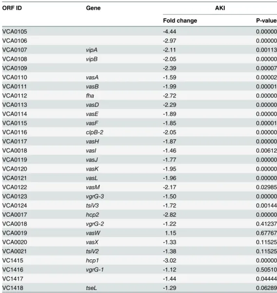
To gain a better understanding of the contribution of VxrB toV.choleraepathogenesis, we per-formed high throughput transcriptome sequencing (RNA-seq) analysis to identify theV. cho-leraegenes controlled by VxrB. We used cells grown under virulence inducing AKI conditions, to mimic the intestinal conditions encountered when we know VxrB is important. 149 genes showed statistically significant differences in gene expression between the wild type and mu-tant (S2andS3Tables). Of these, 80 genes were expressed to greater levels in theΔvxrBmutant relative to the wild type (S2 Table), while 69 were expressed to lower levels in theΔvxrBmutant relative to wild type (S3 Table). Of particular interest was the observation that message abun-dance of most of the T6SS genes in both the large cluster (VCA0105-VCA0124) and the two auxiliary clusters (VCA0017-VCA0022 and VC1415-VC1421) were less in the VxrB mutant relative to wild type (Table 1) (S5 Fig). This finding suggests that VxrB activates expression of the T6SS genes.
VxrB regulates production of T6SS
To further analyze the role ofvxrBin T6SS expression and function, we compared the levels of the major T6SS structural component, Hcp, between wild type andΔvxrBmutantV.cholerae. Quantitative real-time PCR analysis ofhcprevealed that the transcript abundance ofhcpwas decreased by 3.7-fold under AKI conditions and 4.1- fold under LB conditions in theΔvxrB
mutant relative to wild type (Fig 4A). This finding supports that VxrB regulates expression of
hcpand is consistent with the RNA-seq analysis. Additionally the levels of the Hcp protein in
We also determined that complementation of thevxrBmutation (ΔvxrB-Tn7vxrB) restored Hcp to wild-type levels. Because we found lower amounts of Hcp in the supernatant as well as in whole cells, this finding suggests that VxrB is needed to express and secrete Hcp. As negative controls, we included aΔhcp1Δhcp2mutant because it is unable to produce the Hcp proteins
Fig 3. Role of thevxrABCDEoperon in colonization.(A) The top panels shows a schematic
representation of the genomic organization of thevxrABCDElocus The bottom panel shows the results of RT-PCR analysis designed to determine ifvxrABCDEgenes are cotranscribed. RT-PCR primers designed to amplify intergenic regions are indicated. Products were detected for VCA0565-66 (product 1), VCA0566-67 (product 2), VCA0667-68 (product 3), and VCA0568-69 (product 4)V.choleraegenomic DNA template was used for PCR to evaluate primers and amplified product sizes. RT-PCR reaction without reverse
transcriptase (-RT) was used as a negative control. (B) Ability of A1552ΔvxrA,ΔvxrB,ΔvxrC,ΔvxrD,ΔvxrE, andΔvxrB-Tn7vxrBcomplementation strains to colonize the infant mouse intestine was analyzed using a competition assay with wild-type. Each symbol represents the CI in an individual mouse; horizontal bars indicate the median.*, p<0.05;**, p<0.01 by the Wilcoxon Signed Rank Test as compared to the CI of wt.
doi:10.1371/journal.ppat.1004933.g003
[11,50]. As expected, no Hcp production was observed in this mutant. Furthermore, comple-mentation ofhcp1in theΔhcp1Δhcp2mutant partially restored Hcp levels (Fig 4B). Overall these findings suggest that Hcp production is decreased inΔvxrBmutant.
Next we analyzed whether VxrB was needed for T6SS function, by examining T6SS-mediat-ed interbacterial killing. Killing assays between theV.choleraeand the targetE.coliK-12 strain MC4100 showed that wild-typeV.choleraedecreased the numbers ofE.colicompared to con-trol experiments. This killing was dependent on the T6SS, as shown by greater numbers of
E.coliobtained when incubated withV.choleraeΔhcp1Δhcp2mutant andΔvasHmutants, consistent with the findings reported by Ishikawaet al. (Fig 4C) [50]. This phenotype was complemented by introduction of eitherhcp1orhcp2into the Tn7 site on the chromosome. Consistent with our transcriptional and protein analysis presented above, we found thatΔvxrB
mutants mediated lessE.colikilling. These findings suggest that T6SS regulation by VxrB con-tributes to interbacterial killing.
Since VxrB regulates T6SS expression and is required to for intestinal colonization, we next asked whether the T6SS itself is required for intestinal colonization. We performedin vivo Table 1. Expression of Type VI secretion genes in theΔvxrBmutant relative to wild type.
ORF ID Gene AKI
Fold change P-value
VCA0105 -4.44 0.00000
VCA0106 -2.97 0.00000
VCA0107 vipA -2.11 0.00113
VCA0108 vipB -2.05 0.00000
VCA0109 -2.39 0.00007
VCA0110 vasA -1.59 0.00002
VCA0111 vasB -1.99 0.00001
VCA0112 fha -2.72 0.00000
VCA0113 vasD -2.29 0.00000
VCA0114 vasE -1.89 0.00000
VCA0115 vasF -1.85 0.00001
VCA0116 clpB-2 -2.05 0.00000
VCA0117 vasH -1.87 0.00000
VCA0018 vasI -1.46 0.00612
VCA0119 vasJ -1.77 0.00000
VCA0120 vasK -1.95 0.00000
VCA0121 vasL -1.96 0.00000
VCA0122 vasM -2.17 0.02985
VCA0123 vgrG-3 -1.50 0.00000
VCA0124 tsiV3 -1.72 0.00144
VCA0017 hcp2 -2.82 0.00000
VCA0018 vgrG-2 -1.22 0.41237
VCA0019 vasW 1.15 0.67767
VCA0020 vasX -1.33 0.11525
VCA0021 tsiV2 -1.38 0.11525
VC1415 hcp1 -3.02 0.00000
VC1416 vgrG-1 -1.12 0.50510
VC1417 -1.44 0.04444
VC1418 tseL -1.29 0.06289
Fig 4. Analysis of Hcp production and secretion in thevxrBmutant.(A) Quantitative real-time PCR analysis ofhcpusing total RNA isolated from wild-type andΔvxrBgrown in AKI and LB. Experiments were performed using two independent biological replicates, each performed in quadruplicate. The Pfaffl method was used to compare expression levels ofhcpto 16s rRNA and relative expression was calculated by comparing expression inΔvxrBwith that of wt.*, p<0.05 by Student’s t-test. (B) Hcp production and secretion
was analyzed in whole cells and culture supernatants in wild type,ΔvxrB,ΔvxrB::Tn7vxrB,Δhcp1Δhcp2, and
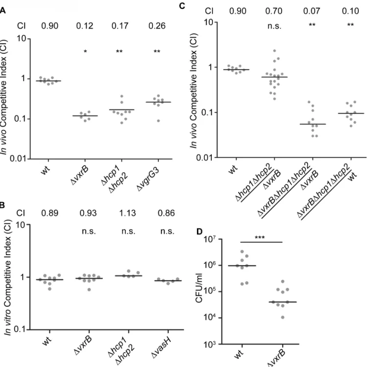
competition assays of a T6SS null mutant (Δhcp1Δhcp2) against wild type in the infant mouse model. We found that thein-vivoCI forΔhcp1Δhcp2was 0.17 (Fig 5A). In addition,ΔvgrG3
also had anin-vivoCI of 0.26 suggesting that T6SS components are important for intestinal colonization (Fig 5A). This suggests that structural components of the T6SS are needed to colo-nize the intestine. Furthermore, this finding also suggests that the colonization defect associat-ed with theΔvxrBmutant could be caused by diminished T6SS production. To evaluate this possibility, we tested thein vivocompetition ofΔhcp1Δhcp2againstΔvxrBand found that these strains competed nearly equally with each other (Fig 5C). Furthermore, in-vivo CI of
ΔvxrBΔhcp1Δhcp2triple mutant againstΔvxrBwas 0.07 andΔvxrBΔhcp1Δhcp2against wt was 0.10. This finding suggests that the colonization defect byΔvxrBwas not solely due to altered expression of T6SS genes and other factors regulated by VxrB also contribute to colonization. It is also likely that T6SS expression is not completely abolished by thevxrBmutation. Indeed, western analysis (Fig 4B) shows that invxrBmutant Hcp production is reduced but not completely eliminated. Similarly in vitro killing assay shows thatvxrBmutant’s interbacterial killing ability is not identical to that of the strain lacking T6SS.
We next asked whether VxrB plays a role in growth in vitro, by performing anin vitro com-petition assay.ΔvxrBmutants grew equally well as wild type, suggesting that neither had a competitive advantage over the otherin vitro(Fig 5B). This outcome suggests that there may be anin vivosignal produced in the infant mouse that triggers T6SS activity and colonization. We also performed single-strain colonization assays in the infant mouse model withΔvxrB. There was a 12.7-fold decrease in colonization forΔvxrBcompared to wild type (Fig 5D). This finding suggests that the colonization defect byΔvxrBwas not solely dependent on wild type, and possibly could be caused by competition with the normal flora or ability of the mutant to adapt to the infection microenvironment.
Discussion
Systematic mutational phenotypic characterization of TCSs has been performed in only a few bacteria, includingVibrio fischeri,E.coli,Bacillus subtilis,Streptococcus pneumoniae, and En-terococcus faecalis[51–55]. In this study, we systematically analyzed the role of allV.cholerae
TCS in colonization of the infant mouse small intestine and identified the RRs that play roles in mouse intestinal colonization. Specifically,ΔVC0719 (phoB),ΔVC1021 (luxO),ΔVC1213 (varA),andΔVC2135 (flrC), andΔVCA0566 (vxrB) exhibited intestinal colonization defects whileΔVC1050,ΔVC1086, andΔVC1087 showed enhanced colonization. Many of the RRs had either no statistically significant defect or minor defects in the infant mouse colonization assay. It remains possible, however, that these RRs have a role in colonization in other infection models.
In vivotranscriptome analysis has been performed on different strains ofV.choleraein the infant mouse and rabbit ileal loop infection models. The analysis of the whole genome expres-sion ofV.choleraeO1 El Tor C6706 cells accumulating in the ceca of orally infected infant
assay) were loaded onto a SDS 13% polyacrylamide gel. Prior to TCA precipitation and total protein quantification, 100μg/ml of BSA was added to the supernatant. After the blot was probed for Hcp, the blot was stripped of all antibodies using Western Blot Stripping Buffer (GM Biosciences, MD) and the same blot was used to probe for CRP and BSA in the whole cell and supernatant, respectively. The data shown is representative of the results of three independent experiments. (C) Interbacterial killing was analyzed by mixingV.choleraestrains and preyE.colistrain MC4100 in a 10:1 ratio, followed by incubation on LB agar plates for 4 hours at 30°C and determination of survivingE.coliMC4100. The data represent averages and standard deviations of three independent experiments.*, p<0.05 by the Student’s t-test as compared to the values for interbacterial killing of the wild-type strain.
Fig 5. Analysis of the role of T6SS in colonization.(A) Ability ofΔvxrB,ΔvxrB-Tn7vxrB,Δhcp1Δhcp2,ΔvgrG3, to colonize the infant mouse intestine was analyzed using a competition assay with isogenic wild-type strain.*, p<0.05;**, p<0.01 by the Wilcoxon Signed Rank Test as compared to wt (CI of 0.90).
(B)In vitrocompetition assay ofΔvxrB,Δhcp1Δhcp2, andΔvasH. Strains were grown in LB at 30°C for 20 hours. Statistical analysis was carried out using Wilcoxon Signed Rank Test, comparing the CI of each strain to the CI of wtlacZ+/ wt lacZ-(shown as wt) (CI of 0.98). (C) Ability ofΔhcp1Δhcp2to colonize
the infant mouse intestine was analyzed using a competition assay withΔvxrB. (D) Ability of wild type andΔvxrBto colonize the infant mouse intestine in single-strain infections. Each symbol represents the CI in an individual mouse; horizontal bars indicate the median.***, p<0.001 by the Student’s t-test as
compared to the values for colonization of the wild-type strain. n.s. indicates mutants were not significantly different from the wt.
doi:10.1371/journal.ppat.1004933.g005
rabbits and the intestines of orally infected infant mice revealed that expression of the genes en-coding RRs is altered duringin vivogrowth conditions as compared toin vitrogrowth in nutri-ent broth and thatin vivoexpression of TCS also differed between the model systems [56]. In the infant rabbit infection model, expression of seven RR (VC1081, VC1082, VC1155,vieA, VC2702 (cbrR), VCA0210, and VCA1105) was increased and 1 RR (carR) was decreased by more than 2-fold significantly in comparison toV.choleraecells grownin vitroin nutrient broth. In the infant mouse infection model, expression of 17 RR (vpsR, VC1050, VC1081, VC1082, VC1086, VC1087, VC1155, VC1522,flrC,cbrR,ompR,dct-D2,vxrB,uhpA,vpsT, VCA1086, and VCA1105) and 9 RR (qstR,phoB, VC1348, VC1638,vieB,cpxR,ntrC, VCA0532,
pgtA) were either decreased and increased significantly by more than 2-fold, respectively, in comparison toV.choleraecells grownin vitroin nutrient broth [56].vxrAandvxrBtranscript levels were decreased 2 and 3-fold, respectively, in the experiments reported by Mandlik and colleagues, but did not reach statistical significance [56]. This work all used theV.choleraeO1 El Tor strain C6706, and so it is yet unknown whethervxrBexpression is similarly regulated in the O1 El Tor A1552 strain used here.
There have been two other studies that analyzedV.choleraeinfection phenotypes on a glob-al scglob-ale, glob-although they did not specificglob-ally target RR. Together, these studies and ours suggests there is a set of genes required for intestinal colonization across multiple models. Fuet al. used random transposon mutants coupled with insertion site sequencing (Tn-seq) in a rabbit model [30]. They identified insertions in two genes—VC1021 (luxO) and VC1155—that showed 8-15-fold reduction in colonization, while strains harboring insertions into RRs VC1348,vieA,
vieB,arcA, VCA0256,uhpA, andpgtAhad less than a 5-fold reduction in colonization (p<0.001). Another Tn-seq study using the infant rabbit model identified defects associated
withluxOandarcAas above, and additionallyphoBandvarA[57]. Combining the results of these studies with ours identifiesluxO,phoB, andvarA, as required forin vivofitness, and oth-ers that are variably identified. Because the Tn-seq work used transposon libraries, it is not known whether all RR were eliminated, so it is possible that their studies missed some critical RR. There are hints in their data, however, that thevxrlocus is necessary in these other models as well. While Fuet al. did not identifyvxrAorvxrBmutants, they did determine that a strain with an insertion into VCA0567 (vxrC) exhibited a 9-fold reduction in colonization (p<0.0001) [30]. Additionally, Kampet al. found that a strain with a transposon insertion in
VCA0565 (vxrA) had a disadvantage in fitness (mean fitness value of 0.6) when the bacteria from rabbit cecum fluid was placed into pond water for 48 hours at 30°C [57]. Collectively, these studies suggest that the Vxr genes play important roles inV.choleraecolonization and environmental dissemination.
Our study revealed that the RR VxrB plays a significant role in colonization andin vitro
inter-bacterial competition through its ability to regulate expression of T6SS genes. Neither
Expression and production of T6SS are tightly regulated at the transcriptional and posttran-scriptional levels in a variety of bacterial systems [12,13,59]. Environmental signals such as iron limitation, thermoregulation, salinity, envelope stress, indole, and growth on surfaces reg-ulate T6SS expression [59]. InV.choleraeA1552, the strain used here, T6SS genes are express-ed when cell are grown in high-osmolarity and low temperature conditions [50]. A recent study revealed that theV.choleraeA1552 T6SS genes are part of the competence regulon and their expression is induced when the bacterium grows on chitinous surfaces in a TfoX-, HapR-, and QstR-dependent manner [60]. Our work presented here identified VxrB as a regulator of the T6SS large gene cluster and the two auxiliary clusters. The predicted cognate HK of VxrB, VxrA, does not exhibit similarity to previously characterized sensory domains. The signals that govern expression and activity of VxrAB and how the VxrAB TCS is integrated into the T6SS regulatory network ofV.choleraeare yet to be determined. We determined that while the wild-type strain has a competitive advantagein vivooverΔvxrB, neither strain had a competitive ad-vantage over the otherin vitro. Furthermore, single infection studies showed thatΔvxrBhad a significant colonization defect compared to wild type, suggesting that VxrB could be involved in competition with normal flora and thatΔvxrBcould have a reduced fitness in infection envi-ronment. These observations also suggest that there may be anin vivosignal produced in the infant mouse that triggers T6SS activity and colonization. Our studies thus provided significant new insights into the regulation of T6SS inV.choleraeand provided further support that the T6SS is critical forV.choleraevirulence.
Materials and Methods
Ethics statement
All animal procedures used were in strict accordance with the NIHGuide for the Care and Use of Laboratory Animals[61] and were approved by the UC Santa Cruz Institutional Animal Care and Use Committee (Yildf1206).
Bacterial strains, plasmids, and culture conditions
The bacterial strains and plasmids used in this study are listed inS1 Table.Escherichia coli
CC118λpir strains were used for DNA manipulation, andE.coliS17-1λpir strains were used for conjugation withV.cholerae. In-frame deletion mutants ofV.choleraewere generated as described earlier [62]. AllV.choleraeandE.colistrains were grown aerobically, at 30°C and 37°C, respectively, unless otherwise noted. All cultures were grown in Luria-Bertani (LB) broth (1% Tryptone, 0.5% Yeast Extract, 1% NaCl), pH 7.5, unless otherwise stated. LB agar medium contains 1.5% (wt/vol) granulated agar (BD Difco, Franklin Lakes, NJ). AKI medium contains 0.5% NaCl, 0.3% NaHCO3, 0.4% Yeast Extract, and 1.5% Peptone, as previously described [63]. Antibiotics were used at the following concentrations: ampicillin 100μg/ml; rifampicin 100μg/ ml; gentamicin 50μg/ml; streptomycin 50μg/ml.
DNA manipulations
An overlapping PCR method was used to generate in-frame deletion constructs of each RR genes using previously published methods [62]. Briefly, a 500–600 bp 5’flanking sequence of the gene, including several nucleotides of the coding region, was PCR amplified using A and del-B primers. del-C and del-D primers were used to amplify the 3’region of the gene including 500–600 bp of the downstream flanking sequence. The two PCR products were joined using the splicing overlap extension technique [64,65] and the resulting PCR product, which lacks 80% of amino acids, was digested with two restriction enzymes and ligated to similarly-digested
pGP704sacB28 suicide plasmid. Construction ofvxrBplasmid harboring point mutations were performed using a similar technique [66] with the following alterations: primers containing the new sequence harboring the point mutations were used in place of the del-B and del-C primers. The deletion constructs were sequenced (UC Berkeley DNA Sequencing Facility, Berkeley, CA) and the clones without any undesired mutations were used. The deletion constructs are listed in
S1 Table.
Generation of in-frame deletion mutants and Tn7 complementation
strains
The deletion plasmids were maintained inE.coliCC118λpir. Biparental matings were carried out with the wild typeV.choleraeand anE.coliS17λpir strain harboring the deletion plasmid. Selection of deletion mutants were done as described [64] and were verified by PCR. The Tn7 complementationV.choleraestrains were generated by triparental matings with donorE.
coliS17λpir carrying pGP704-Tn7 with gene of interest, helperE.coliS17λpir harboring pUX-BF13, andV.choleraestrains. Transconjugants were selected on thiosulfate-citrate-bile salts-sucrose (TCBS) (BD Difco, Franklin Lakes, NJ) agar medium containing gentamicin at 30°C. The Tn7 complementationV.choleraestrains were verified by PCR.
Intestinal colonization assay
Anin vivocompetition assay for intestinal colonization was performed as described previously [46]. Briefly, each of theV.choleraemutant strains (lacZ+) and the fully virulent reference strain (lacZ-otherwise wild-type)) were grown to stationary phase at 30°C with aeration in LB broth. Mutant strains and wild-type were mixed at 1:1 ratios in 1x Phosphate Buffered Saline (PBS). The inoculum was plated on LB agar plates containing 5-bromo-4-chloro-3-indoyl-β -D-galactopyranoside (X-gal) to differentiate wild-type and mutant colonies and to determine the input ratios. Approximately, 106–107cfu were intragastrically administered to groups of 5–7 anesthetized 5-day old CD-1 mice (Charles River Laboratories, Hollister, CA). After 20 hours of incubation, the small intestine was removed, weighed, homogenized, and plated on appropriate selective and differential media to enumerate mutant and wild-type cells recovered and to obtain the output ratios.In vivocompetitive indices were calculated by dividing the small intestine output ratio by the inoculum input ratio of mutant to wild-type strains. For sin-gle strain infections, 107cfu of each strain, including otherwise wild type (lacZ-) strain, were intragastrically administrated to 5-day old CD-1 mice. After 20 hours of incubation, the small intestine was harvested and plated on selective media as previously described above. Statistical analyses for competition infections were performed using Wilcoxon Signed Rank Test. Statisti-cal analyses were performed using Prism 5 software (GraphPad Software, Inc., San Diego, CA) using Wilcoxon Signed Rank Test. P values of<0.05 were determined to be statistically
significant.
Reverse transcription-PCR
Quantitative real time (qRT) PCR
For qRT-PCR expression analysis, RNA was isolated as described below. cDNA was synthe-sized using iScript cDNG Synthesis Kit (Bio-Rad, Hercules, CA) from 1μg of total RNA. Real-time PCR was performed using a Bio-Rad CFX1000 thermal cycler and Bio-RAD CFX96 real-time imager with specific primer pairs (designed within the coding region of the target gene) and SsoAdvanced SYBR green supermix (Bio-Rad, Hercules, CA). Results are from two inde-pendent experiments performed in quadruplicate. All samples were normalized to the expres-sion of the housekeeping gene 16S using the Pfaffl method [67]. Relative expression was calculated by normalizing expression atΔvxrBby that of wt. Statistical analysis was performed using two-tailed student’s t test.
RNA isolation
V.choleraecells were grown aerobically overnight in LB at 37°C, then diluted 1:100 in fresh 10 ml AKI media in borosilicate glass test tubes (diameter, 15mm; height, 150 mm) and incubated at 37°C without shaking for 4 hours. After 4 hours, 10 ml cultures were transferred to 125 ml flasks (for maximal aerated growth on an orbital shaker (250 rpm) for 2 hours. Aliquots (2 ml) of the cultures were collected and centrifuged for 2 min at room temperature. The cell pellets were immediately resuspended in 1 ml of TRIzol (Invitrogen, Carlsbad, CA) and stored at -80°C. Total RNA was isolated according to the manufacturer’s instructions. To remove con-taminating DNA, total RNA was incubated with RNase-free DNase I (Ambion, Grand Island, NY), and an RNeasy mini kit (Qiagen, Valencia, CA) was used to clean up RNA after DNase digestion. Five micrograms of total RNA was treated with a MICROBExpress Kit (Ambion, Grand Island, NY) to remove ribosomal RNA, and the efficiency was confirmed by Bioanalyzer analysis (Agilent Technologies, Santa Clara, CA). Three biological replicates were generated for each condition.
cDNA library construction and Illumina HiSeq sequencing
Libraries for RNA-seq were prepared using NEBNext Ultra Directional RNA Library Prep Kit for Illumina (New England Biolabs, Ipswich, MA). Twelve indexed samples were sequenced per single lane using the HiSeq2500 Illumina sequencing platform for 50 bp single reads (UC Davis Genome Center, UC Davis, CA) and subsequently analyzed and visualized via the CLC Genomics Workbench version 7.5 (Qiagen, Valencia, CA). Samples were mapped to theV. cho-leraegenome N16961. Differentially regulated genes were identified as those displaying a fold change with an absolute value of 1.5-fold or greater. Statistical significance was determined by Empirical analysis of Digital Gene Expression (edgeR) test where p<0.05 was deemed
signifi-cant [68].
Analysis of Hcp production and secretion
V.choleraestrains were grown to an OD600 of 2.0, and the culture (25 ml) was centrifuged at 20,000 g for 10 min to obtain whole cell pellets. The culture supernatant containing secreted proteins were filtered through 0.22μmembranes (Millipore, Billerica, Massachusetts) and se-creted proteins in the culture supernatant were precipitated with 13% trichloroacetic acid (TCA) overnight at 4°C, pelleted by centrifugation at 47,000 g for 30 min at 4°C, wash with ice cold acetone and resuspended in 1x PBS containing Complete protease inhibitor (Roche, Basel, Switzerland). Bovine serum albumin (BSA, 100μg/ml) was added to the culture supernatant prior to TCA precipitation as a control. Protein pellets from whole cell were suspended in 2% sodium dodecyl sulfate (SDS) and protein concentrations were estimated using a Pierce BCA
protein assay kit (Thermo Scientific, Rockford, IL). Equal amounts of total protein (20μg) were loaded onto a SDS 13% polyacrylamide gel electrophoresis (SDS-PAGE). Western blot analyses were performed as described [69] using anti-Hcp polyclonal antiserum provided by the Sun Wai [28], anti-CRP (Neoclone Inc., Madison, WI), and anti-BSA (Santa Cruz Biotech, Santa Cruz, CA). OneMinute Western Blot Stripping Buffer (GM Biosciences, Frederick, MD) was used to remove the Hcp antibodies and the same blot was used again to probe for CRP or BSA. These experiments were conducted with at least three biological replicates.
Bacterial killing assay
Killing assays were performed as described previously [20]. Briefly, bacterial strains were grown overnight on LB plates and resuspended in LB broth containing 340 mM NaCl, asV.
choleraestrain A1552 displayed enhanced interbacterial virulence towardsE.coliunder high osmolarity [50].V.choleraeandE.coliMC4100 were mixed at a 10:1 ratio and 25μl was spot-ted onto LB agar plates containing 340 mM NaCl and incubaspot-ted at 37°C for 4 hours. Spots were harvested, serially diluted, and plated onto LB plates containing 50μg/ml of streptomycin to enumerate survivingE.coliprey cells.
In vitro
competition assay
The following assay was performed similarly as the intestinal colonization assay except no ani-mal models were used. TheV.choleraemutant strains with wild-typelacZallele (lacZ+) and reference strain (lacZ-) were grown to stationary phase at 30°C with aeration in LB broth. Mu-tant strains and wild-type were mixed at 1:1 ratios in 1x PBS. The inoculum was plated on LB agar plates containing X-gal to differentiate colonies formed by the wild-type and mutant strains and to determine the input ratios. The inoculum (50μl) was spotted on to a LB agar plate and incubated at 37°C. After 20–24 hours of incubation, the 50μl spots were scraped off the agar plate and resuspended in 1x PBS. The resuspension was serially diluted and plated on appropriate selective and differential media to enumerate mutant and wild type cells recovered and to obtain the output ratios.In vitrocompetitive indices were calculated by dividing the out-put ratio by the inoculum inout-put ratio of mutant to wild type strains. Statistical analyses were performed using Wilcoxon Signed Rank Test.
Supporting Information
S1 Fig. Confirmation of the predicted operon structure ofvxrABCDE.RNAseq track reads
from wild-type sample. Red and green lines indicate the directionality of the read tracks. Im-ages were prepared by CLC bio version 7.5.1 (Qiagen, Valencia, CA).
(TIF)
S2 Fig. Multiple sequence alignment of VxrA.Amino acid sequence alignment of the HK, VxrA, toV.cholerae V52,V.fischeri MJ11,V.harveyi ATCC BAA-1116,V.parahaemolyticus RIMD 2210633, andV.vulnificus YJ016using ClustalW. Numbers above the sequence corre-spond to the amino acid number of each protein.
(TIF)
S3 Fig. Multiple sequence alignment of VxrB and VxrC.(A) Amino acid sequence alignment of the RR, VxrB, and (B) VxrC toV.cholerae V52,V.fischeri MJ11,V.harveyi ATCC BAA-1116,V.parahaemolyticus RIMD 2210633, andV.vulnificus YJ016using ClustalW. Numbers above the sequence correspond to the amino acid number of each protein.
S4 Fig. Multiple sequence alignment of VxrD and VxrE.(A) Amino acid sequence alignment of VxrD, and (B) VxrE toV.cholerae V52,V.fischeri MJ11,V.harveyi ATCC BAA-1116,V.
parahaemolyticus RIMD 2210633, andV.vulnificus YJ016using ClustalW. Numbers above the sequence correspond to the amino acid number of each protein.
(TIF)
S5 Fig. Expression analysis of the T6SS gene clusters.(A) Schematic representation of the major T6SS large gene cluster and auxiliary clusters 1 and 2. (B) RNAseq data showing the cov-erage of cDNA reads in wild type (wt) andΔvxrBmutant (vxrB) over the large cluster and the two auxiliary clusters. Images were prepared by CLC bio version 7.5.1 (Qiagen, Valencia, CA). (TIF)
S1 Table. Bacterial strains and plasmids used in this study. (DOCX)
S2 Table. Genes positively regulated by VxrB under AKI conditions. (DOCX)
S3 Table. Genes negatively regulated by VxrB under AKI conditions. (DOCX)
Acknowledgments
We thank Sun Nyunt Wai for providing T6SS mutants and anti-Hcp antibodies, UC Davis ge-nome sequencing facility and David Bernick for help with the RNA-seq analysis, William Sause for help with infection studies, Loni Townsley for help with qRT-PCR analysis, and the Yildiz lab members for their valuable discussions and reading the manuscript.
Author Contributions
Conceived and designed the experiments: ATC KMO FHY. Performed the experiments: ATC. Analyzed the data: ATC KMO FHY. Wrote the paper: ATC KMO FHY.
References
1. Ali M, Lopez AL, You YA, Kim YE, Sah B, Maskery B, et al. The global burden of cholera. Bulletin of the World Health Organization. 2012 Mar 1; 90(3):209–18A. doi:10.2471/BLT.11.093427PMID:22461716
2. Kaper JB, Morris JG Jr., Levine MM. Cholera. Clin Microbiol Rev. 1995 Jan; 8(1):48–86. PMID:
7704895
3. DiRita VJ, Parsot C, Jander G, Mekalanos JJ. Regulatory cascade controls virulence inVibrio cholerae. Proc Natl Acad Sci U S A. 1991 Jun 15; 88(12):5403–7. PMID:2052618
4. Higgins DE, Nazareno E, DiRita VJ. The virulence gene activator ToxT fromVibrio choleraeis a mem-ber of the AraC family of transcriptional activators. J Bacteriol. 1992 Nov; 174(21):6974–80. PMID:
1400247
5. Higgins DE, DiRita VJ. Transcriptional control of toxT, a regulatory gene in the ToxR regulon ofVibrio cholerae. Mol Microbiol. 1994 Oct; 14(1):17–29. PMID:7830555
6. Hase CC, Mekalanos JJ. TcpP protein is a positive regulator of virulence gene expression inVibrio cho-lerae. Proc Natl Acad Sci U S A. 1998 Jan 20; 95(2):730–4. PMID:9435261
7. Krukonis ES, Yu RR, Dirita VJ. TheVibrio choleraeToxR/TcpP/ToxT virulence cascade: distinct roles for two membrane-localized transcriptional activators on a single promoter. Mol Microbiol. 2000 Oct; 38-(1):67–84. PMID:11029691
8. Kovacikova G, Lin W, Skorupski K.Vibrio choleraeAphA uses a novel mechanism for virulence gene activation that involves interaction with the LysR-type regulator AphB at thetcpPHpromoter. Mol Micro-biol. 2004 Jul; 53(1):129–42. PMID:15225309
9. Skorupski K, Taylor RK. A new level in theVibrio choleraeToxR virulence cascade: AphA is required for transcriptional activation of thetcpPHoperon. Mol Microbiol. 1999 Feb; 31(3):763–71. PMID:
10048021
10. Kovacikova G, Skorupski K. Regulation of virulence gene expression inVibrio choleraeby quorum sensing: HapR functions at theaphApromoter. Mol Microbiol. 2002 Nov; 46(4):1135–47. PMID:
12421317
11. Pukatzki S, Ma AT, Sturtevant D, Krastins B, Sarracino D, Nelson WC, et al. Identification of a con-served bacterial protein secretion system inVibrio choleraeusing theDictyosteliumhost model system. Proc Natl Acad Sci U S A. 2006 Jan 31; 103(5):1528–33. PMID:16432199
12. Ho BT, Dong TG, Mekalanos JJ. A view to a kill: the bacterial type VI secretion system. Cell host & mi-crobe. 2014 Jan 15; 15(1):9–21.
13. Silverman JM, Brunet YR, Cascales E, Mougous JD. Structure and regulation of the type VI secretion system. Annu Rev Microbiol. 2012; 66:453–72. doi:10.1146/annurev-micro-121809-151619PMID:
22746332
14. Pukatzki S, Ma AT, Revel AT, Sturtevant D, Mekalanos JJ. Type VI secretion system translocates a phage tail spike-like protein into target cells where it cross-links actin. Proc Natl Acad Sci U S A. 2007 Sep 25; 104(39):15508–13. PMID:17873062
15. Dong TG, Ho BT, Yoder-Himes DR, Mekalanos JJ. Identification of T6SS-dependent effector and im-munity proteins by Tn-seq inVibrio cholerae. Proc Natl Acad Sci U S A. 2013 Feb 12; 110(7):2623–8. doi:10.1073/pnas.1222783110PMID:23362380
16. Brooks TM, Unterweger D, Bachmann V, Kostiuk B, Pukatzki S. Lytic activity of theVibrio choleraetype VI secretion toxin VgrG-3 is inhibited by the antitoxin TsaB. J Biol Chem. 2013 Mar 15; 288(11):7618–
25. doi:10.1074/jbc.M112.436725PMID:23341465
17. Ma AT, McAuley S, Pukatzki S, Mekalanos JJ. Translocation of aVibrio choleraetype VI secretion ef-fector requires bacterial endocytosis by host cells. Cell host & microbe. 2009 Mar 19; 5(3):234–43.
18. Russell AB, LeRoux M, Hathazi K, Agnello DM, Ishikawa T, Wiggins PA, et al. Diverse type VI secretion phospholipases are functionally plastic antibacterial effectors. Nature. 2013 Apr 25; 496(7446):508–12. doi:10.1038/nature12074PMID:23552891
19. Miyata ST, Kitaoka M, Brooks TM, McAuley SB, Pukatzki S.Vibrio choleraerequires the type VI secretion system virulence factor VasX to killDictyostelium discoideum. Infect Immun. 2011 Jul; 79-(7):2941–9. doi:10.1128/IAI.01266-10PMID:21555399
20. Miyata ST, Unterweger D, Rudko SP, Pukatzki S. Dual expression profile of type VI secretion system immunity genes protects pandemicVibrio cholerae. PLoS Pathog. 2013; 9(12):e1003752. doi:10.1371/ journal.ppat.1003752PMID:24348240
21. Unterweger D, Miyata ST, Bachmann V, Brooks TM, Mullins T, Kostiuk B, et al. TheVibrio cholerae
type VI secretion system employs diverse effector modules for intraspecific competition. Nature com-munications. 2014; 5:3549. doi:10.1038/ncomms4549PMID:24686479
22. Basler M, Pilhofer M, Henderson GP, Jensen GJ, Mekalanos JJ. Type VI secretion requires a dynamic contractile phage tail-like structure. Nature. 2012 Mar 8; 483(7388):182–6. doi:10.1038/nature10846
PMID:22367545
23. Zheng J, Ho B, Mekalanos JJ. Genetic analysis of anti-amoebae and anti-bacterial activities of the type VI secretion system inVibrio cholerae. PloS one. 2011; 6(8):e23876. doi:10.1371/journal.pone. 0023876PMID:21909372
24. Kitaoka M, Miyata ST, Brooks TM, Unterweger D, Pukatzki S. VasH is a transcriptional regulator of the type VI secretion system functional in endemic and pandemicVibrio cholerae. J Bacteriol. 2011 Dec; 193(23):6471–82. doi:10.1128/JB.05414-11PMID:21949076
25. Bernard CS, Brunet YR, Gavioli M, Lloubes R, Cascales E. Regulation of type VI secretion gene clus-ters by sigma54 and cognate enhancer binding proteins. J Bacteriol. 2011 May; 193(9):2158–67. doi:
10.1128/JB.00029-11PMID:21378190
26. Dong TG, Mekalanos JJ. Characterization of the RpoN regulon reveals differential regulation of T6SS and new flagellar operons inVibrio choleraeO37 strain V52. Nucleic Acids Res. 2012 Sep; 40-(16):7766–75. doi:10.1093/nar/gks567PMID:22723378
27. Zheng J, Shin OS, Cameron DE, Mekalanos JJ. Quorum sensing and a global regulator TsrA control expression of type VI secretion and virulence inVibrio cholerae. Proc Natl Acad Sci U S A. 2010 Dec 7; 107(49):21128–33. doi:10.1073/pnas.1014998107PMID:21084635
29. Ma AT, Mekalanos JJ.In vivoactin cross-linking induced byVibrio choleraetype VI secretion system is associated with intestinal inflammation. Proc Natl Acad Sci U S A. 2010 Mar 2; 107(9):4365–70. doi:
10.1073/pnas.0915156107PMID:20150509
30. Fu Y, Waldor MK, Mekalanos JJ. Tn-Seq analysis of Vibrio cholerae intestinal colonization reveals a role for T6SS-mediated antibacterial activity in the host. Cell host & microbe. 2013 Dec 11; 14(6):652–63.
31. Beier D, Gross R. Regulation of bacterial virulence by two-component systems. Curr Opin Microbiol. 2006 Apr; 9(2):143–52. PMID:16481212
32. Krell T, Lacal J, Busch A, Silva-Jimenez H, Guazzaroni ME, Ramos JL. Bacterial sensor kinases: diver-sity in the recognition of environmental signals. Annu Rev Microbiol. 2010; 64:539–59. doi:10.1146/ annurev.micro.112408.134054PMID:20825354
33. Galperin MY. Diversity of structure and function of response regulator output domains. Curr Opin Micro-biol. 2010 Apr; 13(2):150–9. doi:10.1016/j.mib.2010.01.005PMID:20226724
34. Gao R, Stock AM. Biological insights from structures of two-component proteins. Annu Rev Microbiol. 2009; 63:133–54. doi:10.1146/annurev.micro.091208.073214PMID:19575571
35. Laub MT, Goulian M. Specificity in two-component signal transduction pathways. Annual review of ge-netics. 2007; 41:121–45. PMID:18076326
36. Jang J, Jung KT, Park J, Yoo CK, Rhie GE. TheVibrio choleraeVarS/VarA two-component system con-trols the expression of virulence proteins through ToxT regulation. Microbiology. 2011 May; 157(Pt 5):1466–73. doi:10.1099/mic.0.043737-0PMID:21330435
37. Zhu J, Miller MB, Vance RE, Dziejman M, Bassler BL, Mekalanos JJ. Quorum-sensing regulators con-trol virulence gene expression inVibrio cholerae. Proc Natl Acad Sci U S A. 2002 Mar 5; 99(5):3129–
34. PMID:11854465
38. Tischler AD, Camilli A. Cyclic diguanylate regulatesVibrio choleraevirulence gene expression. Infect Immun. 2005 Sep; 73(9):5873–82. PMID:16113306
39. Tischler AD, Lee SH, Camilli A. TheVibrio cholerae vieSABlocus encodes a pathway contributing to cholera toxin production. J Bacteriol. 2002 Aug; 184(15):4104–13. PMID:12107127
40. Pratt JT, Ismail AM, Camilli A. PhoB regulates both environmental and virulence gene expression in
Vibrio cholerae. Mol Microbiol. 2010 Sep; 77(6):1595–605. doi:10.1111/j.1365-2958.2010.07310.x
PMID:20659293
41. Sengupta N, Paul K, Chowdhury R. The global regulator ArcA modulates expression of virulence fac-tors inVibrio cholerae. Infect Immun. 2003 Oct; 71(10):5583–9. PMID:14500477
42. Bilecen K, Fong JC, Cheng A, Jones CJ, Zamorano-Sanchez D, Yildiz FH. Polymyxin B Resistance and biofilm formation inVibrio choleraeis controlled by the response regulator CarR. Infect Immun. 2015 Jan 12.
43. Butler SM, Camilli A. Both chemotaxis and net motility greatly influence the infectivity ofVibrio cholerae. Proc Natl Acad Sci U S A. 2004 Apr 6; 101(14):5018–23. PMID:15037750
44. Correa NE, Lauriano CM, McGee R, Klose KE. Phosphorylation of the flagellar regulatory protein FlrC is necessary forVibrio choleraemotility and enhanced colonization. Mol Microbiol. 2000 Feb; 35-(4):743–55. PMID:10692152
45. Butler SM, Camilli A. Going against the grain: chemotaxis and infection inVibrio cholerae. Nature re-views Microbiology. 2005 Aug; 3(8):611–20. PMID:16012515
46. Taylor RK, Miller VL, Furlong DB, Mekalanos JJ. Use ofphoAgene fusions to identify a pilus coloniza-tion factor coordinately regulated with cholera toxin. Proc Natl Acad Sci U S A. 1987 May; 84(9):2833–
7. PMID:2883655
47. Galperin MY. Structural classification of bacterial response regulators: diversity of output domains and domain combinations. J Bacteriol. 2006 Jun; 188(12):4169–82. PMID:16740923
48. Delgado J, Forst S, Harlocker S, Inouye M. Identification of a phosphorylation site and functional analy-sis of conserved aspartic acid residues of OmpR, a transcriptional activator forompFandompCin
Escherichia coli. Mol Microbiol. 1993 Dec; 10(5):1037–47. PMID:7934854
49. Ma S, Selvaraj U, Ohman DE, Quarless R, Hassett DJ, Wozniak DJ. Phosphorylation-independent ac-tivity of the response regulators AlgB and AlgR in promoting alginate biosynthesis in mucoid Pseudo-monas aeruginosa. J Bacteriol. 1998 Feb; 180(4):956–68. PMID:9473053
50. Ishikawa T, Sabharwal D, Broms J, Milton DL, Sjostedt A, Uhlin BE, et al. Pathoadaptive conditional regulation of the type VI secretion system inVibrio choleraeO1 strains. Infect Immun. 2012 Feb; 80-(2):575–84. doi:10.1128/IAI.05510-11PMID:22083711
51. Hussa EA, O'Shea TM, Darnell CL, Ruby EG, Visick KL. Two-component response regulators of Vibrio fischeri: identification, mutagenesis, and characterization. J Bacteriol. 2007 Aug; 189(16):5825–38. PMID:17586650
52. Kobayashi K, Ogura M, Yamaguchi H, Yoshida K, Ogasawara N, Tanaka T, et al. Comprehensive DNA microarray analysis ofBacillus subtilistwo-component regulatory systems. J Bacteriol. 2001 Dec; 183-(24):7365–70. PMID:11717295
53. Lange R, Wagner C, de Saizieu A, Flint N, Molnos J, Stieger M, et al. Domain organization and molecu-lar characterization of 13 two-component systems identified by genome sequencing ofStreptococcus pneumoniae. Gene. 1999 Sep 3; 237(1):223–34. PMID:10524254
54. Throup JP, Koretke KK, Bryant AP, Ingraham KA, Chalker AF, Ge Y, et al. A genomic analysis of two-component signal transduction inStreptococcus pneumoniae. Mol Microbiol. 2000 Feb; 35(3):566–76. PMID:10672179
55. Yamamoto K, Hirao K, Oshima T, Aiba H, Utsumi R, Ishihama A. Functional characterization in vitro of all two-component signal transduction systems fromEscherichia coli. J Biol Chem. 2005 Jan 14; 280-(2):1448–56. PMID:15522865
56. Mandlik A, Livny J, Robins WP, Ritchie JM, Mekalanos JJ, Waldor MK. RNA-Seq-based monitoring of infection-linked changes inVibrio choleraegene expression. Cell host & microbe. 2011 Aug 18; 10-(2):165–74.
57. Kamp HD, Patimalla-Dipali B, Lazinski DW, Wallace-Gadsden F, Camilli A. Gene fitness landscapes of
Vibrio choleraeat important stages of its life cycle. PLoS Pathog. 2013; 9(12):e1003800. doi:10.1371/ journal.ppat.1003800PMID:24385900
58. Kelley LA, Sternberg MJ. Protein structure prediction on the Web: a case study using the Phyre server. Nature protocols. 2009; 4(3):363–71. doi:10.1038/nprot.2009.2PMID:19247286
59. Bernard CS, Brunet YR, Gueguen E, Cascales E. Nooks and crannies in type VI secretion regulation. J Bacteriol. 2010 Aug; 192(15):3850–60. doi:10.1128/JB.00370-10PMID:20511495
60. Borgeaud S, Metzger LC, Scrignari T, Blokesch M. Bacterial evolution. The type VI secretion system of
Vibrio choleraefosters horizontal gene transfer. Science. 2015 Jan 2; 347(6217):63–7. doi:10.1126/ science.1260064PMID:25554784
61. National Research Council (U.S.). Committee for the Update of the Guide for the Care and Use of Labo-ratory Animals., Institute forLaboratoryAnimal Research (U.S.), National Academies Press (U.S.). Guide for the care and use of laboratory animals. 8th ed. Washington, D.C.: National Academies Press; 2011. xxv, 220 p. p.
62. Lim B, Beyhan S, Meir J, Yildiz FH. Cyclic-diGMP signal transduction systems inVibrio cholerae: modu-lation of rugosity and biofilm formation. Mol Microbiol. 2006 Apr; 60(2):331–48. PMID:16573684
63. Iwanaga M, Yamamoto K, Higa N, Ichinose Y, Nakasone N, Tanabe M. Culture conditions for stimulat-ing cholera toxin production byVibrio choleraeO1 El Tor. Microbiology and immunology. 1986; 30-(11):1075–83. PMID:3543624
64. Fong JC, Yildiz FH. TherbmBCDEFgene cluster modulates development of rugose colony morphology and biofilm formation inVibrio cholerae. J Bacteriol. 2007 Mar; 189(6):2319–30. PMID:17220218
65. Lefebvre B, Formstecher P, Lefebvre P. Improvement of the gene splicing overlap (SOE) method. Bio-Techniques. 1995 Aug; 19(2):186–8. PMID:8527132
66. Beyhan S, Bilecen K, Salama SR, Casper-Lindley C, Yildiz FH. Regulation of rugosity and biofilm for-mation inVibrio cholerae: comparison of VpsT and VpsR regulons and epistasis analysis ofvpsT,
vpsR, andhapR. J Bacteriol. 2007 Jan; 189(2):388–402. PMID:17071756
67. Pfaffl MW. A new mathematical model for relative quantification in real-time RT-PCR. Nucleic Acids Res. 2001 May 1; 29(9):e45. PMID:11328886
68. Robinson MD, McCarthy DJ, Smyth GK. edgeR: a Bioconductor package for differential expression analysis of digital gene expression data. Bioinformatics. 2010 Jan 1; 26(1):139–40. doi:10.1093/ bioinformatics/btp616PMID:19910308