Arq Neuropsiquiatr 2011;69(1):91-99
Relationship between luid-attenuated
inversion-recovery (FLAIR) signal
intensity and inlammatory mediator’s
levels in the hippocampus of
patients with temporal lobe epilepsy
and mesial temporal sclerosis
Pedro Paulo Vasconcellos Varella1, Joselita Ferreira Carvalho Santiago1, Henrique Carrete Jr.2, Elisa Mieko Suemitsu Higa3,
Elza Márcia Targas Yacubian1, Ricardo Silva Centeno1,
Luís Otávio Sales Ferreira Caboclo1, Eduardo Ferreira de Castro Neto1,
Mauro Canzian4, Débora Amado1, Esper Abrão Cavalheiro1,
Maria da Graça Nafah- Mazzacoratti1,5
ABSTRACT
We investigated a relationship between the FLAIR signal found in mesial temporal sclerosis (MTS) and inflammation. Twenty nine patients were selected through clinical and MRI analysis and submitted to cortico-amygdalo-hippocampectomy to seizure control. Glutamate, TNFα, IL1, nitric oxide (NO) levels and immunostaining against IL1β and CD45 was performed. Control tissues (n=10) were obtained after autopsy of patients without neurological disorders. The glutamate was decreased in the temporal lobe epilepsy (TLE) -MTS group (p<0.001), suggesting increased release of this neurotransmitter. The IL1β and TNFα were increased in the hippocampus (p<0.05) demonstrating an active inflammatory process. A positive linear correlation between FLAIR signal and NO and IL1β levels and a negative linear correlation between FLAIR signal and glutamate concentration was found. Lymphocytes infiltrates were present in hippocampi of TLE patients. These data showed an association between hippocampal signal alteration and increased inflammatory markers in TLE-MTS.
Key words: FLAIR hippocampal signal, cytokines, NO, glutamate and hippocampal sclerosis.
Correlação da intensidade do sinal em FLAIR e os níveis de mediadores inflamatórios no hipocampo de pacientes com epilepsia do lobo temporal e esclerose mesial temporal
RESUMO
Este estudo foi delineado para investigar a presença de relação entre a intensidade de sinal em FLAIR e níveis de citocinas, óxido nítrico (NO) e glutamato no hipocampo de pacientes com epilepsia do lobo temporal refratária, associada com esclerose mesial (TLE-MTS). Vinte e nove pacientes foram selecionados através de análise clínica e de ressonância magnética (RM) que foram submetidos a cortico-amigdalo-hipocampectomia para o controle das crises. Os níveis de glutamato foram avaliados por HPLC, as citocinas TNFα e IL1β por
Correspondence
Maria da Graça Naffah Mazzacoratti Rua Botucatu 862
04023-900 São Paulo SP - Brasil E-mail: [email protected]
Support
CInNAPCe (FAPESP), CAPES, CNPq, MCT (Instituto Nacional de Neurociência Translacional (INNT)
Received 20 January 2010 Received in final form 23 July 2010 Accepted 2 August 2010
ELISA e os níveis de NO via NO system. Avaliamos também por imuno-histoquímica a expressão de IL1β e CD45 em tecidos controles e com esclerose. Tecido controle foi obtido após autópsia de indivíduos mortos sem disfunções inflamatórias e neurológicas (n=10). A concentração de glutamato se mostrou reduzida no tecido TLE-MTS (p<0,001) sugerindo aumento na liberação desse neurotransmissor. TNFα e IL1β também apresentaram níveis elevados no hipocampo dos pacientes (p<0,05), demonstrando um processo inflamatório crônico. Houve uma correlação linear positiva entre a intensidade do sinal em FLAIR e os níveis de NO e IL1β. Em contraste, uma correlação linear negativa foi encontrada entre a intensidade do sinal em FLAIR e níveis de glutamato no hipocampo com esclerose. Infiltrado linfocitário hipocampal também foi visualizado pela imuno-marcação com CD45 em pacientes com TLE-MTS. Esses dados mostraram uma associação entre alteração de sinal na RM e marcadores inflamatórios em pacientes com TLE-MTS.
Palavras-chave: sinal hipocampal em FLAIR, citocinas, NO, glutamato, esclerose hipocampal.
Temporal lobe epilepsy (TLE) represents a severe common syndrome in patients presenting refractory sei-zures. Atrophy and/or signal changes of the hippocampus have been described as the most reliable magnetic reso-nance imaging (MRI) inclusion criteria in mesial temporal sclerosis (MTS). hese alterations are the most frequent pathological indings presented by these patients1. In ad-dition ipsilateral temporal pole signal and volume abnor-malities are described as part of the spectrum of MRI fea-tures in cases of MTS2. he hippocampal sclerosis (HS) is also known as Ammon’s horn sclerosis and includes se-lective hippocampal cell loss, gliosis and synaptic reor-ganization as the mossy ibers sprouting3,4. Indeed, HS is seen as loss of volume, decreased T1 and increased T2 signals on views through the structure, associated with ipsilateral temporal lobe atrophy and signiicant asymme-try of hippocampal volume5,6. he predominant cell loss occurs in pyramidal neurons of the stratum pyramidale of Ammon’s horn. Neuronal population of the stratum oriens survives in most sclerotic hippocampi, along with interneurons scattered throughout the neuron-depleted pyramidal layer. In the dentate gyrus, many of the gran-ule cells are lost and neurons in the grangran-ule cell layer ap-pear to be more dispersed. hus, several lines of evidence point to sclerotic hippocampus as the major structure in-volved in chronic seizures observed in MTS, for review, see7. As reported by Bernasconi et al.8 the hippocampal head is more atrophic than hippocampal body and tail, between other structures also involved such as amygda-la, entorhinal and perihinal cortices.
The immune system and associated inflammatory process play important function in the protection and repair of several tissues, thus constituting an adaptive, beneicial endogenous response. In contrast, immune re-sponse activation can also be a direct or indirect cause of neuronal dysfunctions. During inlammatory process the communication between cells of the immune system
oc-curs either via direct cell-to-cell contact or via soluble fac-tors called cytokines such as IL1β, IL6 and TNFα9. Dele-terious efects of cytokines on neuronal survival are like-ly to be mediated by their ability to provoke an extracel-lular increase of glutamate by acting on the mechanisms of its release and reuptake, potentiating the function of ionotropic glutamate receptors10, enhancing the produc-tion of mediators of oxidative stress11.
In the same direction, nitric oxide (NO) production increases in neurodegenerative diseases as consequence of oxidative stress. NO synthesis is activated in cerebral diseases by the release of glutamate combined with inhi-bition of glutamate removal, which leads to N-methyl-D-aspartate (NMDA) receptor over activation and excess Ca++ inlux into neurons12. It is believed that the toxic ef-fect of NO results from the action of its downstream me-tabolite, ONOO-, which is a highly reactive, formed when NO reacts with superoxide radicals, which also regulate excitotoxicity and induce oxidative DNA, damage13. Exci-totoxicity due excessive glutamate receptors activation is also associated with osmotic imbalance, when countered by an inlux of Na+, Cl– and water. his situation conduc-es to cell edema with eventual plasma membrane rupture and cell death12,14-16.
In this context, the present work was delineated to study a possible correlation between the alteration of the signal in the head, body and tail of the hippocampus ob-tained after luid-attenuated inversion-recovery (FLAIR) MRI analysis and the levels of IL1β, TNFα, glutamate and NO in patients with TLE and MTS submitted to corti-co-amygdalo-hippocampectomy to seizure control. hus, we hypothesize that the inlammatory process associated with excitotoxicity in the hippocampus could be respon-sible for the alteration of signal found in MRI analysis of patients with TLE
an-alyzing the intensity of the ipsilateral signal (hippocam-pal sclerosis) versus the contralateral signal, from each patient. The relationship between the levels of IL1β, TNFα, glutamate and NO with the type of sclerosis us-ing Blumcke citeria17 was also investigated. Furthermore, immuno-histochemistry against IL1β, TNFα and CD45 in autopsy/control and sclerotic tissues was also performed to localize and identify cellular and humoral components of immune system.
METHOD
All experiments were performed under approval from the Institutional Ethics Committee of the Universidade Federal de São Paulo (UNIFESP). Surgical specimens from patients with intractable epilepsy were submitted to standard cortico-amygdalo-hippocampectomy at the Hospital São Paulo (UNIPETE/SPDM-UNIFESP-EPM, Brazil). All cases showing neoplasm, vascular malforma-tions, post-traumatic and ischemic lesions on preopera-tive MRI were excluded. Selected patients (n=29) had de-tailed anamnesis, video-EEG recordings and MRI stud-ies and after these procedures the epileptogenic zone was delineated. he age of the patients with TLE and HS var-ied from 19 to 56 years (36.9±9 years) and this group was composed by 72.4% of women and 27.6 % of men.
he drugs used by these patients for seizure control include: carbamazepine, phenobarbital, diphenylhydanto-in, clobazam, valproate and topiramate. Control hippo-campi were obtained from brains showing no evidence of pathology on the basis of gross and routine histolog-ical examination removed from autopsies (less than 6h of postmortem period). he age of the subjects at death varied from 38 to 84 years (67±12 years) (n=10). his last group was also composed by 60% of women and 40% of men (autopsy/control).
Thus, the levels of IL1β, TNFα, glutamate and NO were measured in the head of hippocampi and correlat-ed with the FLAIR MRI signal, obtaincorrelat-ed in the head, body and tail of this structure. We also compared IL1β, TNFα, glutamate concentration in the hippocampi of patients with HS with those found in control tissues, in similar hippocampal areas, removed during the autopsy of pa-tients, without neurological disease (autopsy/control). We did not compare NO levels from autopsy/control tissue and those obtained from hippocampi of our patients since autopsy tissue is not an appropriate control. he post-mortem levels of NO increases after death due to poly-amines production and anoxia14,15 and several works have reported arterial synthesis of NO after death16.
he autopsies were made by a pathologist from Path-ological Anatomic Department Incor, FMUSP, especially trained for this purpose. hus, the pathologist was able to remove the same structures as performed by the
neuro-surgeon. Using this procedure, similar hippocampal areas from epileptic patients and autopsied subjects could be compared. All patients and families had signed the con-sent term authorizing the tissues utilization.
MRI acquisition
Patients with clinical diagnostic of TLE and con-trol subjects were underwent imaging with a 1.5-T Gy-roscan (Philips Medical System, Eindhoven, he Neth-erlands) using a standard head coil. he protocol includ-ed the following: sagittal T1-weightinclud-ed (repetition time [TR]=433 milliseconds, echo time [TE]=13 milliseconds, ield of view [FOV]=25 cm, 6 mm slice thickness, matrix size=256×512); axial turbo-spin echo FLAIR-weighted (TR=4535 milliseconds, TE=100 milliseconds, FOV=23 cm, 6 mm slice thickness, matrix size=256×512); axial gradient-echo FLAIR*-weighted (TR=707 milliseconds, TE=23 milliseconds, lip angle of 15º, FOV=23 cm, 6 mm slice thickness, matrix size=205×256); inversion recov-ery (IR) T1-weighted (TR=5620 milliseconds, TE=17 mil-liseconds, inversion time TI=400 milliseconds) and lu-id attenuated inversion recovery (FLAIR) (TR=8000 mil-liseconds, TE=150 milmil-liseconds, TI=2350 milliseconds) with the same section thickness, FOV and matrix (3 mm, 23 cm, 256×512) in the coronal planes perpendicular to the long axis of hippocampus, including the totality of the temporal lobe; fast ield echo (FFE) T1-weighted (TR=30 milliseconds), TE=4.6 milliseconds, one acquisition aver-age pulse sequence, lip angle=45º, FOV=23 cm, 1.5 mm slice thickness with no gaps, matrix size=228×512, in the coronal plane. he control group underwent only the cor-onal FLAIR and FFE T1-weighted sequences.
MRI quantitative analysis – hree FLAIR (20% gap, 3 mm thickness) slices including hippocampal head, body and tail on the coronal plane (perpendicular to the hip-pocampal axis) were taken from both groups. Hipocam-pal signal was accessed using Leonardo, Syngo MR 2004A (Siemens Medical Solutions, Erlangen, Germany). he signal was also measured using the same procedure in the pons of each patient, from both groups. This pro-cedure was done to “normalize” variations between nals in diferent MRI as internal standard. hus, inal sig-nal from hippocampal head, body and tail was ever con-sidered as the ratio between its proper signals and those obtained from the pons of the same subject. All hippo-campal and pons delimitations were made by the same neuroradiologist.
Glutamate quantiication
method described by Cavalheiro18 with few modiications as follow: tissue was ultrasonically homogenized in a per-chloric acid 0.1 M, 0.02% sodium disulide and homoser-ine 10 mg/ml solution (as internal standard). he homog-enate was done in a proportion of 15 μL solution for each milligram of wet tissue. The samples were kept at 0ºC overnight and then centrifuged at 11.000 g at 4ºC for 50 min. he supernatant was iltered and submitted to a re-action with O-phthaldialdehyde (OPA) and then injected into HPLC systems. Amino acid reaction was done dis-solving 27 mg of OPA in 1mL of methanol, adding 5 µL of 2-mercaptoetanol and 9 mL of sodium tetraborate 0.1 M (pH 9.3) solution.
Before sample analysis a solution was prepared with 1 mL of stock solution with 2 mL of sodium tetraborate 0.1 M. he pre column derivatization was completed by reacting 100 µL of this solution with 50 µL of amino acid standard solution for exactly 2 minutes before the injec-tion on analytic column.
An isocratic HPLC system coupled to a luorescence detector, a sample injector of 20 µl for HPLC and a col-umn RP-18 50×4.6 mm were utilized (Chromolith Speed-ROD, Merch, Darmstadt). he mobile phase consisted of sodium phosphate bufer 0.05 M (pH 5.95) with methanol 11.5%. he low rate was 3.5 ml/minute, detection with excitation of 348 nm and emission of 460 nm in the lu-orescence detector were employed. For standard chro-matogram amino acid of known concentration were as-sayed and the retention time of each amino acid was ver-iied for no overlapping after sample delivery.
Nissl staining and immuno-histochemistry Brain tissues removed during surgery or autopsy were rapidly immersed in bufered (pH=7.4) 1% paraformalde-hyde, for 24h at 4oC. Collected hippocampi were sliced in 0.5 cm sections through longitudinal axis. All tissues were then dehydrated by an ethanol series, followed by xylol (100%) before inclusion in parain. he parain included tissue was sliced (3 μm) in microtome (Leica) and kept in silane-covered slides. After parain removal (n=3), sec-tions from the hippocampus were submitted to the classi-cal Nissl staining (NS). Cresyl-violet staining was done to assess the specimen orientation and to check the localiza-tion and the extent of the lesion, being possible the classi-ication of the sclerosis types as described by Blumcke17. In summary, ive distinct pathological patterns were rec-ognized after histological analysis: no MTS=“no hippo-campal cell loss , MTS type 1a=classic hippohippo-campal scle-rosis , MTS type 1b=severe hippocampal sclescle-rosis , MTS type 2=CA1 sclerosis, and MTS type 3=end folium scle-rosis. Adjacent sections were selected for immuno-his-tochemistry (n=3).
Parain was removed from slices using 100% xylol and
endogenous peroxidases were blocked with 3% H2O2, for
15 min. After this procedure, the slices were submitted to 10 min of heating in microwave oven. Tissues were blocked using 5% bovine albumin during 90 min and in-cubated in humid chamber with primary monoclonal an-ti-human IL1β, TNFα (1:50) (BD Bioscience, Minneap-olis, USA) and CD45 (1:500) (DAKO clone PD7/26) an-tibodies for 48 hours in the presence of 1% milk. Follow-ing three washes in PBS, the sections were incubated with the secondary antibody (anti-mouse (1:500) from Calbio-chem, Merck KGaA, Darmstadt, Germany). Immunode-tection was performed using the Vectastain ABC Elite Kit (Vector Burlingame, CA, USA) and the complex antigen-antibody was visualized using diaminobenzidine in PBS and H2O2 (1 µL/mL). Tissue sections were mounted on
glass slides and the material was examined with a micro-scope using bright-ield illumination.
Quantiication of IL1β and TNFα
Tissues were homogenized in 0.01M tris-HCl pH 7.6, 0.001M EDTA, nonidet P-40) 10% glycerol, 0.1M NaCl bufer, containing a cocktail of protease inhibitors (Sig-ma Aldrich Corporation, St Louis, USA). he tissues were homogenized by ultrasound (Virsonic 60, Virtis) and stored at –80oC until assay. Total protein was quantiied using method described by Lowry19. A standard curve of both cytokines was done to ind the liner range of Elisa (BD Bioscience OprEITM) method. After this procedure, diferent amount of cerebral tissues were assayed and 80 pg/ml of protein from all sample homogenates were used due to its inclusion in liner range of the method. All as-says were done in duplicate. he asas-says were done accord-ing to method described by manufacturer Kits (Human IL – 1 ELISA Kit BD OpteiaTM e Human TNF ELISA Kit BD OpteiaTM). he optical density was measure at 450 nm in an Elisa reader equipment µQuant Biotek (USA) and af-ter using the software GEN 5 the concentration of IL1β and TNFα in each sample was determined. he concen-trations were expressed in pg/mg protein.
Nitric oxide quantiication
After tissue removal all hipocampi (control and epi-leptic) were separated and frozen in liquid nitrogen in less of 20 seconds. he samples were homogenized in Tris-HCl bufer containing 0.2M of KCl being submitted to a centrifugation at 10 000g during 10 min at 4oC. To 1 mL of supernatant were added 2 ml of NaOH 0.5M and 2 mL of ZnSO4 10%. his mixture was homogenized
described20. his method is highly sensitive and 1 pMol can be detected. he concentrations in all samples were expressed as µMol/mg wet tissue.
Statistical analysis
To compare IL1β, TNFα and glutamate concentra-tion between epileptic and control groups Student t test was employed. To analyze the existence of correlation between two diferent variables the linear correlation of Pearson was employed. For this purpose the software Sta-tistical Package for Social Sciences (SPSS) version 11.0 for Windows was used. A p<0.05 value was accepted as signiicant.
RESULTS
he Table summarizes all clinical data concerning to patients submitted to cortico-amygdalo-hippocampecto-my at the Hospital São Paulo (UNIPETE/SPDM) including sex, age at surgery, presence of initial event, duration of epilepsy, brain side involved, presence of hyperintense sig-nal in temporal pole structures, Engel’s scale after surgery and type of sclerosis, according to Blumcke classiication17. In this context, the Engel’s distribution between pa-tients was 55.2% as IA, 10.3% as IB, 24.1% as IIIA, 3.4% as IVA, 3.6% as IIIB and 3.4 as IVC showing that a great ma-jority of patients presented improvement in their quality of life after surgery, due to minor seizure episodes.
Table. Clinical data of patients submitted to cortico-amygdalo-hippocampectomy.
No Sex Age Initial event Age of begin Duration/years Side Temporal pólo hypersignal Engel scale Sclerosis type
1 F 38 + 10 28 L – IB –
2 M 39 + 7 32 L + IA –
3 F 38 + 26 12 L + IA –
4 M 28 – 25 3 L – IIIA IA
5 F 38 – 7 31 L + IA IB
6 M 28 + 14 14 R – IA IB
7 F 29 + 24 5 L – IA IB
8 F 31 + 15 16 R – IVA –
9 F 19 – 8 11 L + IA IA
10 F 20 – 15 5 L – IIIA AT
11 M 39 – 15 14 L + IA IB
12 F 39 – 22 17 L – IA AT
13 M 41 + 23 18 R – IVC IB
14 M 46 + 12 34 L – IIIA IB
15 F 30 – 5 25 L + IIIB IA
16 M 38 – 33 5 R – IIIA –
17 M 32 – 7 25 L – IB IA
18 F 26 – 21 5 L – IA –
19 F 30 – 8 22 R + IIIA IA
20 F 41 – 1,5 40 L – IA IA
21 F 39 – 26 13 L – IIIA AT
22 F 51 + 16 35 L – IA –
23 F 35 + 7 28 R – IA –
24 F 31 + 0.8 30 R – IA IB
25 F 48 – 6 42 L – IA –
26 F 56 – 27 29 L – IIIA AT
27 F 44 – 13 31 L – IB –
28 F 46 – 23 23 L – IA IA
29 F 49 – 7 42 R + IA –
After Nissl staining, the types of hippocampal sclero-sis were identiied as shown in Fig 1, panel A, B, C and D. he Fig 1A and B shows the stained tissue from patients presenting HS type 1B. Fig 1 C shows an atypical HS (AT) and in the Fig 1D we can visualize an unclassiied HS due to damage of the tissue during surgery.
he quantiication of IL1β by ELISA method in scle-rotic and autopsy/control hippocampi showed values of 117.6±15.4 and 92.3±28.8 pg/mg protein (p=0.023)
re-Fig 1. Hippocampal sclerosis. Nissl staining of hippocampus of patients with TLE and HS [A] Patient number 5 with HS Type 1B (scale bar 1000 μm). [B] Patient number 13, HS Type 1B (scale bar 1000 μm; Cresil). [C] Pa-tient number 12, atypical HS; [D] HS without classiication.
Fig 2. Immunohistochemistry (IL1β). IL1β immunohistochemistry of patients 5 and 9 and autopsy/control tissue. [A] Patient num-ber 9, CA2-CA3 regions (scale bar 100 μm). [B] Patient number 5, CA2- CA3 regions (scale bar 100 μm; [C] Autopsy/control tissue
CA2- CA3 regions (scale bar 100 μm; IL1-β).
CA2 and CA3 regions from the hippocampus of two dif-ferent patients. Panel C shows an autopsy/control tissue submitted to the same staining procedure. As can be vi-sualized, there is an increased expression of IL1β in HS, when compared with the same regions of autopsy/con-trol tissue. Immuno-histochemistry done with TNFα did not show diference between autopsy/control and sclerot-ic hippocampus (data not shown).
he amino acids levels in sclerotic and autopsy/con-trol tissues demonstrated decreased concentration of glu-tamate in patients (4.76±1.17 ng/mg wet tissue), when compared with autopsy/control tissues (6.31±0.83 ng/mg wet tissue, p=0.00051).
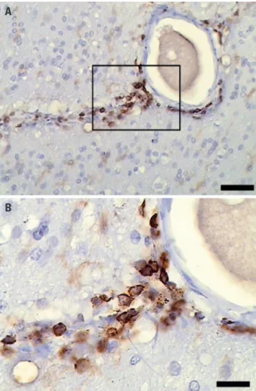
A presence of perivascular lymphocytes infiltrate could also be identiied in Fig 3, when the tissues were submitted to immunostaining against CD45 antibody. his iniltrate could not be visualized in autopsy/control tissues.
A positive correlation was found between the level of IL1β and the signal presented in the tail of hippocam-pus as shown in Fig 4 (top panel) (p=0.033, Pearson
co-eicient=0.436). In contrast, a negative correlation was found between glutamate levels and FLAIR MRI signal intensity in hippocampal tail (p=0.025, Pearson coei-cient=0.457) as shown in Fig 4 (lower panel).
No relationship between the levels of TNFα and FLAIR MRI signal intensity in head, body and tail of the hippocampus of patients with TLE was found, showing no association between these variables (data not shown). In addition, a positive correlation was found analyzing NO concentration and the FLAIR MRI signal intensity in the head (Pearson coeicient=0.498, p=0.013); body (Pearson coeicient=0.505, p=0.012) and tail of the hip-pocampus of patients with epilepsy (Pearson coeicient= 0.532, p=0.007) (Fig 5).
DISCUSSION
he present work showed changes in the concentra-tion of diferent inlammatory mediators such as IL1β and TNFα in the hippocampus of patients with MTS. In addi-tion, a decreased glutamate concentration was also found, when compared with autopsy/control tissues. he
mea-Fig 3. Imminohistochemistry (CD45). [A] Immunohistochemistry against CD45 showing lymphocytes in peri-vascular fashion (scale bar 50 μm, CD45). [B] CD-45 ampliication (scale bar 10 μm).
Fig 4. Correlation between Hippocampal FLAIR signal and IL1β
and glutamate concentrations. [A] Hippocampal FLAIR signal ver-sus IL1β concentration. [B] Hippocampal FLAIR signal versus glu-tamate concentration.
2.2
2.0
1.8
1.6
1.4
1.2
1.0
2.2
2.0
1.8
1.6
1.4
1.2
1.0
FL
A
IR
HPC s
igna
l
FL
A
IR
HPC s
igna
l
IL β concentration
GLU concentration
100 110 120 130 140 150 160
1 2 3 4 5 6 7
A
surement of cytokines NO and glutamate was done in the head of the hippocampus since Bernasconi et al.8 de-scribed this region as that responsible for the major sig-nal alteration on MRI. We also found a linear relation-ship between cytokines and NO levels with the signal on FLAIR MRI in head, body and tail of the hippocampus.
hese data suggest the presence of an inlammatory process in the hippocampus, associated with increased release of glutamate, which is indicative of a persistent
excitotoxicity. Our data are supported by previous re-port21, which demonstrated increased extracellular gluta-mate levels (increased release), using microdialysis probes in humans with TLE. A deiciency in glutamine synthe-sis in astrocytes, found by several authors22, can also ex-plain the high levels of glutamate in extracellular space, suggesting increased release of this amino acid. Working with synaptosomes from hippocampus of patients with TLE and MTS, was found23 increased basal release of glu-tamate and GABA, data that could support our indings. hus, the decreased concentration found in sclerotic hip-pocampus could be associated to increased release of this neurotransmitter, as already reported by several authors. Cytokines, a diverse group of polypeptides that are generally associated with inlammation, immune activa-tion, cell diferentiation or death include interleukins, in-terferons, tumor necrosis factors, chemokines and growth factors24. Cytokines can exert direct action on neurons, indirect action via glial cell, direct action on brain vas-culature and on physiological parameters such as re-gional blood low and temperature. IL1 β and TNFα act on distinct cell-surface receptors but share some com-mon signaling mechanisms related to neurodegenera-tion. IL1 β can induces cell death11, as well as cyclooxy-genase 2 (COX2) and oxide nitric synthase overexpres-sion (iNOS)24. In addition, seizures stimulate increase in the levels of IL1α, IL1β, IL6 and TNFα in the brain25. he mechanism by which IL1 afect seizure activity is not known, but Allan26 showed that the association between administration of AMPA receptor agonist and IL1 pro-duce widespread cortical cell death. his fact is not ob-served when AMPA or IL1 are administered alone26. his fact indeed support the hypothesis that suggest the par-ticipation of increased release of glutamate associated with the increased levels of cytokines, in the process of cell death, observed in the hippocampus of patients with TLE and MTS. Glutamate during excitotoxic phenom-enon stimulates AMPA and NMDA receptors produc-ing excessive depolarization of postsynaptic membrane, which results in an osmotic imbalance when countered by an inlux of Na+, Cl– and water12.
Glial cells can also secrete IL1β and TNFα, which in turn, can act on these cells in an autocrine manner. IL1β and TNFα induce the proliferation of astrocytes, a pro-cess known as astrogliosis11, which is also found in hip-pocampal sclerosis6.
he NO level from sclerotic hippocampus was corre-lated with FLAIR hyperintense signal in the head, body and tail of this structure, supporting the hypothesis of participation of NO production in the hippocampal scle-rosis. Neuronal NOS is localized in the hypothalamic su-praoptic and paraventricular nuclei, both of which are mainly involved in the neurosecretory activity of this
Fig 5. Hippocampal FLAIR signal and NO concentration. [A] FLAIR signal from hippocampal head versus NO concentration. [B] FLAIR signal from hippocampal body versus NO concentration. [C] FLAIR signal from hippocampal tail versus NO concentration.
2.0
1.8
1.6
1.4
1.2
FL
A
IR
HPC s
igna
l
No concentration (Head)
10 20 30 40 50 60 70 80
2.2
2.0
1.8
1.6
1.4
1.2
1.0
FL
A
IR
HPC s
igna
l
No concentration (Body)
10 20 30 40 50 60 70 80
2.2
2.0
1.8
1.6
1.4
1.2
1.0
FL
A
IR
HPC s
igna
l
No concentration (Tail)
10 20 30 40 50 60 70 80
A
B
area. NO is able to interact with many intracellular targets to trigger an array of signal transduction pathways, result-ing in stimulatory or inhibitory output signals. However, it becomes noxious if it is produced in excess27. Recently the term “nitrosative stress” has been used to indicate the cellular damage that is elicited by excess of NO and reac-tive nitrogen species28. In addition, NO has been shown to stimulate the release of noradrenaline and glutamate in the hippocampus29. his data can support our hypothesis concerning to increased release of this neurotransmitter since we found low levels of glutamate in sclerotic tissues, when compared with autopsy/control tissues. Taken to-gether, these data support the idea that the hyperintense signal on FLAIR MRI could be the result of increased ex-pression of cytokines and NO associated with increased excitotoxicity in TLE.
he correlation between ipsilateral versus contralat-eral signals showed that although the hyperintense sig-nal was major in the sclerotic side the contralateral hip-pocampus is frequently involved since it also presented increased FLAIR signal.
As lymphocytes were also visualized in epileptic tissue we can suppose that both humoral and cellular immune systems are activated in TLE associated with HS. his data were previously reported by other authors9,30-32, who dem-onstrate the presence of cytokines, complement activation as well and NO release in the blood after an ictal event. Taken together, these data show the participation of markers of inlammatory process in the physiopathology and in FLAIR signal related to temporal lobe epilepsy, as-sociated to hippocampal sclerosis.
he authors thank to Hilda Reis and Margaret Gori Mouro for their technical assistance as well as all clini-cians involved in the treatment of the patients.
REFERENCES
1. Choi D, Na DG, Byun HS, et al. White-matter change in mesial tempo-ral sclerosis: correlation of MRI with PET, pathology, and clinical features. Epilepsia 1999;40:1634-1641.
2. Mitchell LA, Jackson GD, Kalnins RM, et al. Anterior temporal abnormality in temporal lobe epilepsy: a quantitative MRI and histopathologic study. Neurology 1999;52:327-336.
3. Babb T. Pathological indings in epilepsy. 1 ed. New York: Raven Press; 1987. 4. Mathern GW, Adelson PD, Cahan LD, Leite JP. Hippocampal neuron damage
in human epilepsy: Meyer’s hypothesis revisited. Prog Brain Res 2002;135: 237-251.
5. Cendes F, Andermann F, Gloor P, et al. MRI volumetric measurement of amygdala and hippocampus in temporal lobe epilepsy. Neurology 1993;43: 719-725.
6. Cook MJ. MRI and epilepsy surgery. J Clin Neurosci 1996;3:325-326. 7. de Lanerolle NC, Lee TS. New facets of the neuropathology and molecular
proile of human temporal lobe epilepsy. Epilepsy Behav 2005;7:190-203.
8. Bernasconi N, Bernasconi A, Caramanos Z, Antel SB, Andermann F, Arnold DL. Mesial temporal damage in temporal lobe epilepsy: a volumetric MRI study of the hippocampus, amygdala and parahippocampal region. Brain 2003;126:462-469.
9. Vezzani A, Granata T. Brain inlammation in epilepsy: experimental and clinical evidence. Epilepsia 2005;46:1724-1743.
10. Viviani B, Bartesaghi S, Gardoni F, et al. Interleukin-1beta enhances NMDA receptor-mediated intracellular calcium increase through activation of the Src family of kinases. J Neurosci 2003;23:8692-8700.
11. Wang CX, Shuaib A. Involvement of inlammatory cytokines in central ner-vous system injury. Prog Neurobiol 2002;67:161-172.
12. Dong XX, Wang Y, Qin ZH. Molecular mechanisms of excitotoxicity and their relevance to pathogenesis of neurodegenerative diseases. Acta Pharmacol Sin 2009;30:379-387.
13. Brown GC, Bal-Price A. Inlammatory neurodegeneration mediated by ni-tric oxide, glutamate, and mitochondria. Mol Neurobiol 2003;27:325-355. 14. Clarkson AN, Liu H, Pearson L, et al. Neuroprotective efects of spermine
following hypoxic-ischemic-induced brain damage: a mechanistic study. FASEB J 2004;18:1114-1116.
15. Gilad GM, Gilad VH, Casanova MF, Casero Jr RA. Polyamines and their me-tabolizing enzymes in human frontal cortex and hippocampus: prelimi-nary measurements in afective disorders. Biol Psychiatry 1995;38:227-234. 16. Bloch-Boguslawska E. [The efect of the time of death on the reactivity
of rat caudal artery regulated by NA with a simultaneous use of NOS, CG and COX inhibitors. Part I]. Arch Med Sadowej Kryminol 2006;56:136-143. 17. Blumcke I, Pauli E, Clusmann H, et al. A new clinico-pathological
classii-cation system for mesial temporal sclerosis. Acta Neuropathol 2007;113: 235-244.
18. Cavalheiro EA, Fernandes MJ, Turski L, Nafah-Mazzacoratti MG. Spontane-ous recurrent seizures in rats: amino acid and monoamine determination in the hippocampus. Epilepsia 1994;35:1-11.
19. Lowry OH, Rosebrough NJ, Farr AL, Randall RJ. Protein measurement with the Folin phenol reagent. J Biol Chem 1951;193:265-275.
20. Hampl V, Walters CL, Archer SL. Determination of nitric oxide by the chemi-luminescence reaction with ozone. In: Feelisch M, Stamler JS, (Eds). Meth-ods in nitric oxide research. Chichester: John Wiley & Sons; 1996:310-318. 21. Cavus I, Pan JW, Hetherington HP, et al. Decreased hippocampal volume
on MRI is associated with increased extracellular glutamate in epilepsy pa-tients. Epilepsia 2008;49:1358-1366.
22. Eid T, Thomas MJ, Spencer DD, et al. Loss of glutamine synthetase in the human epileptogenic hippocampus: possible mechanism for raised extra-cellular glutamate in mesial temporal lobe epilepsy. Lancet 2004;363:28-37. 23. Hoogland G, Hens JJ, De Wit M, et al. Glutamate and gamma-aminobutyric
acid content and release of synaptosomes from temporal lobe epilepsy pa-tients. J Neurosci Res 2000;60:686-695.
24. Allan SM, Rothwell NJ. Cytokines and acute neurodegeneration. Nat Rev Neurosci 2001;2:734-744.
25. Jankowsky JL, Patterson PH. The role of cytokines and growth factors in sei-zures and their sequelae. Prog Neurobiol 2001;63:125-149.
26. Allan SM, Rothwell NJ. Cortical death caused by striatal administration of AMPA and interleukin-1 is mediated by activation of cortical NMDA recep-tors. J Cereb Blood Flow Metab 2000;20:1409-1413.
27. Calabrese V, Mancuso C, Calvani M, Rizzarelli E, Butterield DA, Stella AM. Nitric oxide in the central nervous system: neuroprotection versus neuro-toxicity. Nat Rev Neurosci 2007;8:766-775.
28. Guix FX, Uribesalgo I, Coma M, Munoz FJ. The physiology and pathophysi-ology of nitric oxide in the brain. Prog Neurobiol 2005;76:126-152. 29. Lonart G, Wang J, Johnson KM. Nitric oxide induces neurotransmitter
re-lease from hippocampal slices. Eur J Pharmacol 1992;220:271-272. 30. Aronica E, Boer K, van Vliet EA, et al. Complement activation in
experi-mental and human temporal lobe epilepsy. Neurobiol Dis 2007;26:497-511. 31. Bauer S, Cepok S, Todorova-Rudolph A, et al. Etiology and site of temporal
lobe epilepsy inluence postictal cytokine release. Epilepsy Res 2009;86:82-88. 32. Carmeli E, Beiker R, Morad M. Nitric oxide and interlukin-6 levels in
![Fig 1. Hippocampal sclerosis. Nissl staining of hippocampus of patients with TLE and HS [A] Patient number 5 with HS Type 1B (scale bar 1000 μm)](https://thumb-eu.123doks.com/thumbv2/123dok_br/17264871.246564/6.955.61.568.93.445/hippocampal-sclerosis-nissl-staining-hippocampus-patients-patient-number.webp)
![Fig 5. Hippocampal FLAIR signal and NO concentration. [A] FLAIR signal from hippocampal head versus NO concentration](https://thumb-eu.123doks.com/thumbv2/123dok_br/17264871.246564/8.955.61.435.88.890/hippocampal-flair-signal-concentration-flair-signal-hippocampal-concentration.webp)
