Helicobacter pylori
Pei-Yu Chiou1, Cheng-Hung Luo1, Kai-Chih Chang2*, Nien-Tsung Lin1,3*
1Institute of Medical Sciences, Tzu Chi University, Hualien, Taiwan,2Department of Laboratory Medicine and Biotechnology, Tzu Chi University, Hualien, Taiwan, 3Department of Microbiology, Tzu Chi University, Hualien, Taiwan
Abstract
In the model organism Escherichia coli, Min proteins are involved in regulating the division of septa formation. The computational genome analysis ofHelicobacter pylori, a gram-negative microaerophilic bacterium causing gastritis and peptic ulceration, also identified MinC, MinD, and MinE. However, MinC (HP1053) shares a low identity with those of other bacteria and its function inH. pylori remains unclear. In this study, we used morphological and genetic approaches to examine the molecular role of MinC. The results were shown that anH. pylorimutant lacking MinC forms filamentous cells, while the wild-type strain retains the shape of short rods. In addition, a minC mutant regains the short rods when complemented with an intactminCHpgene. The overexpression of MinCHp inE. colidid not affect the growth and cell morphology. Immunofluorescence microscopy revealed that MinCHp forms helix-form structures in H. pylori, whereas MinCHplocalizes at cell poles and pole of new daughter cell inE. coli. In addition, co-immunoprecipitation showed MinC can interact with MinD but not with FtsZ during mid-exponential stage ofH. pylori. Altogether, our results show that MinCHp plays a key role in maintaining proper cell morphology and its function differs from those of MinCEc.
Citation:Chiou P-Y, Luo C-H, Chang K-C, Lin N-T (2013) Maintenance of the Cell Morphology by MinC inHelicobacter pylori. PLoS ONE 8(8): e71208. doi:10.1371/ journal.pone.0071208
Editor:Dipshikha Chakravortty, Indian Institute of Science, India
ReceivedApril 17, 2013;AcceptedJuly 3, 2013;PublishedAugust 1, 2013
Copyright:ß2013 Chiou et al. This is an open-access article distributed under the terms of the Creative Commons Attribution License, which permits unrestricted use, distribution, and reproduction in any medium, provided the original author and source are credited.
Funding:This work was supported by a grant from Tzu Chi University (TCIRP 95009-04). The funders had no role in study design, data collection and analysis, decision to publish, or preparation of the manuscript.
Competing Interests:The authors have declared that no competing interests exist. * E-mail: [email protected] (K-CC); [email protected] (N-TL)
Introduction
Helicobacter pylori, the etiologic agent of human gastritis, peptic ulceration, and gastric carcinoma, infects at least half of the world’s population with the organism being highly restricted to the gastric mucosa of humans [1]. During infection, the major actively replicating forms ofH. pyloricells are spiral-shaped, but they can convert to cocci under environmental stresses, such as starvation and antibiotic treatment. The coccoid form is viable, but not culturable in vitro. It is less virulent than the spiral form; however, it is thought to be crucial in disease transmission and insensitive to antibiotic treatment [2]. Therefore, cell shape is an important pathogenicity factor for H. pylori. So far, the maintenance and establishment of the spiral structure inH. pyloriis known to occur through peptidoglycan relaxation and an intracellular scaffold [3,4,5,6]. While cell division accuracy is crucial for maintaining the shape of some bacteria [7], little is known forH. pylori.
The process of cell division involves the spatial and temporal regulation of the septum formation by the cytoskeletal proteins [7]. In Escherichia coli, precise formation of septum between the two newly segregated sister chromosomes is initiated by FtsZ, which assembles into a ring and recruits a number of proteins, such as FtsA, ZipA, and ZapA, to form the septal ring [8]. In addition, Min proteins are required for the correct division site selection, which prevent polar divisions by blocking the Z ring assembly at cell poles [8]. The functions of FtsZ andmin operon have been characterized in many bacteria [9,10,11,12,13]. Although the task of these proteins, which are involved in cell division, is almost identical in reported bacteria, the components and precise
regulation mechanisms in preventing polar division of Min systems appears to be different among prokaryotic cells. For example, the Min system, which consists of 3 proteins, MinC, MinD, and MinE, and which is composed of an operon in E. coli, can oscillate periodically between the 2 poles of a cell, and MinE destabilized, but only 2 of them, MinC and MinD, are present inBacillus subtilis. The function of MinE is substituted by an unrelated DivIVA protein, which targets MinCD in division sites and retains them at the cell poles. However, we found that the homologs ofminC,D, and E are present, but not contained, in an operon in the sequenced genomes of H. pylori. Moreover, the amino acid sequences of MinC show a low identity with the corresponding proteins of reported bacteria. So far, whetherminCplays any role in cell division ofH. pyloriremains unclear. Therefore, studying MinC’s functions is vital for understanding the cell division and shape-determining factors ofH. pylori. In this study, we generated a
minCmutant to study its biological characters. Our results show thatminCplays a crucial role in maintaining the cell morphology and the movement capabilities ofH. pylori.
Materials and Methods
Bacterial Strains, Plasmids and Growth Conditions
Bacterial strains and plasmids used in this study are listed in Table 1. Strains ofE. coliwere cultivated in Luria-Bertani (LB) (Difco Laboratories, Detroit, MI) solid and liquid media at 37uC.
H. pyloriNCTC 11637 was used to construct mutants. H. pylori
(BAP) containing Columbia agar base (Becton Dikinson, Franklin Lakes, NJ, USA) and 5% horse blood or in Brucella broth (Becton Dikinson) containing 5% fetal bovine serum (FBS). Bacterial growth was measured by monitoring OD600, while live cells were determined by viable count on BAPs. When required, antibiotics were supplemented: ampicillin (Ap, 100mg/mL), chloramphenicol (Cm, 30mg/mL), and kanamycin (Kan, 50mg/mL).
DNA Techniques
The methods described by Sambrook et al [14] were used for preparation of chromosomal DNAs, restriction digestion, DNA ligation and E. coli transformations. Plasmids were isolated by using High-Speed Plasmid Mini Kit (Geneaid, Taipei, Taiwan). Natural transformation of H. pylori was performed as described elsewhere [15,16].
Cell Length Determination, Immunostaining, and Image Acquisition
H. pylorifrom overnight liquid cultures was inoculated into fresh Brucella broth to obtain an initial OD600of 0.05 and grown to an OD600of 0.6 to 0.8. Cells were examined microscopically on poly L-lysine-treated slides with a thin layer of 1% agarose in LB. Cell length was measured as the axis length from one pole to the other of the cells captured in microscope, using ImageJ version 1.46 (http://rbs.info.nih.gov/ij/). Average cell length was determined using at least 2 independent measurements, each on 200 cells. DNA was stained with 49, 6-diamidino-2-phenylindole (DAPI; Sigma, St. Louis, MO) at a final concentration of 1mg/mL and
membrane was stained with FM4-64 (Molecular Probes/Invitro-gen) at a concentration of 1mg/mL. Bacterial viability was
determined by staining the cells with SYTO9/propidium iodine
Table 1.Strains and plasmids used in this study.
Strain or plasmid Genotype or description Source
Strains
E.coli
Top10 F-mcrD(mrr-hsdRMS-mcrBC) 80
lacZDM15lacXD74recA1deoRaraD139D(ara-leu) 7697galUgalK
rpsL (StrR)endA1nupG
Invitrogen
BL21(DE3) F–dcm ompT hsdS(r
B–mB–)gall(DE3) Stratagene
MG1655 wild-type [30]
H. pylori
NCTC 11637 wild-type, containing the entireminC ATCC 43504
PY1 11637,minC::cat This study
PY2 PY1,hp0405::PflaA-minCHpkan This study
PY2-5 PY1,hp0405::PflaA-minCEckan This study
PY3 11637,hp0405::PflaA-minCHpkan This study
PY3-1 11637,hp0405::PflaA-minCEckan This study
Plasmids
pAV35 chloramphenicol acetyltransferase (cat) cassette; Cmr [18]
pJMK30 kanamycin resistance (kan) cassette; Kanr [18]
pUC18 Cloning vector, Apr Fermentas
pOC10 Cloning vector derived from pOK12, replacement of thekangene with thecatgene from pAV35, Cmr [19]
pTZ57R/t T/A Cloning vector, Apr Fermentas
pET30a Cloning and expression vector, Kanr Novagen
pBAD33 pBAD expression; pACYC184 ori; Cmr [31]
pCHL2 hp0405::PflaAkan,Kanr, Cmr This study
pCPY001 pUC18 containingminCHp, Apr This study
pCPY002 pCPY001 withcatinserted into the unique SphI site ofminCHp, Apr, Cmr This study
pCPY003 pCHL2 containingminCHpunderflaApromoter,hp0405::PflaA-minCHpkan,Kanr, Cmr This study
pCPY004 pET30a containing 66his-minCHp,Kanr This study
pCPY005 pET30a containing 66his-minCEc,Kanr This study
pCPY006 pCHL2 containingminCEcunderflaApromoter,hp0405::PflaA-minCEckan,Kanr, Cmr This study
pCPY007 pET30a containing 66his-ftsZ,Kanr This study
pCPY008 pET30a containing 66his-minD,Kanr This study
pCPY009 pBAD33 containingminCHp, Cmr This study
pCPY010 pBAD33 containingminCEc, Cmr This study
pOC0405 pOC10 containinghp0405, Cmr This study
pTZ-PflaA pTZ57R/t containingH. pyloriflagella promoter, Apr This study pTZ-PflaAKm pTZ-PflaA containingkanfrom pJMK30, Apr, Kanr This study
(PI) (LIVE/DEAD BacLight kit, Molecular Probes/Invitrogen) at 24, 48, and 72 h, followed by fluorescence microscopic observa-tion of the stained cells. Images were obtained with a Nikon E800 microscope using a 1006Objective with A = 1.45 and processed using Adobe Photoshop CS3.
The subcellular localization of MinCHp was carried out using immunofluorescence (IF) microscopy [17]. Bacteria were spread on a clean glass slide and allowed to dry briefly. Bacteria on the glass slides were fixed with methanol at room temperature for 15 min, followed by incubation with 0.1% Triton X-100 in PBS for 1 h. The bacteria were treated with 100mg/mL of lysozyme
and 5 mM EDTA in PBS for 1 h at room temperature. Prior to IF staining, bacteria were incubated with 10% (w/v) bovine serum albumin (BSA) in PBS for 30 min at 37uC to block nonspecific binding. Three PBS washes were performed following each incubation or treatment. After incubation for 1 h with anti-MinCHp (1:200), the slides were washed 5 times with PBS containing 0.05% Tween 20 (PBST). Incubation using FITC-conjugated anti-rabbit IgG (1:500) (Santa Cruz, CA, USA) diluted in blocking buffer was carried out for 30 min at 37uC. The cells were washed 3 times with PBST. The nucleoids were stained with DAPI at a final concentration of 0.5mg/mL in H2O. The cells were washed once in H2O. The images of the bacteria were subsequently visualized with a Nikon E800 microscopy.
Sequencing and Identification of theminCGene
The oligonucleotide primers used in this study are listed in Table 2. Primers HP1054-F and HP1052-R for a PCR corresponded to the nucleotide (nt) 2924 to 2946, relative to the hp1054 start codon, and nt 2269 to 2248, relative to the termination codon ofhp1052, respectively. A PCR was performed to amplify the fragment, using theH. pyloriNCTC 11637 genomic DNA as the template. The amplicon was purified using the Gel/ PCR DNA Fragments Extraction Kit (Geneaid, Taipei, Taiwan) and directly sequenced using a 3730 DNA analyzer (Applied Biosystems, CA, USA). The sequence analysis was performed using NCBI packages.
Construction and Complementation of aH. pylori minC
Mutant
TheH. pylori minCwas a PCR amplified from the strain NCTC 11637 genomic DNA using primers minCN and minCC. The PCR product was cloned into the SphI site of pUC18 to generate pCPY001. To create an insertional mutant ofminCinH. pylori, the chloramphenicol acetyltransferase (cat) cassette form pAV35 (kindly provided by J. M. Ketley) [18] was digested with PvuII and inserted into the unique SphI restriction site 27 bp from the start site of minCHp in pCPY001 to generate pCPY002. The
chromosomal minC locus was disrupted through a homologous recombination upon transforming strain NCTC 11637 with pCPY002. A transformant, selected on BAPs supplemented with Cm, was designated H. pylori PY1. The resulting PY1 was confirmed by a PCR analysis using the same primers of minCN and minCC.
To complementH. pyloriPY1, plasmid pCHL2 was constructed in several steps to be used as a vector. (1) The hp0405 gene, amplified using PCR with the primers 0405-F and 0405-R, was cloned into the EcoRV site of pOC10 [19] and the fragment between StuI-HincII restriction sites was subsequently removed, which resulted in the vector pOC0405. (2) TheH. pyloriflagella promoter, PflaA, was amplified using a PCR with primers pflaF and
pflaR and ligated with a pTZ57R/t vector to generate pTZ-PflaA. (3) Thekancassette of pJMK30 (provided by J. M. Ketley) [18] was cloned into the XbaI site of PflaA to generate pTZ-PflaAKm. (4) PflaA and kan were subsequently cloned into the
EcoRI site of pOC0405, generating plasmid pCHL2. The obtained pCHL2 vector possessed a unique StuI for cloning genes of interest between PflaAandkan.
Regarding complementation,minCHpwas PCR-amplified from
theH. pyloriNCTC 11637 genomic DNA, using the primer pair minCN/minCC and was cloned into the unique StuI restriction site of pCHL2 to generate the construct pCPY003. The plasmids were then transformed into mutant PY1 and NCTC 11637 through natural transformation and were selected on BAPs supplemented with Kan. The ectopic integration of the cloned
minCin the strain PY2 (PY1-complemented strain) was verified with the PCR using the primers minCN and minCC, in which an amplicon of 1.3 kb (theminCgene plus acatgene) and 0.6 kb (the
minCgene only) were obtained. The ectopic integration of cloned
minCHpin the strain NCTC 11637 was the designatedH. pyloriPY3
and was verified with PCR analysis using a pair of primers pflaF-minCC. minCEc was PCR-amplified from pCPY005 using the
primer pair NHis-F/minCec-R and cloned into the unique StuI restriction site of pCHL2 to generate the construct pCPY006. Subsequently, the plasmids were introduced into strain PY1 and NCTC 11637 through natural transformation (described above) and transformants were designatedH. pylori PY2-5 and PY3-1, respectively. Finally, the complemented strains were verified with the PCR analysis using a pair of primers, pflaF-minCec-R.
Plasmids Construction
The minCHp and ftsZ gene were amplified by PCR using the
genomic DNA of NCTC 11637 as the template, with the primers minCN/minCC and FtsZP-F/FtsZP-R as the primers, respective-ly. The products were digested with EcoRI and XhoI and cloned into pET30a cleaved with the same enzymes to yield pCPY004 and pCPY007, respectively. TheminDgene was amplified by the PCR with the primers PminD1-F/PminD2-R and the amplicon was digested with SacI and HindIII. The SacI-HindIII fragment was cloned into pET30a cleaved with the same enzymes to yield pCPY008. Purified MinCHp, FtsZ or MinD proteins fromE. coli strain BL21(DE3) carrying pCPY004, pCPY007, or pCPY008
Table 2.Oligonucleotide primers used in this study.
Name Sequence (59R39)
Size (bps)
Restriction site
HP1054-F CAATCAGGTGTTGTTCCAATTC 1182 HP1052-R TCGCATGGACAGCTGAAAGCAA
minCN GAATTCGTCATGTTAAAAACGAATC 588 EcoRI minCC CTCGAGTTTGCTTCATAATACTTCCTT XhoI 0405-F GGATCCCTTACTCAACCCTAA 1400 0405-R GGATCCTTAAAAATAGTTTTAGC
pflaF TTTATTATAGCCCATTTTCATGCT 127 pflaR AGGCCTCCTTGTTATAAAAAACCCA
FtsZP-F GAATTCATGGTTCATCAATCAGAGAT 1158 EcoRI FtsZP-R CTCGAGTCAGTCTTGCTGGATTCTCA XhoI PminD1-F GAGCTCAGGAATCATATGGCAATA 807 SacI PminD2-R AAGCTTAAAAAAATCAAACAAACTCA HindIII minCec-F GGGATCCATGTCAAACACGCCAAT 696 BamHI minCec-R CGTCGACTCAATTTAACGGTTGAA SalI NHis-F ATATACCATGGGCAGCAGCCATCA
Figure 1. Genomic organization ofmingenes in rod-shaped bacteria.(A) Grey arrows represent the genomic regions surrounding themin
genes. White arrows show the localization ofmingenes. (B) Sequence comparison ofH. pyloriMinC with those of other bacterial MinC protein. The consensus line below the sequence alignment indicates identity (*), strong conservation (:), and weak conservation (.) of amino acid matches. Organisms in the alignment includeH. pyloriNCTC 11637 (KC896795; Hp11637),Escherichia coli(NP_415694.1; Ec),Bacillus subtilis(NP_390678; Bs),
were used to raise rabbit anti-MinCHp, anti-FtsZ, or anti-MinD polyclonal antiserum, respectively (Protech Technology Enter-prise, Taipei, Taiwan). TheminCEcgene was amplified by the PCR
fromE. coliMG1655 genomic DNA using the primers minCec-F and minCec-R. The PCR products were digested with BamHI and SalI and ligated with BamHI-SalI-cleaved pET30a to generate pCPY005. The XbaI-XhoI fragments of pCPY004 and pCPY005 were cloned into XbaI-SalI-cleaved pBAD33 to yield pCPY009 and pCPY010, respectively.
Western Blot Analysis
Bacteria cell liquid cultures were centrifuged at 13,0006gfor 1 min and cell pellets were washed twice in PBS. Pellets were resuspended in sterile water. The bacterial suspension was sonicated using an ultrasonicator (Model XL, Misonix, Farming-dale, NY) to break the bacteria. Total protein concentrations were determined by using the Bio-Rad Dc protein assay kit on samples diluted 20-fold in water and on BSA standards in the same diluted buffer. The equal amounts of cell protein per lane were mixed with sample buffer (62.5 mM Tris-HCl, pH 6.8 containing 5% 2-mercaptoethanol, 2% SDS, 10% glycerol, and 0.01% bromophe-nol blue) and heated in a boiling water bath for 10 min. The samples were subjected to polyacrylamide (12%) gel electropho-resis; the protein bands were subsequently transferred onto the polyvinylidene difluoride (PVDF) membranes and probed with rabbit anti-MinCHp antibodies. A peroxidase-conjugated goat affinity-purified antibody against rabbit immunoglobulin G was used as the secondary antibody (Cell Signaling). Immunoreactive bands were detected using enhanced chemiluminescence (Milli-pore) and X-ray film. Band intensities on blots were measured using ImageJ version 1.46.
Motility Assay
Cells ofH. pylorigrown in liquid cultures were stabbed into a soft agar plate containing 0.35% Bacto-agar in Brucella Broth medium and 5% FBS, followed by the incubation of the culture under microaerobic conditions for 3 to 5 days.
Cell Morphology Analysis
To test the MinC sensitivity ofE. coli, the plasmid pCPY009 or pCPY010 was introduced into E. coli MG1655. Exponentially growing of strain was serially diluted by 10. Then 3ml cultures
from each dilution was spotted on a plate with and without arabinose (Sigma) and incubated overnight at 37uC. To study the effects of overexpression, MinCHp, cells harboring pCPY009 were grown overnight in LB medium at 30uC. An overnight culture was diluted 100-fold in an LB medium supplemented with Cm and grown for 2 h at 30uC. The cell cultures were added at various concentrations (0, 0.002, 0.02, or 0.2%) of arabinose and grown at 30uC for an additional 2 h. The cells were spun down, resuspended in LB medium, mixed 1:1 with 2% LB agarose, spotted onto a coverslip, and observed under a light microscope.
Immunoprecipitation
Liquid cultures of H. pylori were grown to mid-log phase (OD600= 0.6 - 0.8) and harvested by centrifugation at 60006g. Cell-free extracts were prepared by suspending cells in a sonication buffer (20 mM Tris-HCl, pH 8.0 containing 300 mM NaCl) and sonicated for 1 min with an ultrasonicator. The resultant suspensions were then centrifuged at 13,0006g for 20 min at 4uC and the supernatants containing proteins were collected. The supernatants were mixed with 30ml of 20% (w/v) Protein A
Sepharose CL-4B (GE Healthcare) for 1 h to remove nonspecif-ically bound proteins. An aliquot (50ml) of supernatant fraction was used as a control for total protein levels prior to immunopre-Figure 2. The effect of MinCHpprotein on cell length distribution ofH. pylori.(A) Cell length distributions of NCTC 11637 (minC+), PY1 (minC
mutant) and PY2 (minCcomplemented strain). (B to D) Gram-stained microscopic images of the three strains shown in panel A to demonstrate the morphology. (B) NCTC 11637; (C) PY1; (D) PY2. Scale bar, 10mm. (E) SDS-PAGE (left panel) and Western blot (right panel) showing the levels of MinC in strains NCTC 11637, PY1, and PY2. Lanes M, PageRuler prestained protein ladder SM0671 (MBI Fermentas); lanes 1 and 4, NCTC 11637; lanes 2 and 5, PY1; lanes 3 and 6, PY2.
cipitation (IP). Lysate was then incubated with anti-MinCHp, anti-FtsZ, or anti-MinD antibodies coupled to Protein A Sepharose beads and incubated with shaking (40 rpm) at 4uC overnight. Beads containing protein complexes were washed 3 times with IP buffer (0.5% NP-40/PBS) and then boiled in sample buffer for 15 min. Samples were subsequently analyzed by SDS-PAGE, followed by immunoblotting.
Nucleotide Sequence Accession Number
The nucleotide sequence of theH. pyloriNCTC 11637minChas been deposited in the GenBank under accession number KC896795.
Results
minGenes are Present inH. pylori, butminCis not Clustered withminDandminE, and Shares a Low Similarity to other Reported Bacteria
As of March 2013, over 40 strains ofH. pylorigenome have been sequenced and registered in database. Our database search revealed thatminCis separated fromminD-minEin all sequenced
H. pyloristrains, different from other bacterial genera in which the corresponding genes are clustered [12,20]. Also, the same genome organization of min occurs in all sequenced Helicobacter spp. Furthermore, genes flankingminCandminDEare conserved in all sequencedHelicobacterspp.:nlpDandlpxCflankingminCgene and Figure 3. Growth curve, cell viability and fluorescent micrograph ofH. pyloristrains.(A) Cells were grown in Brucella broth medium containing 5% FBS, under microaerobic conditions with gentle shaking at 37uC. Concentrations of the cells were monitored by measuring the turbidity (OD600). (B) Viable cells were counted by serially diluted (1:10) of cultured cells in the fresh medium and plating onto Columbia agar base with 5% defibrinated horse blood. After 3–5 days of incubation, the number of colonies was counted to determine the bacterial proliferation.
-N
-, NCTC 11637; -.-, PY1; -&-, PY2. Error bars represent the standard deviations of the means of triplicate samples. Fluorescent micrograph of NCTC11637 (C, D, and E) and PY1 (F, G, and H) stained with LIVE/DEADHkit at 24, 48, and 72 h, respectively, demonstrated morphology and membrane integrity changes during growth in Brucella broth medium containing 5% FBS. Scale bar, 10mm.
ilvCanddprAflankingminDE(Figure 1A). These findings indicate that these DNA regions are stable.
TheminC,minD,andminEgenes ofH. pyloriencode proteins of 195 aa (22.38 kDa), 268 aa (29.31 kDa), and 77 aa (8.92 kDa), respectively. MinD and MinE show high degrees of identity in amino acid sequence to those from other bacteria (48.5% and 43.3% identical to E. coli and B. subtilisMinD, and 29.9% and 14.3% identical to E. coli and B. subtilis MinE/DivIVA, respectively). However, MinC shares low identity with E. coli
and B. subtilis MinC proteins, 17.4% and 6.2%, respectively. Therefore, this study further investigates the role of MinC inH. pylori.
Because of the genetic variability ofH. pylori, we focused our work on the reference strain NCTC 11637. To investigate theH. pylori minC, the gene was cloned from strain NCTC 11637 by a PCR-based strategy using primers HP1054-F and HP1052-R designed according to the minC flanking sequences of H. pylori
strain 26695 (Table 2) for DNA amplification. Sequencing of the amplicon revealed 1,182 bp containing ORF with 195 aa which shared 87% to 95% identity to the MinC from other sequencedH. pyloristrains and shared 20%, 13%, 13%, and 14% identity with the homologs from E. coli, B. subtilis, Neisseria gonorrhoeae, and
Thermotoga maritime, respectively.
It has been shown that four conserved glycine residues at the MinC C-terminus are essential for MinC functionality as a cell division inhibitor and for the interaction of MinC with other Min proteins in E. coli[21]. Four glycine residues (G104, G122, G129, and G139) were also found at C-terminus of theH. pyloriMinC proteins. In addition, a lysine residue at the N-terminus (K3) and an arginine residue (R102) at the C-terminus were also conserved among all known MinC proteins (Figure 1B). These findings
suggest thatH. pylori MinC is a factor involved its cell division, functioning to interact with MinD.
Mutation ofH. pylori minCcauses Cell Filamentation and Growth and Motility Defects
To investigate the role of MinC, aminCmutant of NCTC 11637 was constructed by a marker exchange and designated as PY1. Light microscopic observation of PY1 after gram-staining showed cells with different sizes. As shown in Figure 2, the cell length ranged from 1.6 to 25.7mm with 64.5% of them falling between 5–10mm. Comparing to that of the wild-type (2.2260.75mm), cell elongation of PY1 was obvious. PY1 exhibited a growth rate similar to that of the wild-type, with a generation time of about 6 h (Figure 3A). The OD600 of the wild-type reached the maximum (1.34) at 24 h, while that of the mutant increased to a maximum of 1.88 at 36 h. The OD600 of both strains declined gradually following cessation of growth. However, the number of viable cells of the wild-type was about 10 times more than that of PY1 at 36 h (5.86108versus 4.56107CFU/ml), and the amount of cell protein Figure 4. Distributions of DNA and membrane inH. pylori.Wild-type and mutant PY1 cells were grown to mid-exponential phase and then stained with DAPI (blue for chromosome) and FM4-64 (red for membrane) and observed by fluorescence microscopy. Scale bar, 5mm.
doi:10.1371/journal.pone.0071208.g004
Figure 5. Bacterial motility assay. The indicated strains were stabbed into semisolid agar medium and incubated at 37uC for 72 h. doi:10.1371/journal.pone.0071208.g005
Figure 6. Cellular localization of MinCHpinH. pylori.Selection of
cells (I and II) were observed by fluorescence microscopy. IF microscopy was applied using anti-MinCHpantibody, followed by visualization with FITC-conjugated rabbit IgG. The DNA was labeled by DAPI. Scale bars, 1mm.
of the wild-type was 1.26 times that of the mutant (3.6 versus 2.85 mg/ml).
To compare the cell morphology and membrane integrity, cells of NCTC 11637 and PY1 grown to 24, 48, and 72 h were stained with LIVE/DEADH kit and examined by fluorescence microsco-py. All the wild-type cells were in rod form till 24 h, a portion of them (ca. 6%) became coccoid form at 48 h, and then almost all cells were in cocoid form at 72 h. In contrast, all PY1 cells maintained the elongated form throughout the experiment
(Figure 3). Both strains appeared alive at 24 h; a large portion of the PY1 cells were dead (75%) at 48 h and almost all of them were dead at 72 h. Compared to the mutant cells, larger portions of the wild-type kept alive at 48 h (63%) and 72 h (48%). Furthermore, PY1 contained clearly segregated nucleoids (Figure 4), indicating that mutation in minC caused no defects in chromosome replication or segregation.
It is known that cell morphology affects the cell motility in numerous bacteria [22]. As motility of H. pylori is crucial in colonizing the gastric mucosa [23], we have evaluated the effects of
minCmutation on the motility in this study. Tests were performed on a soft agar plate, comparing the area of spreading zones between the minC mutant and its parental strain. The results showed that the motility activity is reduced by half in mutant strains. As shown in Figure 5, growth of the wild-type cells resulted in a spreading zone of 15 mm in diameter after 72 h (Figure 5), while that formed by PY1 was about 7 mm in diameter (Figure 5). Since cell division related proteins are not involved in flagellar biosynthesis, it appears that the cellular elongation has reduced the cell motility.
Reversal of mutantminCPhenotype inH. pyloriby Complementation
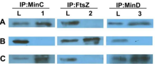
To perform the complementation test, we constructed a vector, pCHL2 (Table 1), that allowed for ectopic integration of the plasmid intoH. pylorichromosome. The integration, targeted at the locus ofhp0405ofH. pylori, was shown to cause no detectable Figure 7. Identification of FtsZ and MinD from co-IP performed
with MinC during mid-exponential cultures of NCTC11637.
Proteins were eluted after co-IP experiments, samples were separated by SDS-PAGE and detected by Western blotting. Western blots were probed with (A) anti-MinC, (B) anti-FtsZ, and (C) anti-MinD. Lanes L, loading control consisting of whole-cell extract; lanes 1, proteins precipitated with anti-MinCHp; lanes 2, proteins precipitated with anti-FtsZ; lanes 3, proteins precipitated with anti-MinD.
doi:10.1371/journal.pone.0071208.g007
Figure 8. The effects of MinCHpand MinCEcproteins on cell length distribution ofH. pylori.(A) Cell length distributions of the PY2-5, PY3,
and PY3-1. (B to D) Differential interference contrast (DIC) microscopic images of the three strains shown in panel A to demonstrate the morphology. (B) PY2-5; (C) PY3; (D) PY3-1. Scale bars, 10mm.
effects on the physiology or morphology of H. pylori [24,25]. A complemented strain, PY2, with the minCHpgene integrated into
the locus of hp0405 of PY1 was constructed. The expression of MinCHpin the complemented strain was confirmed by Western blot analysis (Figure 2E). A densitometry analysis indicated that the expression of MinCHpin PY2 was 1.6 times greater than that of the wild-type NCTC 11637. In addition, about 99% of PY2 cells regained normal cell morphology, exhibited a normal cell length distribution (Figure 2D), and restored their motility (Figure 5). This result suggested that elongation of PY1 cells was significant because of theminCmutation and it was not a polar effect on downstream gene expression.
Cellular Localization of MinC inH. pylori
InE. coliandB. subtilis, MinC is an effector of the Min system responsible for antagonizing cell division and for preventing the sedimentation of FtsZ [11]. However, consequence of minC
mutation may not be the same forH. pylori, because mutation in
minCgene causes the cell to elongate instead of mini-cell formation observed inE. coliandB. subtilis.
To detect the cellular location of MinC, IF microscopy was performed using antibodies against MinC and a secondary antibody tagged to FITC. The results showed that MinC in the mid-log cells assembled into helix-form structures and located mainly in poles (Figure 6).
MinCHpInteracts with MinD but not with FtsZ during Mid-exponential Stage ofH. pylori
To examine whether MinC interacts with MinD and FtsZ inH. pylori, co-IP was performed using antibodies prepared against MinC, FtsZ, or MinD separately, followed by detection of the proteins co-precipitated by Western blotting (Figure 7). Unex-pectedly, the MinD protein was precipitated with MinC, but FtsZ was not (Figure 7, lanes 2 and 3).
The Effects of MinCEcinH. pylori
To test whether MinC ofE. colican complement the deficiency in MinC inH. pylori, theE. coli minCgene was cloned in pCHL2 and introduced into PY1 (forming strain PY2-5) for complemen-tation. Results showed that 81% of the cells had a length shorter than 5mm with an average of 3.24mm, demonstrating that MinCEccould complement the deficiency in MinCHpinH. pylori. To inspect the effects of MinCHpand MinCEconH. pyloricell division, minCHp and minCEc were cloned and inserted into the hp0405locus of NCTC 11637, resulting in strains PY3 and PY3-1, respectively (Table 1). Cells of PY3 had an average length similar to that of the wild-type (2.9961.22mm) and about 7.1% of them
were longer than 5mm (Figure 8A). In contrast, the average cell
length of PY3-1 increased to 5.3163.36mm (Table 3), and the
cells shorter than 2mm decreased to 7% (Table 3).
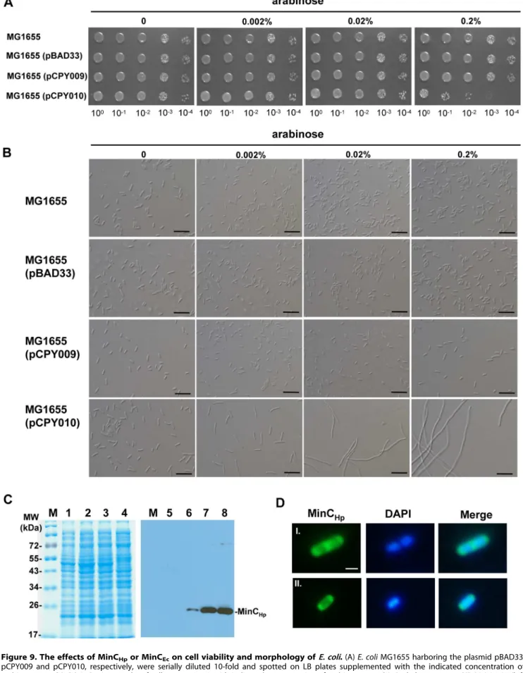
The Effects of MinCHpinE. coli
To inspect the effects of MinCHp and MinCEcon E. colicell division, minCHp and minCEc were cloned in pBAD33 and
introduced into the MG1655, resulting in strains MG1655(pCPY009) and MG1655(pCPY010), respectively. As shown in Figure 9A, growth of the MG1655(pCPY010) cells containing minCEc gene under control of arabinose-inducible
promoter was inhibited by the presence of 0.2% arabinose. But MG1655(pBAD33), carrying the cloning vector only, and MG1655(pCPY009) grew well in the presence of 0.2% arabinose, suggesting that overproduction of MinCHpwas not lethal inE. coli. Light microscopy showed that MG1655 carrying cloning vector only or containing minCHp was similar to the wild-type in
morphology (Figure 9B), but MG1655(pCPY010) formed fila-ments in the presence of 0.002% arabinose. In immunoblotting with anti-MinCHpserum, it was shown that MinCHplevels were elevated with increased concentrations of arabinose (Figure 9C).
IF microscopy showed that MinCHplocalized at both poles of the E. coli cells before septum formation (Figure 9D). Upon septation, the majority of the cells contained intense fluorescence at septum, while some of them still retained small amounts of fluorescence at the poles (Figure 9D), suggesting that MinCHpalso localized at the poles during the late stage of septation inE. coli.
Discussion
In many bacteria, Min proteins are involved in regulation of cell division. It is known that not all threemingenes are ubiquitously present in all microorganisms and the entire minCDE cluster appears to be present only in Gram negative bacteria [26]. In this study, we reveal that H. pylori possesses homologs of minC and
minDE, except that they are in two loci. Our sequence analysis here shows that residues conserved in other bacteria are all present in MinCHp(Figure 1B).
MinC is required for inhibition of septation by FtsZ in many bacteria and deficiency in MinC results in over septation that in turn causes mini-cell formation [8]. In contrast, a MinC deficient mutant ofH. pyloriwas found to form elongated cells in this study. Our observations suggest that MinC of H. pylori is involved in normal septation that is required for normal cell division. To our knowledge, this is the first report that MinC is required for normal septation instead of inhibiting septation.
In E. coli, MinE imparts topological specificity by stimulating MinCD oscillation, thereby ensuring that the concentration of
Table 3.Cell length measurements.
Strain Genotype
Average cell length± SD (mm)
Cell shorter than 2mm (%)
Cell between 2 to 5mm (%)
Cell longer than 5mm (%)
H. pylori
NCTC 11637 wild-type 2.5860.70 17.5 82.5 –
PY1 11637,minC::cat 7.5563.86 1.3 26.1 72.6
PY2 PY1,hp0405::PflaA-minCHpkan 2.7760.80 15.3 84.2 0.5
PY2-5 PY1,hp0405::PflaA-minCEckan 3.2463.03 46.7 34.6 18.7
PY3 11637,hp0405::PflaA-minCHpkan 2.9961.22 16.9 76 7.1
PY3-1 11637,hp0405::PflaA-minCEckan 5.3163.36 6.5 49.3 44.2
Figure 9. The effects of MinCHpor MinCEcon cell viability and morphology ofE. coli.(A)E. coliMG1655 harboring the plasmid pBAD33,
MinCD is highest at the poles [27]. In B. subtilis, MinCDJ are localized at the poles or the site of division through polar targeting by DivIVA [28]. In this study, IF microscopy revealed that MinCHpin the mid-log cells assembled into helix-form structures and located mainly in poles, but do not interact with FtsZ, suggesting that MinCHp-FtsZ interaction is not required for mediation of cell division. It is possible that MinCHpinteracts with other proteins during different stages of cell division inH. pylori.
Several studies have shown that Min proteins can function in heterologous background, for examples, the chloroplasts are enlarged whenminCofE. coliis introduced intoArabidopsis thaliana
[29], cells of B. subtilis transformed with minC of E. coli are elongated [10],E. colicells transformed withminCandminDofN. gonorrhoeae are elongated [13]. In this study, MinCEcprovided in trans resulted in elongation of the wild-type cells and was able to restore the wild-type length to the mutant PY1 (Table 3). It is possible that expression of MinCEcmay prevent the polymeriza-tion of FtsZHp in H. pylori, thereby inhibiting cell division and resulting in cell elongation [26]. In contrast, expression of MinCHp inE. colidid not cause detectable effects on cell morphology. Since the amino acid sequence of FtsZHpshare high degree of similarity with FtsZEc(70%) and MinCHpexhibited no co-IP reaction with FtsZHp, it appears reasonable to predict that MinCHp cannot interact with FtsZEc. Consequently, MinCHpcould not inhibit the
Z-ring formation inE. coli. In addition, based on the observations that i) H. pylori and E. coli FtsZ have different architecture of filaments, ii) the FtsZ-ring positions at both central and acentric regions in H. pylori, and iii) daughter cells show considerably different sizes owing to the asymmetrical division of the cells, Spechtet al. [9] suggest that FtsZ ofH. pylori possesses a unique intrinsic characteristic different from that of E. coliand the cell cycle ofH. pyloriis clearly dissimilar to that of E. coli. Thus, the present observations that i)minCmutation causes cell elongation instead of mini-cell formation, ii) MinC does not interact with FtsZ, and iii) MinCHp causes no effects on cell division when expressed in E. coli have confirmed and extended the previous findings inH. pyloricell division.
Acknowledgments
We thank Y.-H. Tseng for reading the manuscript.
Author Contributions
Conceived and designed the experiments: PYC NTL. Performed the experiments: PYC. Analyzed the data: PYC KCC NTL. Contributed reagents/materials/analysis tools: CHL KCC. Wrote the paper: PYC NTL.
References
1. Dunn BE, Cohen H, Blaser MJ (1997)Helicobacter pylori. Clin Microbiol Rev 10: 720–741.
2. Andersen LP, Wadstrom T (2001)Helicobacter pylori: physiology and genetics; In: Mobley HLT MG, Hazell SL, editor. Washington, DC: ASM press. p.27–38 p. 3. Sycuro LK, Wyckoff TJ, Biboy J, Born P, Pincus Z, et al. (2012) Multiple peptidoglycan modification networks modulate Helicobacter pylori’s cell shape, motility, and colonization potential. PLoS Pathog 8: e1002603.
4. Specht M, Schatzle S, Graumann PL, Waidner B (2011) Helicobacter pylori possesses four coiled-coil-rich proteins that form extended filamentous structures and control cell shape and motility. J Bacteriol 193: 4523–4530.
5. Sycuro LK, Pincus Z, Gutierrez KD, Biboy J, Stern CA, et al. (2010) Peptidoglycan crosslinking relaxation promotesHelicobacter pylori’s helical shape and stomach colonization. Cell 141: 822–833.
6. Waidner B, Specht M, Dempwolff F, Haeberer K, Schaetzle S, et al. (2009) A novel system of cytoskeletal elements in the human pathogenHelicobacter pylori. PLoS Pathog 5: e1000669.
7. Graumann PL (2007) Cytoskeletal elements in bacteria. Annu Rev Microbiol 61: 589–618.
8. Cabeen MT, Jacobs-Wagner C (2010) The bacterial cytoskeleton. Annu Rev Genet 44: 365–392.
9. Specht M, Dempwolff F, Schatzle S, Thomann R, Waidner B (2013) Localization of FtsZ inHelicobacter pyloriand Consequences for Cell Division. J Bacteriol 195: 1411–1420.
10. Pavlendova N, Muchova K, Barak I (2010) Expression ofEscherichia coliMin system inBacillus subtilisand its effect on cell division. FEMS Microbiol Lett 302: 58–68.
11. Lutkenhaus J (2007) Assembly dynamics of the bacterial MinCDE system and spatial regulation of the Z ring. Annu Rev Biochem 76: 539–562.
12. Ramirez-Arcos S, Szeto J, Beveridge T, Victor C, Francis F, et al. (2001) Deletion of the cell-division inhibitor MinC results in lysis ofNeisseria gonorrhoeae. Microbiology 147: 225–237.
13. Szeto J, Ramirez-Arcos S, Raymond C, Hicks LD, Kay CM, et al. (2001) Gonococcal MinD affects cell division inNeisseria gonorrhoeaeandEscherichia coli and exhibits a novel self-interaction. J Bacteriol 183: 6253–6264.
14. Sambrook J FE, Maniatis T (1989) Molecular Cloning: A Laboratory Manual: Cold Spring Harbor Laboratory Press, Cold Spring Harbor, NY.
15. Wang Y, Roos KP, Taylor DE (1993) Transformation ofHelicobacter pyloriby chromosomal metronidazole resistance and by a plasmid with a selectable chloramphenicol resistance marker. J Gen Microbiol 139: 2485–2493. 16. Akopyanz N, Bukanov NO, Westblom TU, Kresovich S, Berg DE (1992) DNA
diversity among clinical isolates ofHelicobacter pylori detected by PCR-based RAPD fingerprinting. Nucleic Acids Res 20: 5137–5142.
17. Lee MJ, Liu CH, Wang SY, Huang CT, Huang H (2006) Characterization of the Soj/Spo0J chromosome segregation proteins and identification of putative parSsequences inHelicobacter pylori. Biochem Biophys Res Commun 342: 744– 750.
18. van Vliet AH, Wooldridge KG, Ketley JM (1998) Iron-responsive gene regulation in aCampylobacter jejuni furmutant. J Bacteriol 180: 5291–5298. 19. Luo CH, Chiou PY, Yang CY, Lin NT (2012) Genome, integration, and
transduction of a novel temperate phage ofHelicobacter pylori. J Virol 86: 8781– 8792.
20. Barak I, Wilkinson AJ (2007) Division site recognition inEscherichia coliand Bacillus subtilis. FEMS Microbiol Rev 31: 311–326.
21. Ramirez-Arcos S, Greco V, Douglas H, Tessier D, Fan D, et al. (2004) Conserved glycines in the C terminus of MinC proteins are implicated in their functionality as cell division inhibitors. J Bacteriol 186: 2841–2855.
22. Young KD (2006) The selective value of bacterial shape. Microbiol Mol Biol Rev 70: 660–703.
23. Ottemann KM, Lowenthal AC (2002)Helicobacter pyloriuses motility for initial colonization and to attain robust infection. Infect Immun 70: 1984–1990. 24. Olson JW, Mehta NS, Maier RJ (2001) Requirement of nickel metabolism
proteins HypA and HypB for full activity of both hydrogenase and urease in Helicobacter pylori. Mol Microbiol 39: 176–182.
25. Olson JW, Agar JN, Johnson MK, Maier RJ (2000) Characterization of the NifU and NifS Fe-S cluster formation proteins essential for viability inHelicobacter pylori. Biochemistry 39: 16213–16219.
26. Rothfield L, Justice S, Garcia-Lara J (1999) Bacterial cell division. Annu Rev Genet 33: 423–448.
27. Hu Z, Mukherjee A, Pichoff S, Lutkenhaus J (1999) The MinC component of the division site selection system inEscherichia coliinteracts with FtsZ to prevent polymerization. Proc Natl Acad Sci U S A 96: 14819–14824.
28. Patrick JE, Kearns DB (2008) MinJ (YvjD) is a topological determinant of cell division inBacillus subtilis. Mol Microbiol 70: 1166–1179.
29. Tavva VS, Collins GB, Dinkins RD (2006) Targeted overexpression of the Escherichia coliMinC protein in higher plants results in abnormal chloroplasts. Plant Cell Rep 25: 341–348.
30. Blattner FR, Plunkett G, 3rd, Bloch CA, Perna NT, Burland V, et al. (1997) The complete genome sequence ofEscherichia coliK-12. Science 277: 1453–1462. 31. Guzman LM, Belin D, Carson MJ, Beckwith J (1995) Tight regulation,
modulation, and high-level expression by vectors containing the arabinose PBAD promoter. J Bacteriol 177: 4121–4130.
MG1655(pCPY009) strain grown without arabinose induction was used as a control (lanes 1 and 5). (D) Selection of cells (I and II) were observed by fluorescence microscopy. IF microscopy to examine the localization of MinCHpin MG1655(pCPY009). Scale bars, 1mm.