ISSN 1996-0808 ©2012 Academic Journals
Full Length Research Paper
Antimicrobial resistance of isolated Streptococcus
pneumoniae in a hospital of the Brazilian public system
Claudia M. F. Pinheiro
1, Edimar C. Pereira
2, Karen S. Ferreira
2, Wagner Batista
2,
Virginia B. C. Junqueira
2, Ligia Ajaime Azzalis
2, Loide Corina Chaves
3, Katya Cristina Rocha
3,
Luiz Carlos de Abreu
3, Alexandre Luiz Affonso Fonseca
2, Vitor E. Valenti
3,4and
Fernando L. A. Fonseca
1,2,3*
1
Setor de Microbiologia do Hospital Municipal de Paulínia, Paulínia, SP, Brasil. 2
Departamento de Farmácia Bioquímica, Universidade Federal de São Paulo, Diadema, SP, Brasil. 3
Laboratório de Escrita Científicaa, Departamento de Morfologia e Fisiologia, Faculdade de Medicina do ABC, Santo André; Brazil.
4Departamento de Fonoaudiologia, Faculdade de Filosofia e Ciências, Universidade Estadual Paulista, UNESP, Marília,
SP, Brasil.
Accepted 25 October, 2011
Streptococcus pneumoniae is the predominant bacterial agent that affects the human population with
pneumonia. This disease is an important cause of death in the elderly and the children under five years old. In this study, 29 strains of invasive S. pneumoniae were isolated from 29 patients of pneumonia, bacteremia and meningitis in the laboratory of the Municipal Hospital in Paulinia, Brazil, from May 2006 to October 2007. Patients’ age ranged from 8 months old to 60 years old. These strains of S.
pneumoniae were isolated from blood, pleural fluid and cerebrospinal fluid (CSF) of patients. After
typing of encapsulated strains of S. pneumoniae through quellung reaction, their resistance to antimicrobial agents was gauged through Disc Diffusion Technique followed by determination of minimum inhibitory concentration (MIC). Among the 29 strains analyzed, 23 were methicillin-sensitive and six were methicillin-resistant and penicillin intermediate resistant. No strain presented full resistance to penicillin. Serotyping was performed only in two samples, which belonged to serotype 18. Our data may alert ambulatory regarding the incidence of pneumococcal strains resistant to the most common drugs due to inappropriate use of antimicrobials and also collaborate to the elaboration of pneumococcal conjugate vaccines specific to each region.
Keywords: Products with antimicrobial action, serotyping, Streptococcus pneumoniae, bacteria, drug
resistance, bacterial.
INTRODUCTION
Pneumonia is the predominant cause of death in children worldwide (Sun et al., 2011). Streptococcus pneuoniae, the pneumococcus is not only the sole cause of lobar pneumonia but also is the commonest bacterial cause of all adult pneumonia and the common cause of bacteremia, sepsis, meningitis, sinusitis, suppurative arthritis, peritonitis and otitis media. Infection of respiratory
*Corresponding author. E-mail: [email protected]. Tel: 4993-5488.
tract leads to high morbidity and mortality in people of all ages, especially the people over 65 years of age and the children under five years of age (Abubakar et al., 2010; Akortha et al., 2010).
1114 Afr. J. Microbiol. Res.
Studies showed that in Brazil the rate of intermediate resistance to penicillin is higher than the full resistance (Levin et al., 1996; Sessegolo et al., 1994).
The quellung reaction has contributed to epidemio-logical studies related to pneumococcal infection. The discovery of invasive serotypes in the community collaborates to the development of vaccines. In Brazil, the Regional Vaccine Project (SIREVA), sponsored by the Pan American Health Organization and the Ministry of Health (National Health Foundation) has indicated variation in the distribution of serotypes and antimicrobial resistance rates among regions of the country and over time (Levin et al., 1996).
In view of the above considerations, this study was undertaken to evaluate the resistance of S. pneumoniae strains to penicillin in different samples.
METHODS
Between May 2006 and October 2007 we analyzed 29 strains of S.
pneumoniae isolated from biological samples (blood, pleural fluid
and cerebrospinal fluid -CSF) of patients with invasive pneumococcal disease, diagnosed through aerobic culture. The study was performed in the Microbiology Department of the Clinical Laboratory of the Municipal Hospital of Paulinia.
The strains were analyzed by conventional laboratory methods, standardized by the Clinical and Laboratory Standard Institute (CLSI). To confirm the identification and serotyping of the samples, the strains were sent to the laboratory for performing the E-test.
Biological materials were sown after collection in sodium thioglycolate, blood agar and chocolate agar. After 12-24 h of incubation at 37°C, there was growth of colonies on t he plates and turbidity in sodium thioglycolate. To test the in vitro antimicrobial susceptibility, we used the disk diffusion method.
Screening for susceptibility to penicillin was made with standardized inoculum (0.5 McFarland scale) through the diffusion method (Bauer Kirky-modified) with oxacillin 1 g of Mueller Hinton plate with 5% defibrinated blood ram. After incubation for 20-24 h in an atmosphere of CO2 at 35°C, it was done the reading of the
diameter of the inhibition halo zone (measured with a ruler and the values in mm) and compared with the CLSI standardized table. If the halo of oxacillin was ≥ 20 mm it was interpreted as penicillin-sensitive if the halo of oxacillin was ≤ 19 mm it was considered as presumed resistant to penicillin, and in this case it was necessary to implement the minimum inhibitory concentration (MIC) of penicillin, to determine if the isolation was penicillin-sensitive, intermediate or resistant (Levin et al., 1996; Zether et al., 2004).
The determination of MIC was performed by the E-test method. This method is based on the diffusion of a continuous concentration gradient of an antimicrobial agent embedded in the tape on the blood agar inoculated with pneumococci and incubated for 20-24 h at 35°C in 5% CO2 atmosphere.
The analysis was performed by observing the amount that corresponded to the intersection of the ellipse areas related to the inhibition of bacterial growth (Zether et al., 2004). The values for interpreting the results of MIC were ≤ 0.06 mg/ml as susceptible, 0.12 to 1.0 mg/ml as resistant intermediate and 2.0 mg/ml as resistant to penicillin full level. As quality control of susceptibility testing we used a strain of Streptococcus pneumonia ATCC 49619 (Levin et al., 1996).
Serotyping was performed through the reaction of the ITB quelling, which is based on the ability of hyperimmune serum to react with the polysaccharide capsule of the bacterium (Levin et al., 1996).
RESULTS
Between May 2006 and October 2007 we identified 29 strains of Streptococcus pneumonia in patients between 8 months old and 60 years old.
Regarding the morphologic evaluation, we noted that the colony of pneumococci on blood agar presented small size, ≥-hemolytic compounds and mucoid profile with concave center. In the Gram staining we observed the presence of Gram positive cocci in pairs (diplococci) with tapered morphology (similar to candle or spear). For differentiation from other ≥-hemolytic streptococcus we considered biochemical evidence: optochin (inhibition zone ≥ 14 mm) and Bili solubility (negative, sensitive and positive, respectively).
We performed an in vitro susceptibility test through the disk diffusion method, which demonstrated sensitivity to the following components: erythromycin, clindamycin, vancomycin, chloramphenicol, ceftriaxone, cefotaxime, cefepime and rifampicin. In relation to oxacillin-sensitive strains we considered it as also sensitive to ampicillin, amoxicillin/ac.clavulanic acid and ampicillin/sulbactam. Resistance to trimethoprim + sulfa occurred in 12 samples, it was not done to cotrimoxazole MIC. There was a significant amount of resistance, which indicates an empirical treatment concern.
We observed that 23 strains were susceptible to penicillin (halo ≥ 20 mm) and six strains were resistant to penicillin intermediate (halo ≤ 19 mm), and no strains showed full resistance to penicillin.
All strains were sent to the Laboratory to perform the E-test and only six strains presented intermediate resistance to penicillin, which indicated that the values obtained in MICs by the E-test method ranged from 0.004 to 0.94 /ml.
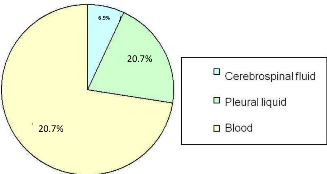
Table 1 presents the types of diseases as well as the number of cases presented at the Municipal Hospital of Paulinia. The most common clinical diagnosis was pneumonia, which was present in 23 cases (79.3%), followed by four cases of bacteremia (13.8%), and meningitis in two cases (6.9%).
Regarding the most common biological specimens investigated, we found that the identification of S.
pneumoniae was higher in the blood (72.4%),
corresponding to 21 samples, followed by six in pleural fluid samples (20.7%) and two samples of CSF, as presented in Figure 1.
Table 1. Number of cases related to the type of disease caused by Streptococcus pneumoniae, present in the
Municipal Hospital of Paulinia.
Age range Pneumonia Bacteremia Meningitis
Children (8 months-8 years) 12 2 0
Adolescents (15-17 years) 2 0 0
Adults (23-60 years) 6 2 2
Elderly (61-65 years) 3 0 0
Total 23 4 2
20.7%
20.7%
6.9%
Figure 1. Number of cases for biological specimens present in the municipal hospital in Paulinia.
Table 2. Age range and sensitivity to Streptococcus pneumoniae oxacillin.
Age range Sensitive (%) Resistent (%) Total (%)
Children (8 months-8 years) 8 (34.8) 6 (100) 14 (48.3)
Adolescents (15-17 years) 2 (8.7) 0 2 (6.9)
Adults (23-60 years) 10 (43.5) 0 10 (34.5)
Elderly (61-65 years) 3 (13) 0 3 (10.3)
Total 23 (79.3) 6 (20.7) 29 (100)
sensitive. For two resistant blood samples, we performed serotyping, which was positive for the serotype 18. The rest were not serotyped.
DISCUSSION
During 15 months we investigated 29 patients admitted at
1116 Afr. J. Microbiol. Res.
According to the literature, some types of pneumonia were associated with various ailments such as urinary tract infection, septicemia, respiratory tract infection, diarrhea and other diseases (Podschum and Ullmann, 1988). Nonetheless, we did not find any association between those variables and the disease in our study.
Most patients had pneumonia as the underlying disease and co-morbidity patients presented adult alcoholism, renal failure, diabetes and cancer and were smokers. The children presented anemia, asthma and disability in swallowing. Community acquired pneumonia is an important cause of morbidity and mortality worldwide. Lower respiratory tract infections, including pneumonia, ranked third amongst the 20 leading causes of death by the World Health Organization (WHO), causing an estimated 429.2 million episodes of illness worldwide in 2004. Lower respiratory tract infections were also the leading cause of burden of disease measured in terms of disability-adjusted life years (defined by WHO as one lost year of ‘healthy’ life, and the burden of disease as a measurement of the gap between current health status and an ideal situation where everyone lives into old age, free of disease and disability) amongst all age groups worldwide, accounting for 94.5 million disability-adjusted life years in 2004 (Song et al., 2011; Abubaka et al., 2011). In this context, previous reports indicated that respiratory infections increase with age. The incidence of Pneumonia was six-fold higher in adults in Finland aged
≥75 years old compared with adults aged 30–44 years old. In rural Thailand, the incidence of pneumonia increased after 55 years old (Olsen et al., 2006). Our data are supported by the literature, since the population investigated in our study was predominantly adult.
For treatment, we used the following drugs: cephalothin/chloramphenicol ampilicila/sub-activity
(Unasyn ®), gatifloxacin, ceftriaxone,
Unasyn®/ceftriaxone, oxacillin/vancomycin, crystalline penicillin, oxacillin and cephalothin. It was not indicated trimethoprim-sulfa treatment, because it showed an important resistance index. For pneumococcal meningitis it was administered ceftriaxone, which is a 3rd generation cephalosporin that has been a good alternative for treatment. In relation to cephalosporin, enterobacter
aerogenes produces an inducible chromosome-encoded
AmpC cephalosporinase. Most AmpC-type β-lactamases naturally produced by Gram-negative organisms hydrolyse amino- and ureido-penicillins, cephamycins (cefoxitin and cefotetan) and, to a lesser extent, oxyimino-cephalosporins (such as ceftazidime) and monobactams (Preston et al., 2000). Zwitterionic cephalosporins (cefepime and cefpirome), together with carbapenems, are usually excluded from the spectrum of activity of AmpC β-lactamases. However, natural cephalosporinases possessing broadened substrate activity have been reported in Enterobacteriaceae, P.
aeruginosa and Acinetobacter baumannii. These extended-spectrum AmpC cephalosporinases confer
reduced susceptibility to all cephalosporins. They differ from regular cephalosporinases by amino acid sub-stitutions, insertions or deletions in four specific regions— the loop, the H-10 helix, the H-2 helix and the C-terminal extremity of the protein—all located in the vicinity of the active site (Rodriguez-Martinez et al., 2009). More-over, an increased resistance to antibiotics has been reported in Pneumoniae as the widespread use of the third generation cephalosporins, β-lactam and broad-spectrum antibiotics (Kamatchi et al., 2009; Zhou et al., 2011).
The mechanism of action of penicillin to inhibit growth of microorganisms includes the inhibition of cell wall synthesis and activation of the endogenous autolytic bacteria. The action of penicillin depends on the cell wall that contains peptidoglycan in its composition. During the process of bacterial replication, penicillin inhibits the enzymes that make the link between the peptide chains and, therefore, prevents the development of the peptidoglycan structure. These enzymes (transpeptidase, carboxypeptidase and endopeptidase) are located just below the cell wall and are called penicillin binding proteins (PBPs). The ability to penetrate the cell wall and the affinity of these proteins with penicillin determine its antibacterial activity. Besides the action on the cell wall, it is considered the action of penicillin in the activation of the endogenous autolytic bacteria, causing their death and subsequent lysis (Laudano, 2011).
As a mechanism of escape from penicillin, pneumococcus changes the structures of PBPs, which are targets for binding of beta-lactam antibiotics. Decreasing the binding affinity of this antibiotic for PBPs a biochemical change occurs which leads to decreased affinity of the drug at the site, preventing the antibiotic to reach that goal (Reinert et al., 2010).
For the treatment of S. pneumoniae with intermediate resistance to penicillin therapy penicillin was recommended as first choice as well as G cefotaxime or ceftriaxone, or alternative drugs such as levofloxacin, grepafloxacin and vancomycin. All antibiotics from the beta-lactam group used for the treatment, present bactericidal activity (Biek et al., 2010). In our study, all susceptibility tests performed in vitro through disk diffu-sion method, showed 100% sensitivity to erythromycin, vancomycin, rifampicin, chloramphenicol, cefotaxime, ceftriaxone and cefepime. Furthermore, all those which were sensitive to oxacillin also showed sensitivity to ampicillin, ampicillin/sulbactam and amoxicillin/ ac.clavulanic. In total, 10 strains, 10 hemoculture and two pleural fluids were resistant to cotrimoxazole (trimethoprim + sulfa) and MIC was not done for this drug as the treatment of these patients was not based on this drug treatment.
antimicrobials is an important factor to be valued in drug therapies (Nagai et al., 2000).
Our investigation points out the importance of the knowledge regarding the antimicrobial resistance and serotyping of pneumococcal strains, in order to monitor the effectiveness of the treatment and profile of the microorganism in this population, comparing them with other profiles in the region. We may help to reveal the best treatment and which type of immunization (vaccine) is more suitable for the region and also advise the limited and prudent use of antibiotics.
Conclusion
Based on our data, we alert the incidence of resistant pneumococcal strains to the most common drugs due to inappropriate use of antimicrobials. Our data collaborate to the formulation of specific pneumococcal conjugate vaccines to each region.
ACKNOWLEDGEMENT
We received financial support from NEPAS.
REFERENCES
Abubakar MS, Fatihu MY, Ibrahim NDG, Oladele SB, Abubakar MB (2010). Camel pneumonia in Nigeria: Epidemiology and bacterial flora in normal and diseased lung. Afr. J. Microb. Res., 4(9):2479 – 2483.
Akortha EE, Ikenebomeh MJ (2010). Nasal colonization of symptomatic pneumonia patients in university of Benin teaching hospital, Benin City, Nigeria by multiple antibiotic resistant Staphylococcus aureus. Afr. J. Microb. Res., 4(6):1071 – 1075.
Biek D, Critchley IA, Riccobene TA, Thye DA (2010). Ceftaroline fosamil: a novel broad-spectrum cephalosporin with expanded anti-Gram-positive activity. J. Antimicrob. Chemother., 65(4):iv9-16. Kamatchi C, Magesh H, Sekhar U, Vaidyanathan R (2009).
Identification of clonal clusters of Klebsiella pneumoniae isolates from Chennai by extended spectrum Beta lactamase genotyping and antibiotic resistance phenotyping analysis. Am. J. Infect. Dis., 5(2): 74-82.
Laudano JB (2011). Ceftaroline fosamil: a new broad-spectrum cephalosporin. J. Antimicrob. Chemother., 66(3):iii11-8.
Levin ASS, Teixeira LM, Sessegolo FF, Baroni AA (1996). Resistence of Streptococcus pneumoniae to antimicrobials in são paulo, brazil: clinical features and serotypes. Rev. Inst. Med. Trop., 38(3):187-192. Nagai K, Matsuo Y, Tsumura N, Sakata Y, Kato H (2000). Antimicrobial
susceptibilities and serotypes of Streptococcus pneumoniae in southwestern japan and correlation of penicillin-binding proteins 2b and 2x mutations in susceptibilities of penicillin g and cefotaxime. Diagn. Microbiol. Infect. Dis., 37(2):107-13.
Musher DM (2011). New modalities in treating pneumococcal pneumonia. Hosp. Pract., 39(2):89-96.
Olsen SJ, Laosiritaworn Y, Siasiriwattana S, Chunsuttiwat S, Dowell SF (2006). The incidence of pneumonia in rural Thailand. Int. J. Infect. Dis., 10(6): 439–445.
Podschum R, Ullmann U (1988). Klebsiella spp. as Nosocomial pathogens: Epidemiology, Taxonomy, Typing methods and pathogenicity factors. Clin. Microbiol. Rev., 11(4): 583-603.
Preston KE, Radomski CC, Venezia RA (2000). Nucleotide sequence of the chromosomal ampC gene of Enterobacter aerogenes. Antimicrob. Agents Chemother., 44(11):3158-62.
Reinert R, Jacobs MR, Kaplan SL (2010). Pneumococcal disease caused by serotype 19A: review of the literature and implications for future vaccine development. Vaccine, 28(26): 4249-59.
Rodriguez-Martinez JM, Poirel L, Nordmann P (2009). Extended-spectrum cephalosporinases in Pseudomonas aeruginosa.
Antimicrob. Agents Chemother., 53(10):1766-71.
Sessegolo JF, Levin ASS, Levy CE, Asensi M, Fackland RR, Teixeira LM (1994). Distribution of serotypes and antimicrobial resistance of
Streptococcus pneumoniae strains isolated in brazil from 1988 to
1994. J. Clin. Microbiol., 32(4):906-11.
Song JH, Thamlikitkul V, Hsueh PR (2011). Clinical and economic burden of community-acquired pneumonia amongst adults in the Asia-Pacific region. Int. J. Antimicrob. Agents, 38(2):108-17. Sun HY, Fujitani S, Quintiliani R, Yu VL (2011). Pneumonia due to
Pseudomonas aeruginosa: part II: antimicrobial resistance, pharmacodynamic concepts, and antibiotic therapy. Chest, 139(4): 1172-85.
Zether EW, Sheibe RM, Dias CAG, Santafé P, Moreira JS, Santos DS, Fritscher CC (2004). The polymerase chain reaction for detection of penicillin resistance in Streptococcus pneumoniae. J. Bras. Pneumol., 30(3): 123-132.
Zhou X, Gao J, Huang Y, Fu S, Chen H (2011). Quinolone resistance in
Escherichia coli and Salmonella spp. isolates from diseased chickens