51 Abstract
Objectives: To determine the prevalence of pneumococcus colonization among HIV-infected outpatients aged 0 to 18 years. To determine the resistance to penicillin of the microorganisms observed, to identify their serotypes, and to determine whether there are associations between known risk factors and colonization in this group.
Material and method: This was an observational and cross-sectional study in which nasopharynx swabs were collected from 112 children on the occasion of their monthly appointments and a questionnaire applied to the mothers. The material collected was processed at the microbiology laboratory of the hospital in accordance with National Committee for Clinical Laboratory Standards (NCCLS) regulations and serotyping was performed at the Centers for Diseases Control and Prevention (CDC). Data were analyzed statistically using the chi-square test and with univariate and multivariate analysis with multiple logistic regression.
Results: The prevalence rate of nasopharyngeal colonization by pneumococci was 28.6%, with a 15.6% rate of resistance to penicillin (6.2% intermediate resistance and 9.4% full resistance). The serotypes identified were 6A, 6B, 7C, 9V, 11A, 13, 14, 15A, 16F, 18C, 19B, 19F, 23B, 23F, and 34. In this population there were no associations between pneumococcal colonization and the risk factors studied.
Conclusions: The prevalence of pneumococcal colonization among HIV-infected children at our service was not higher than prevalence rates observed in healthy children and reported in the literature.
J Pediatr (Rio J). 2006;82(1):51-7: Nasopharyngeal colonization, Streptococcus pneumoniae, HIV-infected children.
Nasopharyngeal colonization
with
Streptococcus pneumoniae
in children
infected with human immunodeficiency virus
Viviane C. Cardoso,1 Maria C. Cervi,2 Otávio A. L. Cintra,2 Adriana S. M. Salathiel,3 Ana C. L. F. Gomes1 Copyright © 2006 by Sociedade Brasileira de Pediatria
ORIGINAL ARTICLE
1. Mestre. Hospital das Clínicas, Faculdade de Medicina de Ribeirão Preto (HCFMRP-USP), São Paulo, SP, Brasil.
2. Doutora/Doutor. HCFMRP-USP, São Paulo, SP, Brasil.
3. Aluna de graduação de Medicina, HCFMRP-USP, São Paulo, SP, Brasil.
Manuscript received May 31 2005, accepted for publication Aug 10 2005.
Suggested citation: Cardoso VC, Cervi MC, Cintra OA, Salathiel AS, Gomes AC. Nasopharyngeal colonization with Streptococcus pneumoniae in children infected with human immunodeficiency virus. J Pediatr (Rio J). 2006;82:51-7.
Introduction
Pneumococcal disease is probably the result of an interaction between bacterial virulence and the defenses of the host, which, in the case of people infected by the a c q u i r e d i m m u n o d e f i c i e n c y v i r u s ( H I V ) , a r e compromised.1 Children infected with HIV are particularly susceptible to systemic disease caused by pneumococcus
and have a risk of invasive disease twelve times that of other children.2
Streptococcus pneumoniae is the most common causal agent of bacterial pneumonia among HIV-infected patients, with disease rates that are 10-100 times greater among infected children than their controls.3 Clinical manifestations are similar to in normal hosts except for the increase in rates of bacteremia and recurrent disease, but mortality from pneumococcal disease is substantial in HIV patients.4 The pattern of invasive bacterial infection is greater among HIV children than their controls, especially after 1 year of age.5
may be that these patients are less able to eliminate the pathogen once bacteremia has occurred. Specific local and systemic defects in the hosts defenses, in particular humoral immunity, may contribute to the elevated incidence of invasive pneumococcal disease.6
The frequency of severe pneumococcal disease in children is of great relevance to public health since the HIV epidemic has coincided with pneumococcal strains resistant to penicillin and a multiplicity of other antibiotics. Responsibility for the increase in pneumococcal disease among HIV positive individuals probably does not lie with increased rates of colonization, but is probably, therefore, the result of systemic immune dysfunction.7
Although the elevated predisposition of HIV-positive children to contract invasive bacterial diseases has been documented,2 there are few studies that describe the differences in colonization patterns and antibiotic susceptibility of the bacteria found in children infected with HIV. Polack et al.8 did not observe differences in rates of colonization by Streptococcus pneumoniae between HIV-positive children, children with as yet indeterminate HIV infections and their uninfected controls (20% against 19%). The age of patients was considered the most important predictor of bacterial colonization in both populations, with the prevalence rates of pneumococcus strains isolated from nasopharynges being five times greater for the age range from 6 months to 2 years than for all other ages. In contrast, Leibovitz et al.9 reported lower colonization rates among HIV-positive Romanian orphans than among controls, but this could also be explained by age differences in the two groups.
Although pneumococci colonizing nasopharynges cannot be considered the direct cause of severe, or even moderate, infections, it is useful to study them in order to assess the prevalence of antimicrobial resistance and the prevalent serotypes, including invasive ones, that cause bacteremia and systemic infection in a given community.10,11
The epidemiological features of pneumococcal disease vary from one country to another and over time, which gives rise to the need for periodic evaluations to establish control strategies. For these reasons it became important for us to investigate the situation at our service where children with HIV infections are cared for. Our objectives were to establish the rate of nasopharyngeal colonization by S. pneumoniae in this population, to determine the pattern of susceptibility to penicillin of the strains found, to identify the pneumococcus serotypes and assess risk factors for colonization in this population.
Methods
Study design
Cross-sectional and observational study.
Population
All children (0-18 years) seen at the Hospital das Clínicas Child and Adolescent Infectology Clinic (CAIC) located at the Ribeirão Preto medical faculty, Universidade de São Paulo (HCFMRP-USP) were included on the study if they had clinical and laboratory diagnoses of HIV based on the 1994 Centers for Diseases Control and Prevention (CDC) criteria.12 During the study period 120 children were being seen at regular monthly appointments. All of the patients had to have the following HIV diagnostic tests: serology by enzyme immunoassay (ELISA) for HIV1/HIV2; agglutination test for HIV1/HIV2; polymerase chain reaction DNA and/or PCR RNA for HIV1. Children who had not had these tests were excluded and so were any with bacterial infections being treated with antimicrobials at the time of data collection or during the previous month. The resulting sample contained 112 children.
Variables studied
General characteristics of the children: age, sex, color, residence, maternal schooling (in years).
Risk factors for pneumococcal infection: attendance at a day care center or school, siblings aged less than 6 years, number of bedrooms, number of people sleeping in the same room as the child, family member with upper airway infection (UAI) during the previous 15 days, previous hospitalization, use of antibiotics during the previous 3 months.
HIV infection: prophylactic antibiotics, vaccination against pneumococcus, use of immunoglobulin, HIV class, use of antiretroviral drugs, CD4 lymphocyte count and viral load.
Pneumococcal colonization: penicillin resistance, test with oxacillin discs, Etest, serotypes.
Ethical considerations
This study was approved by the Committee for Ethics in Research at HCFMRP-USP.
Data collection
was then stored in a biological transport medium for a maximum period of 2 hours and sent to the Microbiology Laboratory at the HCFMRP for microbiological processing according to National Committee for Clinical Laboratory Standards (NCCLS) guidelines.13 Samples identified as S. pneumoniae were submitted for penicillin sensitivity screening tests with 1 µg oxacillin discs. Strains were considered sensitive to penicillin if the growth inhibition halo was > 20 mm and those with halos < 19 mm were considered resistant. Despite the recommendation that only strains with halos < 19 mm should be assessed by quantitative methods, Etests (AB Biodisk, Probac, Brazil) were performed for all strains isolated irrespective of screening results. Inhibition ellipses were observed and the Etest minimum inhibitory concentration (MIC) value was read. This value could vary from 0.002 to 32, and results were defined as follows: < 0.06 µg/ml sensitive to penicillin; 0.1 to 1 µg/ml intermediate penicillin resistance; > 2 µg/ml total penicillin resistance. The isolated strains were then inoculated onto sterile glass with 2 ml of lambs blood, stored in a freezer at -2 to -8 ºC, until they could be sent for serotyping. During storage the strains were periodically re-spotted to guarantee viability. Serotyping was carried out using capsular swelling reaction tests with specific antisera (Staten Seruminstitut, Copenhagen, Denmark) provided by the CDC and employing the Danish nomenclature.
Statistical analysis
The sample size calculation was performed based on a hypothetical rate of 20% of pneumococcus colonization, based on previously published studies, and estimated that swabs would have to be taken from 80 of the 120 children with HIV infections treated at our service. One hundred and twelve of the children were actually enrolled. The characteristics of the study population were analyzed descriptively using frequency tables. Epi-info version 6.04 b was used in order to verify associations or compare proportions between characteristics, employing the chi-square test with significance set at p < 0.05. Risk factors were studied employing univariate analysis to calculate odds ratios (OR) and confidence intervals and then a multivariate analysis was performed with multiple logistic regression with retrograde elimination of variables using Stata 5.0.14
Results
The prevalence rate of nasopharyngeal colonization by Streptococcus pneumoniae in HIV-positive children seen at the CAIC-HCFMRP-USP was 28.6%. Five of the 32 strains isolated proved to be resistant to penicillin (15.6%), with two of these having intermediate resistance while the other three were fully resistant. Twenty-three (71.8%) of
the 32 strains isolated were sent for serotyping; the remaining nine did not remain viable while in storage. Fifteen distinct serotypes were identified: 6A, 6B, 7C, 9V, 11A, 13, 14, 15A, 16F, 18C, 19B, 19F, 23B, 23F and 34. One of the strains was classified as untypeable. The pneumococcus serotypes that were resistant to penicillin were 9V, 13, 14, 18C.
General characteristics of the study population
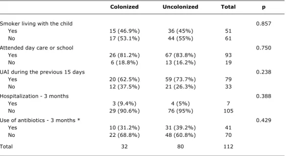
There were no statistical differences between those children who were colonized by pneumococcus and those who werent in any of the variables for the general characteristics of the children or risk factors for pneumococcal colonization (Tables 1 and 2). The mean age of the study group was 83.8 months with a standard deviation of 42.7 (8-221 months). The mode was 72 months and the median 77 months.
Female children accounted for 54.5% of the sample and 45.5% were male; 41.1% were white and 58.9% were not. The great majority of the children were living with their families (85.7%) while 14.3% lived in institutions. A majority of the mothers had received from 1 to 8 years schooling (70.5%). In 45.5% of cases there was an adult smoker living with the child. Ninety-three children were attending day care or school. Seventy-nine (70.5%) of the children or a member of their family had presented an episode of UAI during the 15 days prior to the nasal swab. Just seven children had been hospitalized during the previous 3 months. Thirty-three children had been given antimicrobials during the previous 3 months, with the following mentioned: penicillin (22.3%), macrolide (1.8%), trimethoprim-sulfamethoxazole (SMT-TMP) (1.8%), rifampicin (0.9%), tetracycline (0.9%) and more than one type of antibiotic (1.8%); nine mothers were unable to provide the name of the antimicrobial given their children.
Characteristics of the HIV infections
Neither adjusted nor unadjusted analysis models demonstrated associations between the study variables and risk for nasopharyngeal colonization. There was
only a tendency for the use of immunoglobulin (p = 0.057) to be a risk factor for pneumococcal colonization.
Colonized Uncolonized Total p
Total 32 80 112
Season 0.065
Winter/Fall 8 (25%) 35 (43.8%) 43
Spring/Summer 24 (75%) 45 (56.2%) 69
Age (months) 0.540
0-12 0 (0%) 2 (2.5%) 2
13-24 1 (3.1%) 7 (8.8%) 8
25-36 4 (12.5%) 2 (2.5%) 6
37-48 3 (9.4%) 6 (7.5%) 9
49-60 4 (12.5%) 8 (10%) 12
61-72 2 (6.3%) 8 (10%) 10
73-84 5 (15.6%) 7 (8.8%) 12
85-96 2 (6.3%) 11 (13.8%) 13
97-108 4 (12.5%) 4 (5%) 8
109-120 3 (9.4%) 8 (10%) 11
121-132 1 (3.1%) 6 (7.5%) 7
133-144 3 (9.4%) 4 (5%) 7
145-156 0 (0%) 2 (2.5%) 2
157-168 0 (0%) 2 (2.5%) 2
169-180 0 (0%) 1 (1.3%) 1
205-216 0 (0%) 1 (1.3%) 1
217-228 0 (0%) 1 (1.3%) 1
Age range
< 6 years 14 (43.8%) 33 (41.2%) 47 0.809
> 6 years 18 (56.2%) 47 (58.8%) 65
Gender 0.548
Female 16 (50%) 45 (56.2%) 61
Male 16 (50%) 35 (43.8%) 51
Color 0.430
White 15 (46.9%) 31 (38.8%) 46
Non-white 17 (53.1%) 49 (61.2%) 66
Living 0.348
With family 29 (90.6%) 67 (83.8%) 96
Institution 3 (9.4%) 13 (16.2%) 16
Number of people at home* 0.444
2-4 11 (37.9%) 33 (49.3%) 44
5-10 18 (62.1%) 33 (49.3%) 51
> 10 0 (0%) 1 (1.5%) 1
Number of brothers and sisters* 0.856
None 5 (17.2%) 14 (20.9%) 19
1-2 16 (55.2%) 33 (49.3%) 49
> 2 8 (27.6%) 20 (29.8%) 28
Brothers/Sisters < 6 years 0.369
Yes 13 (40.6%) 40 (50%) 53
No 19 (59.4%) 40 (50%) 59
Number of people in the bedroom 0.691
Up to 3 22 (68.8%) 58 (72.5%) 80
More than 3 10 (31.2%) 22 (27.5%) 32
Mothers schooling 0.403
None 1 (3.1%) 5 (6.3%) 6
1-8 years 26 (81.3%) 53 (66.3%) 79
> 8 yearss 3 (9.4%) 9 (11.3%) 12
Ignored 2 (6.3%) 13 (16.3%) 15
Table 1 - General characteristics of the HIV-infected children seen at the CAIC-HCFMRP-USP (Ribeirão Preto, 2003)
Discussion
The prevalence of nasopharyngeal colonization observed at our service during the study period was 28.6%. This is very different from the results of other evaluations involving seropositive children and adults. Polack et al.8 found a prevalence of colonization of 20% among HIV-positive children and daughters of HIV-positive mothers whose status was not defined, which was not different from their negative controls (19%), suffering from chronic pulmonary diseases. The elevated incidence of pneumococcal disease and prophylaxis with SMT-TMP were not related to nasopharyngeal colonization. A study by Rusen et al.,7 in Kenya, assessed 207 children younger than 5 years, infected with HIV, undetermined and uninfected, by means of several visits (a maximum of five) with clinical assessment and collection of nasal swabs. Colonization was different between groups only when there was concomitant respiratory disease. The rate of colonization among children without respiratory disease varied from 20 to 35%. Colonization was lesser among HIV-positive children (26) and the daughters of HIV mothers (28 with indeterminate status and 68 seronegative) than their controls (85) only when associated with respiratory disease such as coughing, coryza, dyspnea and signs of pneumonia (86% of seven, 60% of 20 against 29% of 31). No differences were observed in terms of colonization when children were asymptomatic (20% of
35, 35% of 94 and 22% of 101). Colonization was not associated with fever, failure to thrive or breastfeeding at the time of the visit, or with previous medication for coughing or the use of antibiotics during the month prior to the visit.7 Another study of pneumococcal colonization involving children infected with HIV and a control group found differences in prevalence, probably related to variations in the age groups of the two, which were not stratified. This research was performed with Romanian orphans, 162 children without HIV infections aged 1 to 38 months and 40 children infected by HIV aged 39 to 106 months. Colonization rates were 30% (12 of 40) of the infected children and 50% (81 de 160) for the uninfected children,9 emphasizing the greater prevalence of colonization among lower age groups. When colonization is studied in the adult population, the prevalence rates are lower than in children, even in HIV populations. In a study by Janoff et al.15 the prevalence rates of pharyngeal colonization were not significantly different between HIV-positive men (14%) and uninfected ones (9%) treated at a sexually transmitted diseases clinic.
All of these data and studies support the concept that, when pneumococcal colonization is being discussed, the factors that interfere with it must be taken into account, such as age, time of year and geographical location. Because of this, we cannot extrapolate data from one population to another.
Colonized Uncolonized Total p
Smoker living with the child 0.857
Yes 15 (46.9%) 36 (45%) 51
No 17 (53.1%) 44 (55%) 61
Attended day care or school 0.750
Yes 26 (81.2%) 67 (83.8%) 93
No 6 (18.8%) 13 (16.2%) 19
UAI during the previous 15 days 0.238
Yes 20 (62.5%) 59 (73.7%) 79
No 12 (37.5%) 21 (26.3%) 33
Hospitalization - 3 months 0.388
Yes 3 (9.4%) 4 (5%) 7
No 29 (90.6%) 76 (95%) 105
Use of antibiotics - 3 months * 0.429
Yes 10 (31.2%) 31 (39.2%) 41
No 22 (68.8%) 48 (60.8%) 70
Total 32 80 112
Table 2 - Risk factors pneumococcus colonization among HIV-infected seen at the CAIC-HCFMRP-USP (Ribeirão Preto, 2003)
UAI = upper airway infection.
The pattern of penicillin resistance found in our population was 15.6%, with intermediate resistance of 6.2% and total resistance of 9.4%. Our analysis of antimicrobial resistance was restricted to penicillin. Our findings were similar to results from a study carried out in São Paulo with children less than 5 years old who exhibited clinical status compatible with acute rhinopharyngitis where penicillin resistance was 15.6%, and no strains had complete resistance.16 There are discrepancies in the rates of resistance found in populations seropositive for HIV; Rusen et al.7 found that 60.8% of 92 strains tested were resistant to penicillin. There was no association between penicillin resistance and HIV infection; while 60% of the strains exhibited intermediate penicillin resistance, high-level resistance was not observed. In observations made by Leibovitz et al.,9 99% of strains isolated were resistant to penicillin, with 74% highly resistant. Those isolated from children infected by HIV and with as yet undetermined status, obtained by Polack et al.8 were less resistant to penicillin than their controls (18% against
50%) and all of the strains isolated by Janoff et al.15 were sensitive to penicillin. Large variations have also been demonstrated in Brazil by studies carried out in different regions. Penicillin resistance among healthy carriers in Fortaleza was 48% and among children with pneumonia it was 50%; with 45% intermediate resistance and 4% total resistance.11
The Etest method was used to determine sensitivity to penicillin irrespective of the oxacillin disc screening results, as recommended by the NCCLS.13 Etest is a combination of dilution and diffusion techniques that has recently come to be used for determining the MIC of several antimicrobials for pneumococcus. Results are interpreted using the same criteria for defining MIC. This is established by the intersection of the line of the bacterial growth inhibition zone with the strip edge. Agreement between the Etest MIC and broth microdilution is better than 80%.17 Resistance was defined using Etest because this is a reliable method when compared with the reference test17-19 and because it is a test that is capable of differentiating Table 3 - Characteristics of HIV infection in children seen at the CAIC-HCFMRP-USP (Ribeirão Preto, 2003)
Colonized Uncolonized Total p
Prophylactic antibiotic * 0.142
Yes 13 (41.9%) 22 (27.5%) 35
No 18 (58.1%) 58 (72.5%) 76
Vaccine
Yes 26 (81.2%) 65 (81.2%) 91 1
No 6 (18.8%) 15 (18.8%) 21
Immunoglobulin 0.053
Yes 12 (37.5%) 16 (20%) 28
No 20 (62.5%) 64 (80%) 84
Clinical classification 0.132
N 5 (15.6%) 5 (6.2%) 10
A 3 (9.4%) 21 (26.3%) 24
B 7 (21.9%) 18 (22.5%) 23
C 17 (53.1%) 36 (45%) 53
Antiretroviral drugs 0.920
Yes 29 (90.6%) 72 (90%) 101
No 3 (9.4%) 8 (10%) 11
CD4 lymphocyte count 0.169
> 25% 13 (40.6%) 44 (55%) 57
< 25% 19 (59.4%) 36 (45%) 55
Viral load* 0.094
< 10,000 18 (60%) 32 (40%) 50
10,000-100,000 10 (33.3%) 38 (47.5%) 48
> 100,000 2 (6.7%) 10 (12.5%) 12
Total 32 80 112
intermediate from total resistance, in contrast with the oxacillin disc test.
Among the serotypes identified in our sample were those known as pediatric serotypes and those that commonly cause invasive disease in children: 6, 14, 19 and 23.11,20-22 With the serotypes identified (6A, 6B, 7C, 9V, 11A, 13, 14, 15A, 16F, 18C, 19B, 19F, 23B, 23F, 34) we can estimate that the 7-valent vaccine should confer around 85.7% protection on our population.
In function of the cross-sectional design of our study we only assessed the carrier status of pneumococcal strains and the resistance profile with respect of penicillin based on a single data collection. There were no samples taken during winter and spring and neither were the same subjects reassessed on separate occasions. This fact did not compromise the research which aimed to get to know the profile at our service. One further aspect to take into account is the small sample size which did not allow for statistical inferences and limited our study to the descriptive type. No associations were found between the classic risk factors for colonization and actual carrier status. It cannot be confirmed, but we suggest that this difficulty may be due to the small sample size. It is possible that, in future studies with larger numbers of participants, associations may be found.
Our findings reinforce the importance of continuous vigilance and the necessity for periodic assessments at any given service of the same sample in order to delineate the situation in which we are working and to provide us with an information base that can be used to guide therapeutic and prophylactic decision-making. Even though it is not possible to extrapolate these data to other groups, this initial evaluation of our service will serve as the basis for future research and for observations of the impact of conjugated vaccines on the history of pneumococcal disease.
Acknowledgements
The authors are grateful to teaching Doctor Lúcia Martins Teixeira at the Universidade Federal do Rio de Janeiro, for serotyping the material; to the Microbiology and Mycology laboratories at HCFMRP-USP and to Micheli Rovanholo for support and technical work.
4. Lesprit P, Decazes JM. Pneumococcal infections in patients with HIV infections. Presse Med. 1997;26:243-7.
5. Andiman WA, Mezger J, Shapiro E. Invasive bacterial infections in children born to women infected with human immunodeficiency virus type 1. J Pediatr. 1994;124:846-52.
6. Janoff EN, Breiman RF, Daley CL, Hopewell PC. Pneumococcal disease during HIV infection. Epidemiologic, clinical, and immunologic perspectives. Ann Intern Med. 1992;117:314-24. 7. Rusen ID, Fraser-Roberts L, Slaney L, Ombette J, Lovgren M, Datta Ndinya-Achola J, et al. Nasopharyngeal pneumococcal colonization among Kenyan children: antibiotic resistance, strain types and associations with human immunodeficiency virus type 1 infection. Pediatr Infect Dis J. 1997;16:656-62. 8. Polack FP, Flayhart DC, Zahurak ML, Dick JD, Willoughby RE.
Colonization by Streptococcus pneumoniae in human immunodeficiency virus-infected children. Pediatr Infect DisJ. 2000;19:608-12.
9. Leibovitz E, Dragomir C, Sfartz S, Porat N, Yagupsky P, Jica S, et al. Nasopharyngeal carriage of multidrug-resistant Streptococcus pneumoniae in institutionalized HIV-infected and HIV-negative children in northeastern Romania. Int J Infect Dis. 1999;3:211-5.
10. Kellner JD, Mcgeer A, Cetron MS, Low DE, Butler JC, Matlow A, et al. The use of Streptococcus pneumoniae nasopharyngeal isolates from healthy children to predict features of invasive disease. Pediatr Infect Dis J. 1998;17:279-86.
11. Rey LC, Wolf B, Moreira JLB, Verhoef J, Farhat CK. S. pneumoniae isolados da nasofaringe de crianças sadias e com pneumonia: taxa de colonização e suscetibilidade aos antimicrobianos. J Pediatr (Rio J). 2002;78:105-12.
12. Centers for Diseases Control and Prevention. Revised classification System of human immunodeficiency virus infection in children less than 13 year of age. MMWR. 1994;43(RR 12): 1-10.
13. National Committee for Clinical Laboratory Standards. Performance standards for antimicrobial susceptibility testing. Supplemental tables, M100-S13. Wayne: National Committee for Clinical Laboratory Standards; 2003.
14. Stata Corporation. Stata reference manual 5.0 Statistics Data analysis. Texas: Stata; 1997.
15. Janoff EN, O´Brien J, Thompson P, Ehret J, Meiklejohn G, Duvall G, et al. Streptococcus pneumoniae colonization, bacteremia, and immune response among persons with human immunodeficiency virus infection. J Infect Dis. 1993;167:49-56. 16. Ferreira LL, Carvalho ES, Berezin EM, Brandileone MC. Colonização e resistência antimicrobiana de Streptococcus pneumoniae isolado em nasofaringe de crianças com rinofaringite aguda. J Pediatr (Rio J). 2001;77:227-34.
17. Mantese OC. Infecção pneumocócica. In: Farhat CC, Carvalho ES, Carvalho LH, Succi RC, editores. Infectologia pediátrica. 2ª ed. São Paulo: Atheneu; 1998. p. 300-12.
18. Baker CN, Stocker SA, Culver DH, Thornsberry C. Comparison of the E Test to agar dilution, broth microdilution and agar diffusion susceptibility testing techniques by using a special challenge set of bacteria. J Clin Microbiol. 1991;29:533-8. 19. Tenover FC, Baker CN, Swenson JM. Evaluation of commercial
methods for determining antimicrobial susceptibility of Streptococcus pneumoniae. J Clin Microbiol. 1996;34:10-4. 20. Porat N, Leibovitz E, Dagan R, Coman G, Sfartz S, Peled N, et
al. Molecular typing of Streptococcus pneumoniae in Northeastern Romania: Unique clones of S.pneumoniae isolated from children hospitalized for infections and from healthy and human immunodeficiency virus-infected children in the community. J Infect Dis. 2000;181:66-74.
21. Ghaffar F, Friedland IR, Mccracken Jr GH. Dynamics of nasopharyngeal colonization by Streptococcus pneumoniae. Pediatr Infect Dis J. 1999;18:638-46.
22. Madhi SA, Petersen K, Madhi A, Wasas A, Klugman KP. Impact of human immunodeficiency virus type 1 on the disease spectrum of Streptococcus pneumoniae in South African children. Pediatr Infect Dis J. 2000;19:1141-7.
References
1. Johnston Jr RB. Pathogenesis of pneumococcal pneumonia.
RID.1991;13(Suppl 6):S509-17.
2. Mao C, Harper M, Mcintosh K, Reddington C, Cohen J, Bachur R, et al. Invasive pneumococcal infections in human immunodeficiency virus-infected children. J Infect Dis. 1996;173:870-6.
3. Bernstein LJ, Ochs HD, Wedgwood RJ, Rubstein A. Defective humoral immunity in pediatric acquired deficiency syndrome. J Pediatr. 1985;107:352-7.
Correspondence: Viviane Cunha Cardoso
Avenida Bandeirantes, 3900, Monte Alegre CEP 14049-900 Ribeirão Preto, SP Brazil Tel.: +55 (16) 602.3317/602.3316 Fax: +55 (16) 602.3306