Submitted28 October 2015 Accepted 27 November 2015 Published17 December 2015 Corresponding author Valerie Booth, [email protected]
Academic editor Ayyalusamy Ramamoorthy
Additional Information and Declarations can be found on page 16
DOI10.7717/peerj.1516 Copyright
2015 Brannan et al.
Distributed under
Creative Commons CC-BY 4.0 OPEN ACCESS
Differential scanning calorimetry of
whole
Escherichia coli
treated with the
antimicrobial peptide MSI-78 indicate a
multi-hit mechanism with ribosomes as a
novel target
Alexander M. Brannan1, William A. Whelan1, Emma Cole1and Valerie Booth1,2 1Department of Biochemistry, Memorial University of Newfoundland, St. John’s Newfoundland and Labrador,
Canada
2Department of Physics and Physical Oceanography, Memorial University of Newfoundland,
St. John’s Newfoundland and Labrador, Canada
ABSTRACT
Differential Scanning Calorimetry (DSC) of intact Escherichia coli (E. coli)was used to identify non-lipidic targets of the antimicrobial peptide (AMP) MSI-78. The DSC thermograms revealed that, in addition to its known lytic properties, MSI-78 also has a striking effect on ribosomes. MSI-78’s effect on DSC scans of bacteria was similar to that of kanamycin, an antibiotic drug known to target the 30S small ribosomal subunit. An in vitrotranscription/translation assay helped confirm MSI-78’s targeting of ribosomes. The scrambled version of MSI-78 also affected the ribosome peak of the DSC scans, but required greater amounts of peptide to cause a similar effect to the unscrambled peptide. Furthermore, the effect of the scrambled peptide was not specific to the ribosomes; other regions of the DSC thermogram were also affected. These results suggest that MSI-78’s effects onE. coliare at least somewhat dependent on its particular structural features, rather than a sole function of its overall charge and hydrophobicity. When considered along with earlier work detailing MSI-78’s membrane lytic properties, it appears that MSI-78 operates via a multi-hit mechanism with multiple targets.
SubjectsBiophysics, Infectious Diseases
Keywords Antimicrobial peptides, Differential Scanning Calorimetry (DSC), Membrane active peptides
INTRODUCTION
driving the interactions of AMPs with the membrane of target pathogens, which can result in the direct killing of the pathogen via membrane disruption, and/or provide a means for the AMP to access intracellular targets.
Much hope has been expressed for the potential of novel AMP-based therapeutics to help address the growing problem of antibiotic resistance to conventional small antibiotics (Rossi et al., 2008;Spellberg et al., 2008;Boucher et al., 2009;Yeung, Gellatly & Hancock, 2011). However, successfully developing AMP-based drugs, in particular for internal use, has proved very challenging (Matsuzaki, 2009;Wimley & Hristova, 2011;
Pasupuleti, Schmidtchen & Malmsten, 2012). One major barrier to the optimization of AMPs for therapeutic use rests with the simplified model membrane systems in which detailed mechanistic studies and AMP sequence optimization are generally performed. By focusing solely on the lipids, studies that are limited to model membrane systems fail to capture the myriad of potentially important interactions that AMPs may have with their target microbes.
An example of the gap that exists between mechanistic studies in model membranes and the conditions under which AMPs actually carry out their function is the observation that AMPs cause leakage in model membranes at protein:lipid (P:L) ratios∼10,000 times lower than the P:L ratio needed to see bacterial growth inhibition (Wimley, 2010). On the one hand, solid state NMR, vesicle leakage and other biophysical experiments aimed at understanding the structural mechanisms of AMP-induced bilayer disruptions employ model membranes and overall AMP:lipid molar ratios of∼1: 200 (Wimley, 2010). On the other hand, the minimal inhibitory concentration (MIC) assays used to define the concentration of AMP needed to prevent bacterial growth are typically performed with overall molar ratios of∼1000 AMP:1 lipid—a difference in AMP:lipid ratio of
by different AMPs does not always correlate with their relative ability to prevent bacterial growth (Friedrich et al., 2000;He, Krauson & Wimley, 2014).
Thus, while AMP-lipid interactions are key in understanding AMP mechanism, it is vital to look beyond model membranes and examine AMPs’ potentially functional interactions with other components of the target cell. While the vast majority of studies of AMP mechanism have focused on lipids, there are indeed AMPs for which non-lipidic intracellular targets are known (Brogden, 2005;Marcos & Gandia, 2009), although AMP-membrane interactions can still be important for allowing AMPs to access the interior of the cell. Known non-lytic mechanisms for AMPs include targeting nucleic acids (Park, Kim & Kim, 1998),chaperones (Kragol et al., 2001), cell wall synthesis (Brotz et al., 1998), protein synthesis (Patrzykat et al., 2002), and enzymatic activity (Andreu & Rivas, 1998), as well as the G-quadruplex in mammalian cancer cells (Jana et al., 2013).
In order to provide a more integrated view of AMP-bacteria interactions, one strategy is to start with the same biophysical methods used to study and optimize AMP inter-actions with model membrane systems, and extend these methods for use with whole bacteria (e.g.,Gee et al., 2013;Maisetta et al., 2013;Hall et al., 2014). In this work, we describe differential scanning calorimetry (DSC) studies of the AMP MSI-78 interacting with whole bacteria.
MSI-78 is a well-studied and clinically tested analogue of magainin-2 (Zasloff, 1987;
Gottler & Ramamoorthy, 2009). It has 22 residues and possesses a+9 net charge, due to the abundance of lysine residues, as well as a large hydrophobic content (55%). Solid state NMR and other biophysical studies indicate that MSI-78 induces positive curvature strain and forms torroidal pores in model lipid bilayer systems (Hallock, Lee & Ramamoorthy, 2003;Ramamoorthy et al., 2006;Ramamoorthy, 2009;Lee et al., 2013;Lee et al., 2015;
Porcelli et al., 2006). The presence of MSI-78 leads to a blue shift in the fluorescence emission maximum of ANS (8-Anilino-1-naphthalenesulfonic acid) treatedE. colicells and also causes the release of fluorescent dye from lipid vesicles (Ramamoorthy et al., 2006), which together suggest that interactions with both the inner and outer membranes are involved in MSI-78’s mechanism. Previous work in our lab (Pius, Morrow & Booth, 2012) extended NMR studies of MSI-78 beyond model lipid systems to wholeE. coli, using solid state NMR. This study suggested (1) a large fraction of MSI-78 does not reach the bilayer and may remain in association with non-lipidic components of the cell envelope and (2) even at relatively low levels of MSI-78, there is a large degree of membrane disruption, suggesting that the peptide may be able to reach the inside of the cells and interact with an intracellular component. Thus, to further probe the interaction of MSI-78 with intact bacteria, we turned to DSC, a method that, in principle, can be used to simultaneously reveal the effect of the peptide on lipid and non-lipidic cell envelope components, as well as intracellular components, of intactE. coli.
2003; Kaletunc et al., 2004; Lepock, 2005;Nguyen, Corry & Miles, 2006; Tunick, Bayles & Novak, 2006; Abuladze et al., 2009;Lee & Kaletunç, 2010), archaeal cells (Milek et al., 2007), endospores (Belliveau et al., 1992), and eukaryotic cells (Lepock et al., 1990;Obuchi et al., 2000). DSC has also been used to probe the mechanisms of numerous AMPs in model membrane systems, including the cathelicidins (Andrushchenko, Vogel & Prenner, 2007), LL-37 (Sevcsik et al., 2007),β-17 (Epand et al., 2003), and MSI-78 (Hallock, Lee & Ramamoorthy, 2003;Ramamoorthy et al., 2006). The aim of our study was to use DSC to reveal the interactions of MSI-78 with components of whole cells, in particular with the non-lipidic cell envelope and intracellular cell components.
MATERIALS AND METHODS
Peptide and bacteria preparation
MSI-78 (Maloy & Kari, 1995) and a scrambled version of MSI-78 were chemically synthesized, purified by HPLC, and desalted, as in (Pius, Morrow & Booth, 2012). Stocks of 60 µLE. coli JM109 (1:1-culture/glycerol) were stored at−80 ◦C until use. 50µL of culture was added to 10 mL of Luria-Bertani (LB) broth in 15 mL conical tubes and placed overnight in an incubator at 37.0 ◦C rotating at 160 rpm. 200
µL of this overnight culture was then used to inoculate 20 mL sterile LB broth in a 100 ml Erlenmeyer. This was grown, under the same conditions described above, until the bacterial culture reached mid-log phase as indicated by an optical density of approximately 0.65 when measured at a wavelength of 600 nm. Ten mL of this mid log culture was transferred to a 50 mL centrifuge tube and centrifuged at 4100 g, at 4 ◦C, for 10 min in a Thermo Sorvall RC6+
Centrifuge, using an f21s (6×50) rotor (Thermo Fisher Scientific Inc., Waltham, MA, USA). The supernatant was discarded and 2 mL of M9 minimal media (Elbing & Brent, 2002) was added and the cells were resuspended by gentle vortexing.
Peptide and antibiotic treatment
Peptides were added as a weight percent of the dry weight of bacterial cells present. Bacterial cell dry weight was calculated in accordance with a previously established standard curve (Pius, Morrow & Booth, 2012) relating the OD600 to the dry weight of cells present, and used to calculate the amount of peptide required for a particular treatment. An appropriate amount of a 1 mg/ml stock of AMP, or antibiotic stock solution, either kanamycin or streptomycin (Sigma-Aldrich CO, St. Louis, MO, USA) was added after resuspension of JM109 cells in M9, followed by gentle inversion (at room temperature) to mix the JM109 cells and peptide.
Differential Scanning Calorimetry (DSC)
a rate of 0.5 ◦C/min, under a pressure of 3 atm. Samples were cooled back to 5 ◦C and a rescan performed. After completion the DSC cell was washed with 1L of distilled water, 1 L of 2% Contrad, and then 1 L of distilled water.
Raw DSC data was plotted using the program DataGraph (Visual Data Tools Inc.). The water-water baselines for our DSC instrument are linear and the sample rescans are also linear with the exception of a few small peaks. Thus, we opted to subtract linear baselines only from the data, rather than employing a more complicated, and potentially artifact-prone baseline subtraction scheme. The baselines we used were linear fits to the high and low temperature extremes of the plotted data and were subtracted from the raw scans. That is to say, for each temperature range of interest and displayed in the figures, a separate linear fit to the endpoints was performed and subtracted from the scan.
Minimal inhibitory concentration assays
Minimal Inhibitory Concentration (MIC) assays were carried out using JM109Escherichia coli. The working concentration was 1.6×10⁵cfu/mL in Mueller Hinton (MH) broth. MSI-78 was dissolved in 20 mM sodium phosphate buffer, pH 7, to give a concentration of 1 mg/mL. 100µL of MSI-78 solution was added to wells B2 and C2 of an untreated, sterile 96-well polypropylene plate. Peptide concentrations ranged from 250µg/mL to 0.49µg/mL. Sodium phosphate buffer controls (v=100µL) were added to wells D2 and E2. 100µL of Solution A (0.04% acetic acid+0.02% bovine serum albumin) was added to rows B2-B11, C2-C11, D2-D11, and E2-E11. Serial dilutions were carried out by addition of 100µL of previous well to subsequent wells in the lettered row (i.e., 100µL of B2 added into B3). 100µL of column 11 was discarded to keep same working volume. 100µL of diluted JM109 (1/200) was added to all wells. Controls included bacteria and MH broth alone. The plates were incubated at 37 ◦C in a shaking incubator set at 140 RPM for 16 h. The MIC was defined as the lowest concentration of peptide that resulted in no bacterial culture growth as judged by visual inspection of the opacity.
Hemolytic assay
Hemolytic assays were performed in triplicate to test MSI-78 and scrambled MSI-78 activity against eukaryotic (human) cells. Erythrocytes were obtained from blood collected in a 60 mL conical tube with an EDTA anti-coagulant. This was then transferred to 15 mL conical tubes, and the level of blood was marked with a permanent marker. The tubes were centrifuged at 3,000 rpm at 4 ◦C for 15 min. The plasma was discarded and erythrocytes were resuspended in 150 mM Tris–HCl buffer to the marked line levels, giving the same concentration of erythrocytes as was present in the previous step. Two more washing steps of centrifugation and cell resuspension were completed. After the final washing, the erythrocytes were resuspended in the appropriate volume of 100 mM sodium phosphate buffer, pH 7. A 1 mL sample of this resulting solution was added to 9 mL of 100 mM sodium phosphate buffer for a dilution of 1:10. This yielded an erythrocyte concentration of approximately 5×10⁸cells/mL.
give the desired concentration in a 1.30 mL sample volume. Then, 200µL of the diluted erythrocytes was added. The final concentrations of the peptides after these dilutions were 24.35 micrograms/mL and 48.7 micrograms/mL for both peptides that were assayed. Two controls were utilized, one exhibiting 100% hemolysis, and one that exhibits 0%. The positive (100%) control was made by adding 200µL of diluted erythrocytes to 1.3 mL of 1 % Triton X-100 detergent solution in 100 mM sodium phosphate buffer. The negative (0%) control was made by adding 200µL of diluted erythrocytes to 1.3 mL of 100 mM sodium phosphate buffer. Solutions were incubated in a water bath for an hour at 37 ◦C. Samples were then centrifuged at 3,000 rpm, 4 ◦C for 15 min to pellet any erythrocytes that were not lysed. The supernatants were collected by pipette into cuvettes and measured at 541 nm on a Genesys 10S UV–VIS Thermo Scientific Spectrometer (Thermo Fisher Scientific Inc., Waltham, MA, USA).
Percent hemolysis was calculated by using the equation:
%Hemolysis=Absorbance of Sample-Absorbance of Negative Control
Absorbance of Positive Control ×100%. (1)
In vitro transcription/translation assay
E. coli JM101 cells were made competent by the rubidium chloride method. These cells were transformed with 4 ng of Luciferase T7 Control DNA plasmid (Promega, Madison, WI, USA) and spread on Luria-Bertani (LB) agar plates containing 100µg/mL ampicillin. After incubation at 37 ◦C for 18–24 h, 10 mL of LB broth containing 100
µg/mL ampicillin was inoculated with a single colony from the overnight plates. Plasmids were isolated and purified with the PureYieldTM Plasmid MiniPrep System (Promega, Madison, WI). Plasmid concentration was measured by using a Thermo Nanodrop Spectrophotometer (Thermo Fisher Scientific, Wilmington, DE, USA) set to measure for nucleic acids (dsDNA).
Figure 1 Differential Scanning Calorimetry scans of untreatedE. coliJM109 cells.Thermograms for seven different samples, are displayed with an arbitrary vertical offset. The initial scans are shown in the upper panel and the rescans are shown below in the same colour as the first scan. A linear baseline has been subtracted from each of the initial scans but the rescans are shown unmodified. The scan shown in black is the most similar scan to the average of all the control scans and was selected to display in the rest of the figures for comparison. Grey dotted vertical lines indicate peaks that were consistently observed in all initial scans.
and 50 µg/mL kanamycin) was measured in duplicate. The results were normalized by setting the activity in water to 1.0.
RESULTS
29 ◦C, 55 ◦C, 70 ◦C, 94 ◦C, 102 ◦C, and 123 ◦C (indicated by the dashed lines inFig. 1). These transitions correspond well with those found by other groups (Mackey et al., 1991;
Lee & Kaletunc, 2002b).Mackey et al. (1991)identified these transitions with the melting of membrane lipids (20–40 ◦C), ribosomal components and tRNA (47–85 ◦C), and DNA and cell wall components at higher temperatures (95–125 ◦C).
After each bacterial sample was scanned a second time (Fig. 1, lower panel), the only features observed were small peaks at 29 ◦C and at 117 ◦C, as well as even smaller peaks at 110 and 125 ◦C. This is consistent with theMackey et al. (1991)identification of the 29 ◦C peak as lipid, as well as their finding that the region from 110 and 125 ◦C included contributions from DNA and carbohydrate transitions. Lipid transitions, as well as some DNA and carbohydrate transitions, are expected to be reversible, but given the crowded environment present in the whole cells, the proteins are expected to denature irreversibly (Lepock, 2005).
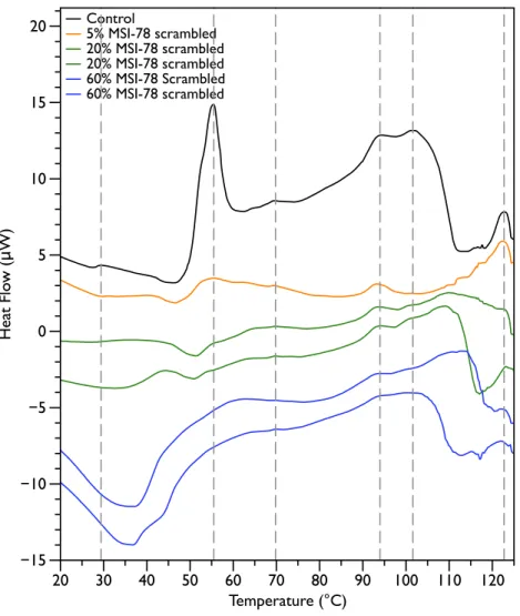
Figure 2shows thermal scans of JM109 cells treated with increasing amounts of MSI-78, ranging from 2.5% to 60%, where the % expressed is the dry weight of peptide compared to the dry weight of the bacteria (as inPius, Morrow & Booth, 2012). As expected for a peptide believed to have a lytic mechanism, the addition of MSI-78 does cause the lipid transition at∼29 ◦C to disappear, even with the smallest amount of MSI-78, consistent with MSI-78 disruption of the lipid bilayer. However, the major change that occurs in the DSC scans with MSI-78 is a decrease in the amplitude of the 55 ◦C peak with increasing peptide, and the concomitant appearance of a new peak at 40 ◦C that increases in amplitude with increasing concentrations of MSI-78. One explanation consistent with this observation is an interaction between MSI-78 and the ribosomes that induces the formation of a less stable ribosome structure that denatures at a lower temperature.
The normal protocol employed for MSI-78 treatment of the cells in this study was to leave the MSI-78 in the samples during the scan—i.e., in general there was no wash step to remove any excess MSI-78 that might remain free in solution rather than bound to the cells. Therefore, two scans were performed in which the cells were treated with MSI-78, but then the cells centrifuged and supernatant discarded to remove any excess MSI-78. The result (Fig. S1) was identical to the scans shown inFig. 2. Hence, it appears that the MSI-78 is able to interact with the cells before the start of the scan.
Figure 2 Differential Scanning Calorimetry scans ofE. coliJM109 cells treated with the AMP MSI-78. JM109 cells were treated with 0 to 60% weight MSI-78 per dry weight of cells.
Table 1 Minimal inhibitory concentration (MIC) data for JM109E. coliwith MSI-78 and scrambled
MSI-78. The experiments were performed in triplicate—identical values for each peptide were obtained each time.
MSI-78 Mean MIC against JM109 (ug/ml)
MSI-78 2.0
MSI-78s 16.5
the appearance of the new peak at 40 ◦C. The exothermic peak at∼37 ◦C likely derives from metabolism of the peptide by the bacteria (Lepock, 2005).
Figure 3 Differential Scanning Calorimetry scans ofE. coliJM109 cells with addition of a scrambled
variant of MSI-78. JM109 cells were treated with a scrambled variant of MSI-78, i.e., a peptide with the same amino acid content as MSI-78 but the primary sequence order scrambled so it cannot form an am-phipathic alpha-helix.
Table 2 Hemolytic Activity of MSI-78 and scrambled MSI-78. The values indicated are the average of absorbance measurements from 3 samples measured at 541 nm.
Sample Average absorbance Standard deviation % Hemolysis
Negative control 0.033 0.0039 N/A
Positive control 0.870 0.064 N/A
24.35µg/mL MSI-78 0.093 0.0059 6.90%
48.7µg/mL MSI-78 0.116 0.010 9.54%
24.35µg/mL scrambled MSI-78 0.0517 0.0074 2.15%
Figure 4 Differential Scanning Calorimetry Scans ofE. coliJM109 cells treated with various
concen-trations of aminoglycoside antibiotics streptomycin and kanamycin. JM109 cells were treated with the antibiotic (A) streptomycin, and (B) kanamycin. Both are inhibitors of the bacterial 30S small ribosomal subunit.
no change in the 55 ◦C peak. With kanamycin (Fig. 4B), there was a large decrease in intensity of the 55 ◦C peak and a concomitant appearance of small peaks at∼47, 52, and 57 ◦C. The new small peaks may correspond to structures with transition temperatures that have been modified by the kanamycin. Or alternatively, at least for the 52 and 57 ◦C peaks, may correspond to transitions present in the untreated cells but ‘‘revealed’’ by the kanamycin-induced reduction in intensity for the strong 55 ◦C peak. In any case, it is apparent that not all ribosome-affecting agents have the same effect on the DSC scan of the bacteria. This is perhaps not surprising, given that kanamycin and streptomycin are known to have different effects on the ribosome—e.g., streptomycin decreases the mobility of selected regions of the 30 s unit (Gromadski & Rodnina, 2004), whereas kanamycin stabilizes peptidyl-tRNA binding (Semenkov et al., 1982).
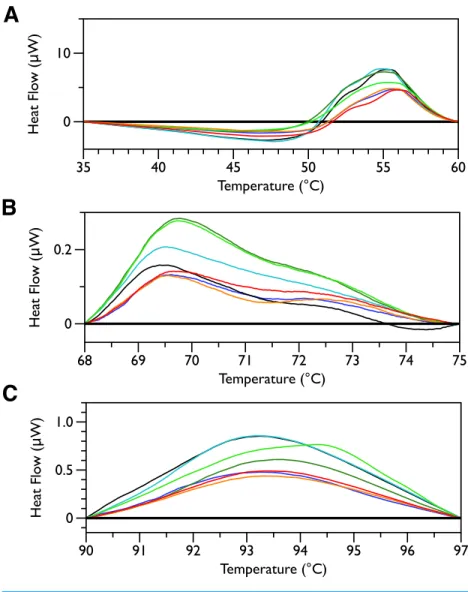
Figure 5 DSC scans of untreatedE. coliJM109 cells with temperature ranges analyzed separately.A linear baseline was fit to each scan over the lower and upper regions of each temperature range and this baseline subtracted from the data (A) 35 to 60 ◦C, (B) 68 to 75 ◦C, and (C) 90 to 97 ◦C.
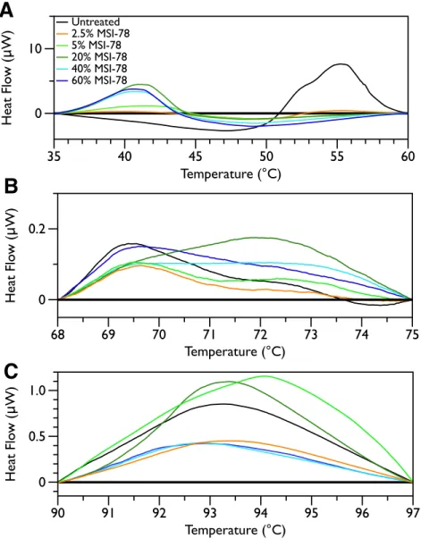
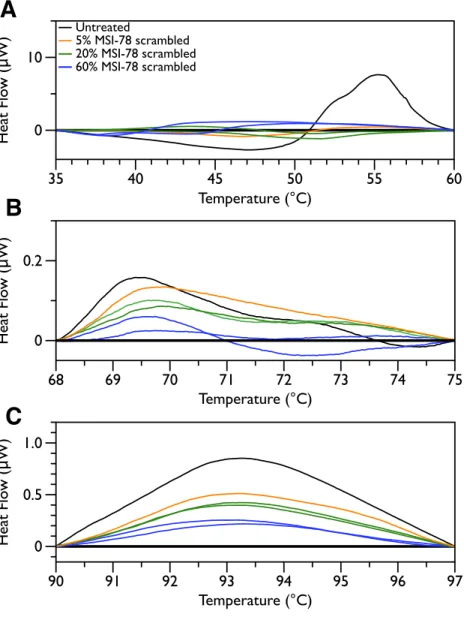
method. Interestingly, the scrambled peptide reduces the intensity of the peaks in the 68 to 75 and 90 to 97 ◦C regions in a manner that increases with increasing peptide concentration (Fig. 7). That MSI-78 has greater effects than the scrambled peptide on the 55 ◦C peak, but smaller effects than the scrambled peptide on the rest of the thermogram, suggests that MSI-78’s action is more specific than the scrambled peptide. Neither streptomycin nor kanamycin showed consistent changes to the 68 to 75 and 90 to 97 ◦C regions (Figs. 4A and4B).
To provide further evidence of MSI-78’s effects on ribosomes, an in vitro
Figure 6 DSC Scans ofE. coliJM109 Cells treated with MSI-78 with temperature ranges analyzed
sep-arately. A linear baseline was fit to each scan over the lower and upper regions of each temperature range and this baseline subtracted from the data (A) 35 to 60 ◦C, (B) 68 to 75 ◦C, and (C) 90 to 97 ◦C. The colours match those fromFig. 2.
DISCUSSION
Figure 7 DSC Scans ofE. coliJM109 Cells treated with scrambled MSI-78 with temperature ranges
analyzed separately.A linear baseline was fit to each scan over the lower and upper regions of each tem-perature range and this baseline subtracted from the data (A) 35 to 60 ◦C, (B) 68 to 75 ◦C, and (C) 90 to 97 ◦C. The colours match those fromFig. 3.
Figure 8 Normalized Luciferase Activity ofin vitroTranscription/Translation Assay of Vector-bound
Luciferase to Test for Inhibition of Protein Synthesis.
Introduction), AMP:lipid ratio may not provide a meaningful comparison. There is indeed precedence for AMP to function via interference with ribosomes, for example the AMP PR-39 (Boman, Agerberth & Boman, 1993) has been shown to inhibit protein synthesis. Furthermore, magainin 2, the AMP on which the MSI-78 sequence is based, has been shown to locate to the cell cytoplasm (Haukland et al., 2001).
In contrast to MSI-78, a scrambled version of MSI-78 showed a weaker effect (Fig. 3) on ribosomes, and unlike MSI-78, also affected other cellular components (Fig. 7). Together, the results with MSI-78 and its scrambled version suggest that MSI-78’s effects are at least somewhat dependent on its particular structural features, rather than a sole function of its overall charge and hydrophobicity. This finding is consistent with earlier studies that showed that disrupting the secondary structure of MSI-78’s related AMP, magainin-2, and analogs also disrupted their function (Chen et al., 1988;Matsuzaki et al., 1997). One of the hopes of our study was to examine if it might be possible to use DSC to see interactions between MSI-78 and the carbohydrate components of the cell envelope. However, our results contained too much sample-to-sample variation in this region of the DSC scans to say definitively if MSI-78 had an effect on these components or not. It would be interesting in future to perform DSC scans of cells treated with a variety of AMPs with previously identified targets including other well-studied AMPs that are thought to be lytic, as well as AMPs that target DNA or cell envelope components.
DSC verifies that MSI-78 does indeed have an intracellular target and that this target is the ribosomes. MSI-78’s ability to permeabilize model andE. colimembranes has previously been well documented ((Hallock, Lee & Ramamoorthy, 2003;Ramamoorthy et al., 2006);
Gottler & Ramamoorthy, 2009;Lee et al., 2013). Putting all the results together, it appears that MSI-78 acts via multiple mechanisms—i.e., by inhibiting ribosomal activity as well as disrupting the cell membrane. Multiple researchers have suggested that at least some AMPs kill target pathogens via a multi-hit mechanism (e.g.,Friedrich et al., 2000;Hancock & Sahl, 2006;Jenssen, Hamill & Hancock, 2006;Marcos & Gandia, 2009;Wimley, 2010;
Nguyen, Haney & Vogel, 2011) and the DSC work is certainly consistent with a multiple-hit mechanism for MSI-78. Our study only examined one AMP, and so extending the results to other AMPs would be speculative. However, what it not speculative is the observation that once an AMP has been found to lyse model or pathogenic membranes, it is rare for that AMP to be subjected to further analysis to identify potential non-lipidic mechanisms of action. One potential consequence of this narrow focus with respect to an AMP’s mechanism is the difficulty encountered in rational design of AMPs for clinical use via optimization of AMP-lipid interactions. The poor correlation between model membrane lytic activity and effectiveness in MIC assays observed for some AMPs (e.g.,Friedrich et al., 2000;He, Krauson & Wimley, 2014) may well be due to the presence of non-lipid targets of an AMP that are not reflected in the model membranes used for AMP optimization. Successful optimization of an AMP for clinical use may well require optimization of AMP interactions with both lipid and non-lipid targets of that AMP.
ACKNOWLEDGEMENTS
The authors would like to express their gratitude to Matthias Mack, for his advice on the transcription assay, Rob Brown for help with the hemolytic assays, and Kapil Tahlan for fruitful discussions.
ADDITIONAL INFORMATION AND DECLARATIONS
Funding
This research was supported by an NSERC Discovery Grant to V. Booth (312676). The funders had no role in study design, data collection and analysis, decision to publish, or preparation of the manuscript.
Grant Disclosures
The following grant information was disclosed by the authors: NSERC Discovery: 312676.
Competing Interests
Author Contributions
• Alexander M. Brannan conceived and designed the experiments, performed the experiments, analyzed the data, wrote the paper, prepared figures and/or tables, reviewed drafts of the paper.
• William A. Whelan and Emma Cole performed the experiments, analyzed the data, reviewed drafts of the paper.
• Valerie Booth conceived and designed the experiments, analyzed the data, wrote the paper, prepared figures and/or tables, reviewed drafts of the paper.
Data Availability
The following information was supplied regarding data availability: Raw data can be found in the Supplemental Information.
Supplemental Information
Supplemental information for this article can be found online athttp://dx.doi.org/10.7717/ peerj.1516#supplemental-information.
REFERENCES
Abuladze MK, Sokhadze VM, Namchevadze EN, Kiziria E, Tabatadze LV, Lejava LV, Gogichaishvili S, Bakradze NB. 2009.Thermal analysis of whole bacterial cells exposed to potassium permanganate using differential scanning calorimetry: a biphasic dose-dependent response to stress.Scientific World Journal9:109–117
DOI 10.1100/tsw.2009.3.
Alpas H, Lee J, Bozoglu F, Kaletunc G. 2003.Evaluation of high hydrostatic pressure sensitivity of Staphylococcus aureus andEscherichia coliO157:H7 by differential scanning calorimetry.International Journal of Food Microbiology87:229–237
DOI 10.1016/S0168-1605(03)00066-7.
Anderson WA, Hedges ND, Jones MV, Cole MB. 1991.Thermal inactivation of Listeria monocytogenes studied by differential scanning calorimetry.Journal of General Microbiology137:1419–1424DOI 10.1099/00221287-137-6-1419.
Andreu D, Rivas L. 1998.Animal antimicrobial peptides: an overview.Biopolymers
47:415–433DOI 10.1002/(SICI)1097-0282(1998)47:6<415::AID-BIP2>3.0.CO;2-D. Andrushchenko VV, Vogel HJ, Prenner EJ. 2007.Interactions of tryptophan-rich
cathe-licidin antimicrobial peptides with model membranes studied by differential scan-ning calorimetry.Biochimica et Biophysica ACTA/General Subjects1768:2447–2458
DOI 10.1016/j.bbamem.2007.05.015.
Bagheri A, Taheri-Araghi S, Ha BY. 2015.How cell concentrations are implicated in cell selectivity of antimicrobial peptides.Langmuir31:8052–8062
DOI 10.1021/acs.langmuir.5b01533.
Bechinger B, Salnikov ES. 2012.The membrane interactions of antimicrobial peptides revealed by solid-state NMR spectroscopy.Chemistry and Physics of Lipids
165:282–301DOI 10.1016/j.chemphyslip.2012.01.009.
Belliveau BH, Beaman TC, Pankratz HS, Gerhardt P. 1992.Heat killing of bacterial spores analyzed by differential scanning calorimetry.Journal of Bacteriology
174:4463–4474.
Boman HG, Agerberth B, Boman A. 1993.Mechanisms of action onEscherichia coliof cecropin P1 and PR-39, two antibacterial peptides from pig intestine.Infection and Immunity 61:2978–2984.
Boucher HW, Talbot GH, Bradley JS, Edwards JE, Gilbert D, Rice LB, Scheld M, Spellberg B, Bartlett J. 2009.Bad bugs, no drugs: no ESKAPE! An update from the Infectious Diseases Society of America.Clinical Infectious Diseases48:1–12
DOI 10.1086/595011.
Brogden KA. 2005.Antimicrobial peptides: pore formers or metabolic inhibitors in bacteria?Nature Reviews Microbiology 3:238–250DOI 10.1038/nrmicro1098. Brotz H, Bierbaum G, Leopold K, Reynolds PE, Sahl HG. 1998.The lantibiotic
mer-sacidin inhibits peptidoglycan synthesis by targeting lipid II.Antimicrobial Agents and Chemotherapy 42:154–160.
Chen HC, Brown JH, Morell JL, Huang CM. 1988.Synthetic magainin analogues with improved antimicrobial activity.FEBS Letters236:462–466
DOI 10.1016/0014-5793(88)80077-2.
Elbing K, Brent R. 2002.Media preparation and bacteriological tools.Current Protocols in Molecular BiologyEpub ahead of print 1 Aug 2002
DOI 10.1002/0471142727.mb0101s59.
Epand RF, Umezawa N, Porter EA, Gellman SH, Epand RM. 2003.Interactions of the antimicrobial beta-peptide beta-17 with phospholipid vesicles differ from membrane interactions of magainins.European Journal of Biochemistry 270:1240–1248
DOI 10.1046/j.1432-1033.2003.03484.x.
Friedrich CL, Moyles D, Beveridge TJ, Hancock RE. 2000.Antibacterial action of structurally diverse cationic peptides on gram-positive bacteria.Antimicrobial Agents and Chemotherapy 44:2086–2092DOI 10.1128/AAC.44.8.2086-2092.2000.
Gee ML, Burton M, Grevis-James A, Hossain MA, McArthur S, Palombo EA, Wade JD, Clayton AH. 2013.Imaging the action of antimicrobial peptides on living bacterial cells.Scientific Reports3:1557DOI 10.1038/srep01557.
Gottler LM, Ramamoorthy A. 2009.Structure, membrane orientation, mechanism, and function of pexiganan–a highly potent antimicrobial peptide designed from magainin.Biochimica et Biophysica ACTA/General Subjects1788:1680–1686
DOI 10.1016/j.bbamem.2008.10.009.
Hall K, Lee TH, Mechler AI, Swann MJ, Aguilar MI. 2014.Real-time measurement of membrane conformational states induced by antimicrobial peptides: balance between recovery and lysis.Scientific Reports4:5479DOI 10.1038/srep05479. Hallock KJ, Lee DK, Ramamoorthy A. 2003.MSI-78, an analogue of the magainin
antimicrobial peptides, disrupts lipid bilayer structure via positive curvature strain. Biophysical Journal84:3052–3060DOI 10.1016/S0006-3495(03)70031-9.
Hancock RE, Sahl HG. 2006.Antimicrobial and host-defense peptides as new anti-infective therapeutic strategies.Nature Biotechnology24:1551–1557
DOI 10.1038/nbt1267.
Haukland HH, Ulvatne H, Sandvik K, Vorland LH. 2001.The antimicrobial peptides lactoferricin B and magainin 2 cross over the bacterial cytoplasmic membrane and reside in the cytoplasm.FEBS Letters508:389–393
DOI 10.1016/S0014-5793(01)03100-3.
He J, Krauson AJ, Wimley WC. 2014.Toward the de novo design of antimicrobial peptides: lack of correlation between peptide permeabilization of lipid vesicles and antimicrobial, cytolytic, or cytotoxic activity in living cells.Biopolymers102:1–6
DOI 10.1002/bip.22281.
Jana J, Kar RK, Ghosh A, Biswas A, Ghosh S, Bhunia A, Chatterjee S. 2013.Human cathelicidin peptide LL37 binds telomeric G-quadruplex.Mol Biosyst.
Jenssen H, Hamill P, Hancock RE. 2006.Peptide antimicrobial agents.Clinical Microbi-ology Reviews19:491–511 DOI 10.1128/CMR.00056-05.
Kaletunc G, Lee J, Alpas H, Bozoglu F. 2004.Evaluation of structural changes induced by high hydrostatic pressure in Leuconostoc mesenteroides.Applied and Environmental Microbiology70:1116–1122DOI 10.1128/AEM.70.2.1116-1122.2004.
Kragol G, Lovas S, Varadi G, Condie BA, Hoffmann R, Otvos LJ. 2001.The antibacterial peptide pyrrhocoricin inhibits the ATPase actions of DnaK and prevents chaperone-assisted protein folding.Biochemistry40:3016–3026DOI 10.1021/bi002656a. Lee DK, Bhunia A, Kotler SA, Ramamoorthy A. 2015.Detergent-type membrane
fragmentation by MSI-78, MSI-367, MSI-594, and MSI-843 antimicrobial peptides and inhibition by cholesterol: a solid-state nuclear magnetic resonance study. Biochemistry54:1897–1907DOI 10.1021/bi501418m.
Lee DK, Brender JR, Sciacca MF, Krishnamoorthy J, Yu C, Ramamoorthy A. 2013. Lipid composition-dependent membrane fragmentation and pore-forming mech-anisms of membrane disruption by pexiganan (MSI-78).Biochemistry.
Lee J, Kaletunc G. 2002a.Calorimetric determination of inactivation parameters of micro-organisms.Journal of Applied Microbiology 93:178–189
DOI 10.1046/j.1365-2672.2002.01677.x.
Lee J, Kaletunc G. 2002b.Evaluation of the heat inactivation ofEscherichia coliand Lac-tobacillus plantarum by differential scanning calorimetry.Applied and Environmental Microbiology68:5379–5386DOI 10.1128/AEM.68.11.5379-5386.2002.
Lee J, Kaletunç G. 2010.Inactivation ofhiiSalmonella Enteritidis strains by combination of high hydrostatic pressure and nisin.International Journal of Food Microbiology
Lepock JR. 2005.Measurement of protein stability and protein denaturation in cells using differential scanning calorimetry.Methods35:117–125
DOI 10.1016/j.ymeth.2004.08.002.
Lepock JR, Frey HE, Heynen MP, Nishio J, Waters B, Ritchie KP, Kruuv J. 1990. Increased thermostability of thermotolerant CHL V79 cells as determined by differential scanning calorimetry.Journal of Cellular Physiology 142:628–634
DOI 10.1002/jcp.1041420324.
Lepock JR, Frey HE, Inniss WE. 1990.Thermal analysis of bacteria by differential scanning calorimetry: relationship of protein denaturationin situto maximum growth temperature.Biochimica et Biophysica ACTA/General Subjects1055:19–26
DOI 10.1016/0167-4889(90)90086-S.
Mackey BM, Miles CA, Parsons SE, Seymour DA. 1991.Thermal denaturation of whole cells and cell components ofEscherichia coliexamined by differential scanning calorimetry.Journal of General Microbiology137:2361–2374
DOI 10.1099/00221287-137-10-2361.
Mackey BM, Miles CA, Seymour DA, Parsons SE. 1993.Thermal denaturation and loss of viability inEscherichia coliand Bacillus stearothermophilus.Letters in Applied Microbiology16:56–58DOI 10.1111/j.1472-765X.1993.tb00341.x.
Mackey BM, Parsons SE, Miles CA, Owen RJ. 1988.The relationship between the base composition of bacterial DNA and its intracellular melting temperature as determined by differential scanning calorimetry.Journal of General Microbiology
134:1185–1195.
Maisetta G, Vitali A, Scorciapino MA, Rinaldi AC, Petruzzelli R, Brancatisano FL, Esin S, Stringaro A, Colone M, Luzi C, Bozzi A, Campa M, Batoni G. 2013. pH-dependent disruption ofEscherichia coliATCC 25922 and model membranes by the human antimicrobial peptides hepcidin 20 and 25.FEBS Journal280:2842–2854
DOI 10.1111/febs.12288.
Maloy WL, Kari UP. 1995.Structure–activity studies on magainins and other host defense peptides.Biopolymers37:105–122DOI 10.1002/bip.360370206.
Marcos JF, Gandia M. 2009.Antimicrobial peptides: to membranes and beyond.Expert Opinion on Drug Discovery4:659–671DOI 10.1517/17460440902992888.
Matsuzaki K. 2009.Control of cell selectivity of antimicrobial peptides.Biochimica et Biophysica ACTA/General Subjects1788:1687–1692
DOI 10.1016/j.bbamem.2008.09.013.
Matsuzaki K, Sugishita K, Harada M, Fujii N, Miyajima K. 1997.Interactions of an antimicrobial peptide, magainin 2, with outer and inner membranes of
Gram-negative bacteria.Biochimica et Biophysica ACTA/General Subjects
1327:119–130DOI 10.1016/S0005-2736(97)00051-5.
Melo MN, Ferre R, Castanho MA. 2009.Antimicrobial peptides: linking partition, activity and high membrane-bound concentrations.Nature Reviews Microbiology
Milek I, Crnigoj M, Ulrih NP, Kaletunc G. 2007.In vivocharacterization of thermal stabilities of Aeropyrum pernix cellular components by differential scanning
calorimetry.Canadian Journal of Microbiology53:1038–1045DOI 10.1139/W07-069. Miles CA, Mackey BM, Parsons SE. 1986.Differential scanning calorimetry of bacteria.
Journal of General Microbiology132:939–952.
Mohácsi-Farkas C, Farkas J, Meszaros L, Reichart O, Andrassy E. 1999.Thermal denaturation of bacterial cells examined by differential scanning calorimetry.Journal of Thermal Analysis and Calorimetry 57:409–414DOI 10.1023/A:1010139204401. Nguyen HT, Corry JE, Miles CA. 2006.Heat resistance and mechanism of heat
inac-tivation in thermophilic campylobacters.Applied and Environmental Microbiology
72:908–913DOI 10.1128/AEM.72.1.908-913.2006.
Nguyen LT, Haney EF, Vogel HJ. 2011.The expanding scope of antimicrobial peptide structures and their modes of action.Trends in Biotechnology29:464–472
DOI 10.1016/j.tibtech.2011.05.001.
Obuchi K, Iwahashi H, Lepock JR, Komatsu Y. 2000.Calorimetric characterization of critical targets for killing and acquired thermotolerance in yeast.Yeast 16:111–119
DOI 10.1002/(SICI)1097-0061(20000130)16:2<111::AID-YEA507>3.0.CO;2-V. Park CB, Kim HS, Kim SC. 1998.Mechanism of action of the antimicrobial peptide buforin II: buforin II kills microorganisms by penetrating the cell membrane and inhibiting cellular functions.Biochemical and Biophysical Research Communications
244:253–257DOI 10.1006/bbrc.1998.8159.
Pasupuleti M, Schmidtchen A, Malmsten M. 2012.Antimicrobial peptides: key compo-nents of the innate immune system.Critical Reviews in Biotechnology32:143–171
DOI 10.3109/07388551.2011.594423.
Patrzykat A, Friedrich CL, Zhang L, Mendoza V, Hancock RE. 2002.Sublethal con-centrations of pleurocidin-derived antimicrobial peptides inhibit macromolecular synthesis inEscherichia coli.Antimicrobial Agents and Chemotherapy46:605–614
DOI 10.1128/AAC.46.3.605-614.2002.
Pedrolli DB, Mack M. 2014.Bacterial flavin mononucleotide riboswitches as targets for flavin analogs.Methods in Molecular Biology1103:165–176
DOI 10.1007/978-1-62703-730-3_13.
Pius J, Morrow MR, Booth V. 2012.(2)H solid-state nuclear magnetic resonance
investigation of wholeEscherichia coliinteracting with antimicrobial peptide MSI-78. Biochemistry51:118–125DOI 10.1021/bi201569t.
Porcelli F, Buck-Koehntop BA, Thennarasu S, Ramamoorthy A, Veglia G. 2006. Structures of the dimeric and monomeric variants of magainin antimicrobial peptides (MSI-78 and MSI-594) in micelles and bilayers, determined by NMR spectroscopy.Biochemistry45:5793–5799DOI 10.1021/bi0601813.
Ramamoorthy A. 2009.Beyond NMR spectra of antimicrobial peptides: dynamical images at atomic resolution and functional insights.Solid State Nuclear Magnetic Resonance35:201–207DOI 10.1016/j.ssnmr.2009.03.003.
MSI-78 and MSI-594 derived from magainin 2 and melittin.Biophysical Journal
91:206–216DOI 10.1529/biophysj.105.073890.
Rossi LM, Rangasamy P, Zhang J, Qiu XQ, Wu GY. 2008.Research advances in the development of peptide antibiotics.Journal of Pharmaceutical Sciences97:1060–1070
DOI 10.1002/jps.21053.
Semenkov Y, Katunin VI, Makarov EM, Kirillov SV. 1982.Quantitative study of kanamycin action on different functions ofEscherichia coliribosomes.FEBS Letters
144:121–124DOI 10.1016/0014-5793(82)80583-8.
Sevcsik E, Pabst G, Jilek A, Lohner K. 2007.How lipids influence the mode of action of membrane-active peptides.Biochimica et Biophysica ACTA/General Subjects
1768:2586–2595DOI 10.1016/j.bbamem.2007.06.015.
Shai Y. 1999.Mechanism of the binding, insertion and destabilization of phospholipid bilayer membranes by alpha-helical antimicrobial and cell non-selective membrane-lytic peptides.Biochimica et Biophysica ACTA/General Subjects1462:55–70
DOI 10.1016/S0005-2736(99)00200-X.
Spellberg B, Guidos R, Gilbert D, Bradley J, Boucher HW, Scheld WM, Bartlett JG, Edwards JJ. 2008.The epidemic of antibiotic-resistant infections: a call to action for the medical community from the Infectious Diseases Society of America.Clinical Infectious Diseases46:155–164DOI 10.1086/524891.
Teixeira P, Castro H, Mohacsi-Farkas C, Kirby R. 1997.Identification of sites of injury in Lactobacillus bulgaricus during heat stress.Journal of Applied Microbiology
83:219–226DOI 10.1046/j.1365-2672.1997.00221.x.
Tunick MH, Bayles DO, Novak JS. 2006.DSC analysis of foodborne bacteria.Journal of Thermal Analysis and Calorimetry83:23–26DOI 10.1007/s10973-005-7042-8. Wimley WC. 2010.Describing the mechanism of antimicrobial peptide action with the
interfacial activity model.ACS Chemical Biology5:905–917DOI 10.1021/cb1001558. Wimley WC, Hristova K. 2011.Antimicrobial peptides: successes, challenges and
unanswered questions.Journal of Membrane Biology 239:27–34
DOI 10.1007/s00232-011-9343-0.
Yeung AT, Gellatly SL, Hancock RE. 2011.Multifunctional cationic host defence peptides and their clinical applications.Cellular and Molecular Life Science.
Zasloff M. 2002.Antimicrobial peptides of multicellular organisms.Nature415:389–395
DOI 10.1038/415389a.