III
a
-Dependent PI4P Is Subverted by HCV and Is Targeted
by a 4-Anilino Quinazoline with Antiviral Activity
Annalisa Bianco1, Veronica Reghellin1, Lorena Donnici1, Simone Fenu1, Reinaldo Alvarez1, Chiara Baruffa2, Francesco Peri2, Massimiliano Pagani1, Sergio Abrignani1, Petra Neddermann1*, Raffaele De Francesco1*
1Department of Genomics and Molecular Biology, Virology Program, Istituto Nazionale Genetica Molecolare (INGM), Milano, Italy,2Department of Biotechnology and Biosciences, University of Milano Bicocca, Milano, Italy
Abstract
4-anilino quinazolines have been identified as inhibitors of HCV replication. The target of this class of compounds was proposed to be the viral protein NS5A, although unequivocal proof has never been presented. A 4-anilino quinazoline moiety is often found in kinase inhibitors, leading us to formulate the hypothesis that the anti-HCV activity displayed by these compounds might be due to inhibition of a cellular kinase. Type III phosphatidylinositol 4-kinasea (PI4KIIIa) has recently been identified as a host factor for HCV replication. We therefore evaluated AL-9, a compound prototypical of the 4-anilino quinazoline class, on selected phosphatidylinositol kinases. AL-9 inhibited purified PI4KIIIaand, to a lesser extent, PI4KIIIb. In Huh7.5 cells, PI4KIIIais responsible for the phosphatidylinositol-4 phosphate (PI4P) pool present in the plasma membrane. Accordingly, we observed a gradual decrease of PI4P in the plasma membrane upon incubation with AL-9, indicating that this agent inhibits PI4KIIIaalso in living cells. Conversely, AL-9 did not affect the level of PI4P in the Golgi membrane, suggesting that the PI4KIIIb isoform was not significantly inhibited under our experimental conditions. Incubation of cells expressing HCV proteins with AL-9 induced abnormally large clusters of NS5A, a phenomenon previously observed upon silencing PI4KIIIaby RNA interference. In light of our findings, we propose that the antiviral effect of 4-anilino quinazoline compounds is mediated by the inhibition of PI4KIIIaand the consequent depletion of PI4P required for the HCV membranous web. In addition, we noted that HCV has a profound effect on cellular PI4P distribution, causing significant enrichment of PI4P in the HCV-membranous web and a concomitant depletion of PI4P in the plasma membrane. This observation implies that HCV – by recruiting PI4KIIIa in the RNA replication complex – hijacks PI4P metabolism, ultimately resulting in a markedly altered subcellular distribution of the PI4KIIIaproduct.
Citation:Bianco A, Reghellin V, Donnici L, Fenu S, Alvarez R, et al. (2012) Metabolism of Phosphatidylinositol 4-Kinase IIIa-Dependent PI4P Is Subverted by HCV and Is Targeted by a 4-Anilino Quinazoline with Antiviral Activity. PLoS Pathog 8(3): e1002576. doi:10.1371/journal.ppat.1002576
Editor:Guangxiang George Luo, University of Kentucky College of Medicine, United States of America
ReceivedAugust 16, 2011;AcceptedJanuary 26, 2012;PublishedMarch 8, 2012
Copyright:ß2012 Bianco et al. This is an open-access article distributed under the terms of the Creative Commons Attribution License, which permits unrestricted use, distribution, and reproduction in any medium, provided the original author and source are credited.
Funding:This work/research was partially supported by the Italian Ministry of Education, University and Research (MIUR) (grant number RBAP10TPXK to INGM). FP acknowledges the Regione Lombardia for financial support (grant number 14546, Network Enabled Drug Design, NEDD). The funders have no role in the study design, data collection and analysis, decision to publish, or preparation of the manuscript.
Competing Interests:The authors have declared that no competing interests exist.
* E-mail: neddermann@ingm.org (PN); defrancesco@ingm.org (RDF)
Introduction
Hepatitis C virus (HCV) is an enveloped, single-stranded RNA virus classified as member of the Hepacivirus genus within the Flaviviridae family. The 9.6 kb positive-sense RNA genome contains a single open reading frame encoding a polyprotein of about 3,000 amino acids, flanked by highly structured 59and 39
untranslated (UTR) regions. Following its release into the cytoplasm of the host cell, viral RNA is translated via an internal ribosome entry site (IRES), giving rise to a single polypeptide that is cleaved into 10 different mature protein products: Core, gpE1, gpE2, p7, NS2, NS3, NS4A, NS4B, NS5A, and NS5B. HCV RNA replication takes place in the cytoplasm, in association with a virus-induced intracellular membrane structure termed ‘‘membra-nous web’’, onto which NS proteins assemble to form the so-called RNA replication complexes.
It is estimated that 3% of the world’s population are chronically infected by the hepatitis C virus (HCV). Most infections become chronic and over time evolve into chronic hepatitis. The most unwanted complication of chronic hepatitis is cirrhosis, a massive liver fibrosis, which can lead to liver failure and hepatocellular carcinoma.
more are being developed [1]. While the initial efforts to the discovery of DAA focused almost exclusively on the best characterized HCV enzymes required for viral replication – the NS3/4A protease and the NS5B polymerase – in the past few years the NS5A viral protein has been attracting more and more attention as a target for drug development [1,2]. NS5A possesses no known enzymatic activity. It is a multifunctional non-structural protein important for viral replication [3–6] as well as viral assembly [7–9]. It is a phosphoprotein consisting of three domains [10]. Domain I is highly conserved and forms a dimeric structure [11,12], whereas domains II and III are believed to adopt a ‘‘natively unfolded’’ conformation [13,14].
In recent years, several anti-HCV compounds identified using cell-based replicon screens were indicated to target NS5A based on the analysis of the mutations associated with emergence of resistance in the replicon system [15–17].
The most studied series of these ‘‘NS5A inhibitors’’ is represented by BMS-790052, an agent that is leading the field, having demonstrated potent antiviral activity in clinical studies [18]. Compounds in this class are characterized by a complex, dimeric or pseudo-dimeric structure and a high molecular weight, when compared with conventional ‘‘drug-like’’ small molecules [17,19]. Resistance mutations against these compounds emerge readily in domain I of NS5A [20], with the most recurrent of these changes corresponding to variant of tyrosine at position 93 [20]. Although direct interaction with purified NS5A has not been demonstrated, compelling reverse genetic experiments [20] as well as molecular models [15,21] strongly support the notion that NS5A is the direct target of these compounds.
A less characterized series of compounds, belonging to a different chemical class, was also initially indicated to target NS5A on the basis of the mutation pattern observed in resistant replicons [21]. The common structural element of this latter class of inhibitors is a 4-anilino quinazoline core. A representative member of this class of compounds is A-831/AZD-2836, an experimental antiviral agent that entered clinical trials but was later discontin-ued due to the lack of adequate exposure [17]. For these agents, the mutations reported to be associated with resistance were found
to be different from those expected for the NS5A inhibitor described above, pointing to a different mechanism of action: a few mutations were found at the C-terminal end of NS5A domain I (E212D, L199F and T200P), whereas most mutations occurred in NS5A domains II and III (P299L, S370P, V388D, V362A, S390G and S370P). Additional mutations were also found in NS4B (S258T) and NS5B (S76A) [17,21,22]. Reverse genetics studies in which these mutations were reintroduced in the replicon, however, did not recapitulate the resistant phenotype observed in the original cellular clones [17], leaving thus the possibility open that these compounds act through a different viral or cellular target.
Interestingly, many kinase inhibitors, including some approved antitumoral drugs (gefitinib, lapatinib, erlotinib) are 4-anilino quinazoline derivatives [23–25]. Altogether, these considerations led us to investigate whether the anti-HCV activity displayed by these compounds might be due to inhibition of a cellular kinase.
Recently, several small-interfering RNA (siRNA) screening campaigns have identified type III phosphatidylinositol 4-kinases (PI4K) as crucial host factors for HCV replication. In particular, PI4KIIIawas found to be required for HCV RNA replication in a cell line- and genotype-independent manner, whereas the requirement for theb isoform was observed to be less dramatic and limited to Con-1 (genotype 1b) replicons [26–29]. It was shown that the catalytic activity of PI4KIIIais required to rescue HCV replication in cells with a stable knock-down of PI4KIIIa. In addition, it has been proposed that NS5A stimulates PI4KIIIa
activity by direct interaction via domain I [30–32]. All these observations taken together made us consider the phosphatidyli-nositol kinases a potential alternative candidate target for 4-anilino quinazoline inhibitors of HCV replication.
In this paper, we present evidence that AL-9, a member of this class of compounds previously reported to target NS5A, inhibits PI4P formation by direct inhibition of phosphatidylinositol 4-kinase IIIa (PI4KIIIa). In addition, we provide evidence that pharmacological inhibition of PI4KIIIa with AL-9 results in altered subcellular distribution of NS5A similar to that observed after RNAi knock-down of the PI4KIIIa mRNA, strongly supporting a mechanism of HCV inhibition mediated by the inhibition of PI4KIIIa. Moreover, we show that HCV subverts components of the phosphatidylinositol-4 phosphate (PI4P) pathway to function in favor of its own life cycle, thereby enriching the PI4P concentration in the membranous web while depleting the plasma membrane PI4P pool.
Results
Compound AL-9 inhibits HCV replication in vitro
AL-9 is a member of 4-anilino quinazoline-containing HCV replication inhibitors described previously ([21]; Figure 1). In order to confirm its anti-HCV activity, we tested the effect of this compound on HCV replication in Huh7.5 cells stably expressing genotype 1b or 2a subgenomic replicons (Con1-SR and JFH-A4, respectively). The EC50values, calculated by measuring viral RNA
after incubation with AL-9 for three days, are reported in Table 1. Replicon EC50 values for AL-9 were found to be 0.29mM and
0.75mM for genotype 1b and 2a, respectively. In order to prove
that AL-9 inhibits HCV replication not only in the context of a HCV subgenomic replicon, but also in the context of the complete viral life-cycle, we determined the inhibitory activity using the J6/ JFH-1 HCV virus. In this case, the EC50value was found to be
1.2mM, a figure comparable with the result obtained with
genotype 2a subgenomic replicon. CC50 values are shown for
Con1-SR, JFH-A4 and Huh7.5 cells, respectively. In summary,
Author Summary
It is estimated that 3% of the world’s population are chronically infected by the hepatitis C virus (HCV). Most infections become chronic and eventually evolve into cirrhosis and hepatocellular carcinoma. Host factors are interesting targets for anti-HCV therapies due to their inherent high genetic barrier to resistance. Recently, phosphatidylinositol 4-kinasea (PI4KIIIa) has been identi-fied as a crucial host factor for HCV replication. Many different pathogens, including HCV, subvert components of the phosphatidylinositol-4 phosphate (PI4P) pathway to function in favor of their own life cycle. In this paper, we show that HCV dramatically alters cellular PI4P metabolism and distribution, resulting in the enrichment of PI4P in the membranous web required for viral replication with a concomitant decrease of PI4P in the plasma-membrane. Moreover, we demonstrate that 4-anilino quinazolines, antiviral agents previously believed to target HCV NS5A, do in fact inhibit PI4P formation by inhibition of PI4KIIIa. This compound class is a promising lead for the development of a novel antiviral therapy based on PI4KIIIa inhibition. Specific PI4KIIIa inhibitors would also be important research tools required for a deeper under-standing of the functions and regulation of PI4P.
AL-9 inhibits HCV across different genotypes with activity in the sub-micromolar to low micromolar range in the absence of significant cytotoxic effects.
AL-9 is an inhibitor of PI4KIIIa
In the following experiment, we investigated whether AL-9 inhibits the purified type III phosphatidylinositol 4-kinases PI4KIIIaand PI4KIIIb(Figure 2). Both enzymes were inhibited by AL-9 with a five-fold preference for PI4KIIIa(IC50of 0.57mM
and 3.08mM, respectively). This result demonstrates that AL-9
inhibits type III PI4 kinases in vitro at concentrations similar to
those required for its anti-HCV activity, displaying a moderate selectivity for theaover thebisoform. We also tested the activity of AL-9 on two class I PI3-kinases (p110aand p110b). While PI3-kinase p110awas inhibited with an IC50of 1.1mM, the potency of
AL-9 for PI3-kinase p110bwas significantly lower (40% inhibition @10mM, data not shown).
Our hypothesis is that AL-9 inhibits HCV replication via
inhibition of PI4KIIIa. Thus, we wanted to assess whether AL-9 also inhibited PI4KIIIain living cells. To this aim, we needed to set up an assay that allowed us to monitor the activity of this kinase in intact cells. PI4KIIIais primarily localized to the ER, whereas PI4KIIIbis localized to the Golgi membranes [33]. It was shown that PI4KIIIb contributes to the synthesis of PI4P at the Golgi membranes [34,35]. Subcellular localization of the enzymes, however, does not always coincide with their function. Thus, PI4KIIIa, considered to be an ER-resident enzyme, has previously been shown to be critical for the generation and maintenance of
the plasma membrane PI4P pool during phospholipase C activation and Ca2 signaling in HEK-293 or Cos-7 cells [35,36]
as well as in resting Cos-7 cells [37]. Whether PI4KIIIa is responsible for the maintenance of the plasma membrane PI4P pool under normal cell culture conditions in hepatoma cells is currently not known. Hammond et al [37] have developed immunocytochemical techniques that enable selective staining of the PI4P pool present in the plasma membrane (plasma membrane staining protocol) or in the intracellular membranes (Golgi staining protocol), respectively. We used this technique, in combination with RNA gene silencing or pharmacological inhibition, to decipher which of the type III enzymes participates in the synthesis of the Golgi- or plasma membrane PI4P-pools in Huh7.5 hepatoma cells.
To address which type III PI4 kinase is responsible for the synthesis of the different cellular PI4P pools, Huh7.5 cells were treated with siRNAs targeting PI4KIIIa, PI4KIIIbor an unrelated siRNA (mock-siRNA) as described in the Materials and Methods section. Immunoblots assays show specific knockdown of PI4KIIIa
or PI4KIIIb by their corresponding siRNAs (Figure 3C). Three days after siRNA treatment, PI4P was revealed either by the plasma membrane staining protocol (Figure 3A, upper panel) or by the Golgi membrane staining protocol (Figure 3A, lower panel). In cells treated with the unrelated siRNA (mock-siRNA), PI4P was detected both in the plasma membrane and in intracellular membranes. Intracellular PI4P was localized primarily in the Golgi membranes, as judged by the colocalization with the Golgi marker giantin. Silencing of PI4KIIIa resulted in a significant decrease of the PI4P level in the plasma membrane. Concomi-tantly with the decrease in the plasma membrane PI4P levels, we consistently observed a pronounced increase of PI4P level in the Golgi membrane following PI4KIIIaknockdown. In the case of
Figure 1. Chemical structure of AL-9.For the synthetic pathway and procedure see Supporting Information.
doi:10.1371/journal.ppat.1002576.g001
Table 1.List of EC50values of AL-9 for different HCV
genotypes.
genotype EC50(mM) CC50(mM)
Con1-SR 1b 0.29 (+/20.09) 29.3 (+/22.8)
JFH-A4 2a 0.75 (+/20.15) 18.9 (+/23.2)
Huh7.5+J6/JFH-1 HCV 2a 1.2 (+/20.37) 25.1 (+/24.6)*
Huh7.5 cells replicating subgenomic replicons of genotype 1b or 2a (Con1-SR and JFH-A4, respectively) or Huh7.5 cells infected with the chimeric virus J6/JFH were treated with AL-9 for three days and intracellular viral RNA was measured by real time PCR. The data are representative of at least three independent experiments, and the standard deviations are shown.
*CC
50measured in uninfected Huh7.5 cells. doi:10.1371/journal.ppat.1002576.t001
PI4KIIIb knockdown, we observed a ,30% decrease of Golgi
membrane PI4P level, whereas the PI4P levels of the plasma membrane remained substantially unaffected (Figure 3B).
These results are in line with the previously reported role for PI4KIIIa in maintaining the PI4P plasma membrane pool [35– 37] and confirm the importance of PI4KIIIbfor the synthesis of at least part of the Golgi membrane PI4P [34,35]. We also observed that decreased expression of PI4KIIIaresulted in an unexpected increase in the level of the Golgi membrane pool (Figure 3A), suggesting a complex level of cross-talk between the cellular type III PI4 kinases in maintaining the physiological PI4P levels at the Golgi membrane, at least in our experimental model.
In order to confirm and extend the results described above, we utilized a known pharmacological inhibitor of the type III PI4 kinases. PIK93 was previously exploited to distinguish between the roles of the two PI4KIII isoforms [38,39]. In particular, a concentration of 0.5mM PIK93 is expected to affect only PI4KIIIb, whereas 30mM PIK93 should inhibit both PI4KIIIband PI4KIIIa.
Thus, Huh7.5 cells were treated with 0.5mM or 30mM PIK93 or with DMSO as control. After two hours of incubation, PI4P was revealed either by the plasma membrane staining protocol (Figure 4A, upper panel) or by the Golgi staining protocol (Figure 4A, lower panel). PI4P levels associated with the Golgi membranes decreased by ,25% after incubation with 0.5mM
PIK93 (Figure 4B). This is in line with PI4KIIIbcontributing to the production of PI4P present in the Golgi membranes (PI4KIIa, another contributor of Golgi-localized PI4P is not inhibited by PIK93 [38,39]). Increasing PIK93 concentration to 30mM further
increased the inhibition of the intracellular membrane PI4P pool, to
,65% (Figure 4B). This could be due to a more complete inhibition
of PI4KIIIb; however, based on this experiment, we cannot rule out a contribution of PI4KIIIaactivity to the maintenance of the Golgi membrane PI4P pool. In contrast to what observed in the Golgi-associated membranes, the plasma membrane PI4P level was not significantly affected upon incubation with 0.5mM PIK93, but decreased by nearly 50% after incubation with 30mM of PIK93 (Figure 4B). Combined with the RNAi experiments described above, these results support the notion that, in Huh7.5 cells, PI4KIIIais involved in the maintenance of the plasma membrane PI4P pool, whereas PI4KIIIbis at least partly responsible for the maintenance of the Golgi membrane PI4P pool.
We then evaluated the PI4K inhibitory activity of AL-9 in Huh7.5 cells using the same methodology. Briefly, Huh7.5 cells were incubated either with DMSO or with increasing concentra-tion of AL-9 (1, 2, 4 or 8mM) for two hours (Figure 5A). Treatment with AL-9 gradually reduced the amount of PI4P in the plasma membrane (Figure 5B). Conversely, the concentration of PI4P in the Golgi-associated membranes remained substantially unaltered up to the highest AL-9 concentration used (Figure 5B). In all, the results described above suggest that AL-9 inhibits PI4KIIIa also in living cells, while not appreciably affecting the activity of PI4KIIIb. This is in line with the selectivity for PI4KIIIaover PI4KIIIbobserved in the biochemical assays.
HCV alters the intracellular and plasma membrane distribution of PI4P
Viral infection induces modification of intracellular membrane structures [40] and, for some RNA viruses including HCV, it has
been shown that these induced membranous structures are highly enriched for PI4P [32,41]. Before testing the activity of AL-9 in HCV-infected cells, we wanted to know what the impact of HCV on cellular membrane structures was, with special regard to the subcellular membrane distribution of PI4P.
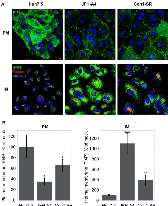
Naı¨ve Huh7.5 cells or cells actively replicating the genotype 2a or 1b HCV subgenomic replicon were investigated for their PI4P concentration in internal membranes or plasma membranes, respectively (Figure 6A). As previously shown, cells expressing the HCV replicon form a membranous web that is highly enriched for PI4P (Figure 6A, lower panel). The level of PI4P in these virus-specific membrane structures is markedly higher in JFH-A4 cells, containing the very efficient genotype 2a JFH-1 replicon, compared to the Con1-SR cells, which are based on the genotype 1b Con1 replicon, possibly mirroring the different RNA replication efficien-cy. It is well established that the kinase responsible for the production of the PI4P pool present in these structures is PI4KIIIa. In the current model, PI4KIIIa interacts with the viral protein NS5A, leading to up-regulation of the kinase activity and accumulation of PI4P in the virus-specific membranous web [30– 32]. Conversely, the results shown in Figures 3–5 suggest that – in absence of viral replication – a major function of PI4KIIIais the synthesis of the PI4P pool in the plasma membrane. We therefore asked ourselves whether the presence of HCV could not only influence distribution and enrichment of PI4P in internal membranes, but also alter the PI4P plasma membrane pool. In Figure 6A, we show that, concomitantly with the increase of PI4P in the internal membranes (lower panel), HCV replication promotes a marked decrease of PI4P concentration in the plasma membrane (upper panel). Relative quantification of the PI4P levels in the different experimental conditions is shown in Figure 6B. This experiment demonstrates that the presence of HCV causes a dramatic change of PI4P localization in cellular membranes, whereby the increase of PI4P concentration in the virus-specific membranous structures appears to be accompanied by a depletion of the PI4P pool normally present in the plasma membrane.
We next investigated whether HCV-associated changes in PI4P distribution could be reverted upon cure of the HCV replicon by specific inhibitors. We treated JFH-A4 cells for two weeks either with the HCV RdRP inhibitor HCV-796 or with the HCV NS3/ 4A protease inhibitor MK-5172 and followed PI4P localization in internal membranes and in the plasma membrane (Figure 7). Independent of the type of inhibitor used, the result shows that the HCV-induced PI4P-enriched membranous web in JFH-A4 cells disappeared upon suppression of HCV replication and that the intracellular PI4P localization returned to the Golgi-localization as observed in the naı¨ve Huh7.5 cells (left column). In parallel, the plasma membrane concentration of PI4P increases to the levels observed in naı¨ve cells (middle column). NS5A staining (right column) as well as real-time RT-PCR (not shown) indicated that the prolonged treatment with HCV-inhibitor led to complete and stable suppression of viral protein expression and undetectable level of HCV RNA. Thus, removal of HCV RNA brings PI4P synthesis and distribution back to a level comparable to naı¨ve Huh7.5 cells. It is worth of note, however, that the previous presence of HCV replicons in the cured cells induced some irreversible morphological changes of unknown nature, such as a smaller cell size.
are shown. Four randomly picked fields were analyzed per each condition, as described in Materials and Methods. Data are presented as averages6
SEM. *, p,0.05; **, p,0.01. (C) Immunoblot analysis of protein expression after RNAi silencing. Lysates prepared from Huh7.5 cells transfected with irrelevant siRNA (mock), PI4KIIIasiRNA (PIKA) or PI4KIIIbsiRNA (PIKB) were analyzed by immunoblotting with PI4KIIIa, PI4KIIIborb-actin antibodies as indicated in the figure. Positions of the protein molecular weight markers are shown on the left side.
Figure 4. Effect of PIK93 on Golgi or plasma membrane PI4P in Huh7.5 cells.(A) Confocal microscopy images of Huh7.5 cells incubated with DMSO (left column), 0.5mM PIK93 (central column) or with 30mM PIK93 (right column) for 2 hours prior to fixation and staining as described in Materials and Methods. PI4P (green) localized to the plasma membrane (PM) was detected using the plasma membrane staining protocol (upper panel) [37]. Nuclei were stained by the Hoechst dye (blue). PI4P in the intracellular membranes (IM) was revealed using the Golgi staining protocol (lower panel). Together with PI4P, Golgi membranes were stained with the Golgi marker giantin (red). Colocalization of PI4P with Golgi membranes results in yellow color (zoomed sections are indicated by a yellow square). (B) Quantification of PI4P levels by immunofluorescence analysis. Changes in mean fluorescence intensity relative to the control (DMSO) are shown. Four randomly picked fields were analyzed per each condition. Normalization was performed as detailed in Materials and Methods. Data are presented as averages6SEM. *, p,0.05; **, p,0.01; ***, p,0.001. doi:10.1371/journal.ppat.1002576.g004
AL-9 inhibits PI4KIIIain HCV-replicating cells
We have shown that PI4KIIIa is inhibited by AL-9 in naı¨ve Huh7.5 cells. As discussed above, in HCV-replicating cells, the kinase activity of PI4KIIIa is up-regulated by a direct protein-protein interaction with the viral protein-protein NS5A [30,32]. In the following experiment (Figure 8), we explored whether AL-9 is able to inhibit PI4KIIIa also in this context. JFH-A4 cells were
incubated with increasing concentration of AL-9 for 4 hours and PI4P concentration in the HCV membranous web was followed by immunostaining (Golgi staining protocol). Treatment of cells with AL-9 lead to clear inhibition of PI4P accumulation in the HCV membranous web. Incubation with 8mM AL-9 depleted as much as 70% of the PI4P present in the intracellular membranes of replicon-containing cells. This result confirms that AL-9 inhibits
Figure 6. HCV impacts subcellular PI4P distribution.(A) Huh7.5 cells, JFH-A4 and Con1-SR cells were analyzed by confocal microscopy for the presence of PI4P (green) in the plasma membranes (PM, upper panel) or in the intracellular membrane (IM, lower panel) using the protocols described in Materials and Methods. Nuclei were stained by the Hoechst dye (blue). For internal membrane staining, giantin (red) was used as a specific marker for Golgi membranes. (B) Quantification of PI4P levels by immunofluorescence analysis. Changes in mean fluorescence intensity relative to the control (Huh7.5 cells) are shown. Four randomly picked fields were analyzed per each condition. Normalization was performed as detailed in Materials and Methods. Data are presented as averages6SEM. *, p,0.05; **, p,0.01; ***, p,0.001.
doi:10.1371/journal.ppat.1002576.g006
Figure 7. Reversibility of HCV-induced changes in PI4P subcellular distribution.JFH-A4 cells were incubated for 14 days with the HCV RdRP inhibitor HCV-796 (2mM) or the HCV NS3/4A protease inhibitor MK-5172 (0.2mM). Cure from HCV was controlled by detection of NS5A with a specific NS5A antibody (red, right column). As control, untreated Huh7.5 cells or JFH-A4 cells were used. Cells were fixed and PI4P (green) was detected in the internal membranes (IM, left column) or in the plasma membrane (PM, central column). For internal membrane staining giantin (red) was used as a specific marker for Golgi membranes. Nuclei were stained by the Hoechst dye (blue).
PI4KIIIa independent of its membranous localization and suggests that this inhibition could be responsible for the observed antiviral effect. Since AL-9 has anti-HCV activity in the concentration range used here, longer incubation of HCV replicons with AL-9 results in inhibition of HCV RNA- and protein-synthesis. As a consequence, the PI4P-enriched HCV membranous web would disintegrate. In this case loss of PI4P in the internal membranes could be not a direct consequence of PI4KIIIa inhibition, but a consequence of disintegration of the HCV membranous web.
In order to rule out this possibility, we checked localization of NS5A, a presumed marker for HCV replication sites, after 4 hours of incubation with AL-9. Localization of NS5A does not change, suggesting that AL-9 does not significantly change the structure of the HCV membranous web upon 4 hours of treatment. Moreover, incubating the same replicon cells for 4 hours with HCV-796, an HCV polymerase inhibitor, did not lead to appreciable depletion of the membranous web PI4P pool indicating that the loss of PI4P in the HCV-induced intracellular membranes is the direct consequence of inhibition of PI4KIIIa, and not the consequence of inhibition of HCV replication. Additional evidence is provided in the experiment below, in which expression of the HCV polyprotein, and consequently formation of a membranous web, was driven by cDNA plasmid rather than by autonomously replicating HCV RNA.
PI4KIIIainhibition by AL-9 alters sub-cellular distribution of NS5A
It was previously shown that knock-down of PI4KIIIa
expression by RNAi resulted in the production of large NS5A clusters. This was achieved in an experimental setting where the HCV polyprotein was expressed from DNA constructs, thus avoiding potential confounding effects due to inhibition of HCV RNA replication [27,32]. We wanted to assess whether pharma-cological inhibition of PI4KIIIa kinase activity would lead to similar effects on NS5A subcellular localization. Thus we followed the effect of AL-9 on NS5A localization after transient DNA transfection in Huh7-Lunet/T7 cells with a plasmid expressing genotype 2a nonstructural proteins NS3-NS5B under the control of a T7 promoter [42]. Cells were treated either with DMSO (upper panels) or with 8mM AL-9 (lower panels) for 2, 8 or
16 hours and localization of NS5A as well as PI4P were followed by indirect fluorescence microscopy (Figure 9). Cells successfully transfected with the HCV polyprotein expressed NS5A and induced the PI4P-enriched membranous web. After 8 hours of treatment with AL-9, changes in NS5A localization in form of larger clusters become visible. At the same time, PI4P concentra-tion in the membranous web started to decrease. After 16 hours of incubation with AL-9, NS5A was concentrated almost exclusively in large clusters. At this time-point, PI4P in the internal membranes had completely vanished.
In summary, this experiment shows that, in cells expressing the HCV polyprotein from cDNA, prolonged treatment with AL-9 results in a redistribution of NS5A into large clustered structures
with high resemblance to the structures previously observed after silencing of the PI4KIIIagene by RNAi [27,32]. Concomitantly, we observed a depletion of the PI4P pool present in the HCV-induced membranous structures. These results indicate that the catalytic activity of PI4KIIIa is directly or indirectly required for the proper localization of HCV NS5A protein into the membranous web. Furthermore, the experiment just described lands additional support to the notion that the antiviral effect of AL-9 is mediated by the inhibition of PI4KIIIa.
Discussion
In the present paper, we show that a compound belonging to the class of 4-anilino quinazoline inhibitors of HCV replication is an inhibitor of PI4KIIIa, a cellular lipid kinase required for viral replication.
PI4KIIIabelongs to the family of type III phosphatidylinositol 4-kinases, enzymes that catalyze the conversion of phosphatidy-linositol to phosphatidyphosphatidy-linositol 4-phosphate (PI4P). PI4P is the most abundant monophosphorylated inositol phospholipid in mammalian cells and the importance of this phospholipid is just started to be unraveled [43]. In addition to playing important roles in intracellular signaling and membrane trafficking, phosphatidy-linositol lipids and their metabolizing enzymes are also exploited by many different viruses in order to transform cellular membranes in structures supporting their replication [40,44,45]. PI4KIIIbwas shown to be a host factor required for enterovirus replication [41], whereas several reports have demonstrated that PI4KIIIais crucial for HCV replication [26–29]. Owing to the importance of this pathway, the need for specific inhibitors of PI4III kinases is increasing. Only recently, some enviroxime-like compounds with antiviral activity against enterovirus have been demonstrated to target PI4KIIIb. One of these agents is a very specific inhibitor of theb-isoform of the type III PI4-kinases [46]. So far, no such compound exists for the PI4KIII-a isoform. A commonly used inhibitor for type III phosphatidylinositol 4-kinases is PIK93, which has originally been designed to inhibit class I PI3-kinases [38]. This compound allows differential inhibition of PI4KIIIbalone or PI4KIIIaand PI4KIIIbtogether depending on the concentration used. In this paper, we show that a 4-anilino quinazoline derivative, termed AL-9 (Figure 1 and Figure S1), is able to inhibit PI4KIIIain a test tube as well as in living cells. AL-9 inhibited purified PI4KIIIa, with a moderate (,5-fold) selectivity over thebisoform (Figure 2). In cell culture,
we observed that treatment with AL-9 efficiently inhibits the maintenance of the plasma membrane PI4P pool in Huh7.5 cells while not significantly affecting the Golgi membrane pool at the highest concentration used (Figure 5). This finding is in line with the moderate selectivity observed in the biochemical assay. Thus, AL-9 represents a lead candidate for the development of more potent and more specific inhibitors of PI4KIIIa.
Anti-HCV compounds of the 4-anilino quinazoline class were previously assumed to exert their antiviral effect via inhibition of the viral protein NS5A. This conclusion rested on analysis of the mutations found in the HCV replicon in association with
Figure 8. AL-9 inhibits PI4KIIIain HCV-replicating cells.(A) JFH-A4 cells were treated with DMSO or AL-9 for 4 hours and internal membranes were stained for PI4P (green) and the Golgi marker giantin (red) using the Golgi staining protocol, as described in Materials and Methods. DMSO or AL-9 concentrations are indicated within the image. Alternatively, cells were stained for NS5A as described in Materials and Methods (indicated as NS5A). Nuclear DNA was stained with Hoechst dye (blue). PI4KIIIa, associated with the HCV-associated membranous web is inhibited by AL-9. The decrease of PI4P is not due to inhibition of the HCV replication indicated by unchanged NS5A expression and localization (lower panel). (B) Quantification of PI4P levels by immunofluorescence analysis. Changes in mean fluorescence intensity relative to the control (DMSO) are shown. Four randomly picked fields were analyzed per each condition. Normalization was performed as detailed in Materials and Methods. Data are presented as averages6SEM. **, p,0.01; ***, p,0.001.
resistance to these agents [21]. Mutations generated against 4-anilino quinazolines were localized mainly in NS5A, in triplets that occurred all in NS5A or appeared concomitant with changed in NS4B or NS5B [17,22] (see also Introduction). Reverse genetic experiments, in which these mutations were reintroduced in the replicon (single, double and triple combinations), however, did not
support a role for these mutations in conferring resistance to 4-anilino quinazolines [17]. In order to assess whether the reported mutations conferred any level of resistance to AL-9, we independently performed reverse genetics studies in which selected mutations triplets, reported to be associated with the higher level of resistance, were reintroduced in a genotype 1b replicon with the
Figure 9. Inhibition of PI4KIIIaby AL-9 induces the formation of large NS5A clusters.Huh7-Lunet/T7 cells were transiently transfected with the plasmid pTM-NS3-5B which expresses the HCV nonstructural proteins under the control of the T7 RNA polymerase promoter. Cells were treated with DMSO (upper panels) or 8mM AL-9 (lower panels) for 2, 8 or 16 hours and were then stained for NS5A (red) and PI4P (green) using the Golgi staining protocol as described in Materials and Methods. Nuclear DNA was stained with the Hoechst dye (blue). Zoomed sections are indicated by a yellow square. Long incubation with AL-9 (8–16 hours) results in increased NS5A clustering and concomitantly a decrease of PI4P in the internal membranes.
doi:10.1371/journal.ppat.1002576.g009
same genetic background as the one reported in the original resistance study (Figure S2). These mutation triplets are: FAG: L199F+V362A+S390G (NS5A), DLD: E212D+P299L+V388D (NS5A), and PPA: T200P+S370P(NS5A)+S76A(NS5B). We ob-served that the replicon containing the first triplet lost the ability to replicate at significant level. For replicons containing the latter two combinations of mutations, RNA replication could be measured, although at a lower level compared to the parental construct (35% and 20%, respectively). These replicons, however, remained equally sensitive to AL-9 as the parental replicon (Figure S2), opening the question as to which really is the target of this compound class. We are currently trying to select HCV replicons resistant to AL-9. So far we were unable to identify mutations that confer resistance to AL-9.
Our new data on AL-9 suggest that inhibition of HCV replication by 4-anilino quinazoline compounds is a consequence of PI4KIIIa inhibition. Our conclusion rests on a number of experimental findings. First of all, we showed that AL-9 is an inhibitor of purified type III PI4 kinases. Furthermore, we clearly demonstrated that AL-9 inhibits PI4KIIIa both in naı¨ve Huh7.5 cells (Figure 5, discussed above) as well in cells harboring actively replicating HCV RNA (Figure 8). In cells where HCV replication occurs, PI4KIIIa interacts physically with HCV NS5A. This interaction, in turn, leads to the stimulation of PI4P synthesis at the HCV replication sites [32]. Treatment of replicon-harboring cells with AL-9 leads to efficient suppression of the PI4P pool at the HCV replication sites and does so independently of inhibition of HCV replication. This indicates that – although the enzymatic activity of PI4KIIIais modulated by the interaction with the HCV protein NS5A – it remains sensitive to the action of the 4-anilino quinazoline inhibitor.
We also investigated whether the dramatic changes observed in PI4P membrane levels by treatment with AL-9 could be associated with alteration in the subcellular distribution of type III PI4 kinases. To this aim, we analyzed the subcellular distribution of the type III PI4 kinases in Huh7.5 or Luc-A4 cells following incubation with AL-9 (Figure S3). We observed no major effect of AL-9 on the localization of either PI4KIIIaor PI4KIIIb, in line with the notion that the observed effects are primarily due to the inhibition of the kinase activity rather than to an altered protein subcellular distribution.
In cells that express the HCV polyprotein from a trans-gene, knock-down of PI4KIIIaby RNAi was previously shown to cause a dramatic change in NS5A subcellular distribution, from a pattern consistent with localization in the membranous web replication complexes to abnormally large cytoplasmic clusters [27,30,32]. In Figure 9, we show that AL-9 treatment of cells ectopically expressing the HCV nonstructural proteins results in a time-dependent depletion of PI4P and a concomitant change of NS5A localization to the large-clustered structures discussed above, reinforcing the notion that the anti-HCV effect of AL-9 and related compounds are likely to be mediated by the inhibition of PI4KIIIa.
We also found that PI3K p110ais inhibited by AL-9 in vitro at concentration similar to those needed to inhibit type III PI4-kinases. However, no Class I PI3-kinase has been shown to influence HCV replication thus inhibition of HCV replication by AL-9 is not due to inhibition of Class I PI3-kinases. So far, the only PI3-kinase that resulted as positive hit for HCV replication inhibition in siRNA screens is PI3-kinase C2 gamma [28]. Future work will have to address whether AL-9 inhibits PI3KC2G in addition to Type III PI4-kinases.
During the characterization of AL-9 we focused our attention on various aspects of PI4P metabolism in Huh7.5 cells with and
without replicating HCV. We observed a typical Golgi localization of PI4P in intracellular membranes of naı¨ve Huh7.5 cells and confirmed a role for PI4KIIIbin maintaining at least part of this pool. In order to get the complete picture we also investigated the PI4P pool present in the plasma membrane. In yeast, Stt4p, the ortholog to the mammalian PI4KIIIa, is localized at the plasma membrane and it is the major contributor for the synthesis of the plasma membrane-localized PI4P [43]. In mammalian cells, the role of PI4KIIIa for the maintenance of the plasma membrane PI4P pool has been demonstrated in HEK-293 and Cos-7 cells [35–37]. Here we demonstrate that liver-derived Huh7.5 cells are endowed with a rich PI4P pool in the plasma membrane and that the enzyme responsible for its maintenance is PI4KIIIa. In HCV-replicating cells, the subcellular PI4P distribution is profoundly altered. As already reported previously, the presence of HCV causes the induction of a membranous web highly enriched for PI4KIIIa-syntesized PI4P. In accordance, several reports demon-strate that NS5A recruits PI4KIIIa to the membranous web by direct protein-protein interaction, thereby stimulating its enzy-matic activity [30–32]. Concomitantly with the induction of highly PI4P-enriched internal membranes, we observe a marked decrease of PI4P in the plasma membrane. One possible explanation could be that – by hijacking PIKIIIa– HCV might be able to enrich PI4P in the virus-induced membranous web not only by directly activating the enzymatic activity of PI4KIIIa recruited into the HCV RNA replication compartment, but also by preventing transport of the PI4KIIIa-synthesized PI4P from the synthesis site to the plasma membrane. How PI4KIIIa, localized at the ER, synthesizes the PI4P pool present in the plasma membrane it is still an enigma. This topological discrepancy can partially be resolved assuming that PI4KIIIa-dependent PI4P production occurs on ER-PM contact sites, that is, sites of close apposition between ER and PM. In yeast it has been demonstrated that a complex interplay between different proteins regulate the PI4P metabolism at the plasma membrane [47]. Among these proteins are Osh, the yeast ortholog of the human OSBP and the ER membrane VAP proteins Scs2 and Scs22, the yeast orthologs of human VAP proteins. Interestingly, h-VAP-33 and OSBP have been shown to be important for HCV replication [48–50]. It may be possible that recruitment of PI4KIIIa to the HCV membranous web through NS5A prevents interaction of PI4KIIIa with its cellular protein partners required to direct PI4P to the plasma membrane. Upon withdrawal of HCV from the cells (Figure 7) PI4KIIIais again free for interaction with the adequate partners. A possible role of PI4KIIIain PI4P trafficking between the plasma- and intracellular membranes is suggested by our finding that RNAi silencing of this PI4 kinase results in decreased concentration of PI4P in the plasma membrane with a concomitant increase in the level of PI4P in the endomembranes (Figure 3). Such a function of PI4KIIIa would have to be independent of the kinase activity, since pharmacolog-ical inhibition (with PIK93 or AL-9) does not recapitulate this phenomenon observed by knocking down the protein expression. In summary, the presence of HCV may change PI4P metabolism not only by activating the catalytic activity of PI4KIIIaby NS5A but also by modulating the PI4P distribution between different membrane compartments. The net result is an enrichment of the PI4P pool in the HCV-induced membranous web with a concomitant depletion of the plasma membrane PI4P pool.
compounds, inhibits PI4KIIIaand to our knowledge, it is the first compound with a clear preference for PI4KIIIa over PI4KIIIb. For this reason, AL-9 offers a good candidate as lead compound for the development of more potent and specific pharmacological inhibitors of PI4KIIIato be used both as important research tools as well as leads for initial drug discovery.
Materials and Methods
Reagents and plasmids
The HCV RNA polymerase inhibitor HCV-796 and the PI kinase inhibitor PIK93 were a gift from Arrow Pharmaceuticals. The HCV protease inhibitor MK-5172 was purchased from Selleck Chemicals. Nucleic acids were manipulated according to standard protocols. Plasmid FBac-His-CD-PI4KA was constructed as follows: the catalytic domain of PI4KIIIawas amplified by PCR using the oligonucleotides 59 -CACTGCGGATCCATAATGGG-GATGATGCAGTGTGTGATTG-39 (sense), 59 -CCTGCGAA-TTCTCAGTAGGGGATGTCATTC-39 (antisense) and the plasmid pEF1A-PIK4CA untagged (a kind gift from G. Randall, Department of Microbiology, University of Chicago) as template. The resulting PCR fragment was subcloned into the vector pCR-Blunt II-Topo (Invitrogen) and finally cloned into the BamH1– XhoI cloning sites of the plasmid vector pFastBac THT-B. The resulting protein expressed from this plasmid contains an N-terminal hexa-histidine tag and starts at PI4KIIIa amino acid G873 (reference sequence NM_058004). pTM-NS3-5B expression vector expressing the HCV genotype 2a nonstructural proteins under the control of the T7 promoter was a generous gift from V. Lohmann (Department of Molecular Virology, University of Heidelberg) [42]. Synthesis of compound AL-9 is described in
Supporting Information.
Cells lines and culture conditions
The human hepatoma-derived cell line Huh7.5 [51] were grown in Dulbecco’s modified Eagle’s medium (DMEM) supple-mented with 10% fetal bovine serum, 100 U/ml penicillin, 100mg/ml Streptomycin and 2 mM L-glutamine; G418 (0.8 mg/ml) was added to cell lines containing the HCV replicon. Stable cell lines expressing HCV genotype 1b or 2a subgenomic replicons were generated by electroporation of in vitro-transcribed RNA into Huh7.5 cells [52] and following selection with G418 (0.8 mg/ml) for three weeks. Con1-SR: Huh7.5 cells replicating the Con1 subgenomic replicon with the adaptive mutations E1202G in NS3 and S2204R in NS5A. JFH-A4: Huh7.5 cells replicating the JFH-1 subgenomic replicon together with the luciferase reporter gene constructed as described previously [53]. JFH-A4 cells were cured from the HCV replicon by two weeks of treatment with the protease inhibitor MK-5172 (0.2mM) or the HCV RNA polymerase inhibitor HCV-796 (2mM), respectively. Huh7-Lunet/T7 cells were a kind gift from V. Lohmann (Department of Molecular Virology, University of Heidelberg, Germany).
Replication and infection assays
For replication assays, JFH-A4 or Con1-SR cells were plated at the density of 36104or 66104cells/well, respectively, in 24-well dishes the day before the experiment. Cells were treated with AL-9 resulting in a final concentration of 1% DMSO in the cell medium. After three days of treatment, RNA was extracted using the RNeasy Mini Kit (Qiagen) and HCV RNA was quantified by real time PCR using the following oligonucleotide and probe set designed for the HCV IRES as described previously [52]: sense (59-GCGAAAGGCCTTGTGGTACT-39), antisense (59
-CACG-GTCTACGAGACCTCCC-39), and probe (59 -CCTGATAGG-GTGCTTGCGAGTGCC-39, 596-carboxyfluorescein [FAM]/39
6-carboxytetramethylrhodamine [TAMRA]). GAPDH mRNA was used as internal control for data normalization.
Production of infectious virus was performed as follows: J6/ JFH-1 chimeric RNA (1-846(J6CF)/847-3034(JFH1) was electro-porated into Huh7.5 cells using the protocol described previously [52]. Briefly, 26106cells were electroporated with 10mg of RNA in a final volume of 200ml and 46106cells were plated in a T-75 flask. Three days post electroporation, medium was harvested and stored at220uC in small aliquots. Calculation of EC50 of AL-9
using the infectious HCV virus was performed as follows: Huh7.5 cells were plated at 46104 cells/well in 24-well plates the day
before infection. Infection was started by addition of 10ml of cell medium containing infectious virus (see above) at an MOI of 50 in a final volume of 400ml. After 6 hours of incubation, medium was
removed and replaced with 400ml of fresh medium containing serial dilutions of AL-9. RNA was collected after 72 hours of incubation and quantified by real time PCR. Cell cytotoxicity (CC50) of AL-9 was calculated using the cell viability assay
CellTiter-Blue (Promega). Huh7.5, JFH-A4, or Con1-SR cells (56103 cells/well in 96-well dishes) were plated the day before
treatment. AL-9 was added and cell viability was measured after four days of treatment.
Expression and purification of the catalytic domain of PI4KIIIa
Recombinant baculovirus was generated with the plasmid FBac-His-CD-PI4KA using the Bac-to-Bac system following the instructions of the manufacturer (Invitrogen). For protein expression, Sf9 cells were infected with recombinant baculovirus at a density of 26106Sf9 cells/ml for 3 days at 20uC. To prepare
cell extract (1.56108 cells), cells were incubated in hypotonic buffer (10 mM HEPES (pH 7.5), 10 mM NaCl, 1 mM Tris(2-carboxyethyl)phosphine (TCEP) and EDTA-free protease inhib-itor cocktail (Complete, Roche) for 30 min in ice and mechanically broken by 20 strokes of a Dounce homogenizer. After homoge-nizing, cells were incubated in lysis buffer (50 mM HEPES (pH 7.5), 500 mM NaCl, 10% glycerol, 1% Triton-X100, 1 mM TCEP and EDTA-free protease inhibitor cocktail (Complete, Roche) for further 30 min in ice and cell extract was cleared by centrifugation for 45 min at 20.000 g. The cleared supernatant was incubated in batch with Ni-Sepharose High Performance (GE Healthcare) for 2 hours at 4uC with continuous shaking. The resin was first washed with 10 resin-volumes of wash buffer (50 mM HEPES (pH 7.5), 10% glycerol, 0.4% Triton X-100, 150 mM NaCl and 20 mM imidazol) followed by elution with wash buffer containing 250 mM imidazole. Active fraction (0.5 ml) were dialyzed against 50 mM HEPES (pH 7.5), 150 mM NaCl, 1 mM DTT, 0.4% Triton X-100 and 10% glycerol and stored at280uC in small aliquots.
In vitro kinase assay
PI4K kinase activity was assayed with the ADP-Glo Kinase Assay (Promega), according to the manufacturer’s instructions. Briefly, 0.5ml of PI4KIIIa-CD or 0.05ml PI4KIIIb (32 ng,
Invitrogen) were preincubated with DMSO or AL-9 in reaction buffer (20 mM Tris (pH 7.5), 5 mM MgCl2, 2 mM DTT, 0.5 mM
EGTA, 0.4% Triton X-100) for 10 min at room temperature in a final volume of 8ml. The reaction was started by addition of 2ml of ATP and PI:PS Lipid Kinase Substrate (Invitrogen) to give a final concentration of 100mM and 150mM, respectively. After
1 hour of incubation at room temperature the reaction was stopped and further processed as described by the manufacturer.
In parallel the reaction was performed without PI:PS substrate in order to detect contaminating ATPase activity present in the protein fractions. This activity was subtracted from the measured kinase activity. Kinase activity of PI3Ka(p110a/p85a) and PI3Kb
(p110b/p85a) was assayed as above using 5 ng or 20 ng, respectively (Millipore). Reaction buffer was changed to 50 mM HEPES pH 7.5, 10 mM MgCl2and 1 mM DTT.
Indirect immunofluorescence
Cells were plated one day before the experiment in 24-well plates (56104 cells/well for Huh7.5 and JFH-A4 cells, 76104 cells/well for Con1-SR and 16105 cells/well for cured JFH-A4
cells). Cells were either untreated or treated with compounds for the time as indicated in the figure legend. PI4P staining of the plasma membrane or internal membranes was performed exactly as described previously [37]. Primary antibodies used were: PI4P IgM (Cat.No. Z-P004, 1:300, Echelon), Giantin anti-body (Cat. No. PRB-114C-200 1:1000, Covance), affinity-purified rabbit anti-NS5A antibody (1:2000) [54], anti-PI4KIIIa kinase (Cat. No. 4902, 1:50, Cell Signaling), anti-PI4KIIIb kinase (Cat. No, 611817, 1:500, BD Transduction). Secondary antibodies used were goat anti-mouse IgM Alexa Fluor 488 (Cat. No. A-21042, 1:600, Invitrogen) and goat anti-rabbit Alexa Fluor 568 (Cat.-No. A-11011, 1:600, Invitrogen). For type III PI4K kinases or NS5A staining, all incubations were performed at room temper-ature. Cells were washed once with PBS and fixed with 300ml of 4% PFA for 15 min. Cells were washed three times with PBS and permeabilized with 500ml of 0.1% Triton X-100 (or 0.5% for
PI4KIIIakinase staining) in PBS for 10 min. Unspecific binding was blocked by incubation with 3% BSA in PBS (for PI4KIIIa
staining no blocking was performed). After incubation with the primary antibody in blocking buffer, cell were washed with PBS and subsequently incubated with goat secondary antibodies conjugated to Alexa-Fluor 568, or Alexa-Fluor 488 at a dilution of 1:600. Nuclei were stained with Hoechst dye 33342 (Sigma; 1:4000). Slides were then mounted with 5ml ProLong Gold Antifade (Invitrogen) and analyzed by using an inverted Leica TCS SP5 scanning laser confocal microscope. Digital images were taken using LAS AF software (Leica) and processed using Volocity software (Perkin Elmer). Quantification of fluorescence intensity was determined from multiple images using Volocity. Relative changes in fluorescence intensity mean values where obtained from four randomly picked fields for each condition (150,300
cells). For plasma membrane staining, total PI4P fluorescence intensity obtained in each condition was normalized to the number of cells present in each field. For the quantification of relative PI4P levels in internal membranes, PI4P fluorescence intensity was normalized using the fluorescence intensity of the Golgi marker giantin. Quantitative immunofluorescence data are presented as means6the standard error of the mean (SEM). For the calculation of statistical significance, a two-tailed, unpaired t-test was performed.
siRNA silencing
36104 Huh7.5 cells/well were seeded in 24-well plates on microscope cover glasses and transfected with 50 nM of siRNAs in serum-free Opti-MEM (Invitrogen) using Lipofectamine RNAi-MAX (Invitrogen), according to the manufacturer’s protocol. For western blot analysis, the transfection reaction was proportionally scaled up to 6-well plates. In order to maximize the silencing efficiency, 24 hours after the first transfection, the cells were subjected to a second round of siRNA transfection. siRNA sequences were the following (59R39sense strand): mock siRNA, 59-GUAUGACCGACUACGCGUA[dT][dT]-39(custom,
Sigma-Aldrich); PI4KIIIa siRNA, 59 -CCGCCAUGUUCUCAGAU-AA[dT][dT]-39 (custom, Sigma-Aldrich); and PI4KIIIb siRNA, 59-GCACUGUGCCCAACUAUGA[dT][dT]-39 (Silencer Vali-dated siRNA s10543; Ambion). Three days after the initial transfection, cells were stained for PI4P as described previously [37], or subjected to western blot analysis. For immunoblot analysis of protein expression, cells were harvested with TEN buffer (10 mM Tris/HCl pH 8.0, 1 mM EDTA, 100 mM NaCl), washed once with PBS and lysed with 2X protein sample buffer (125 mM Tris-HCl pH 6.8, 10 mM EDTA, 0,003 gr bromophenol blue, 20% glycerol, 4% SDS and 10% b-mercaptoethanol; 200ml). The samples were then sonicated, heated at 95uC and loaded onto 7.5% polyacrylamide-SDS page (Criterion, Biorad). After electrophoresis proteins were transferred to a nitrocellulose membrane and unspecific binding was blocked by PBS supplemented with 0.5% Tween (PBS-T) and 5% milk. Membranes were then incubated overnight at 4uC with primary antibodies (anti-PI4KIIIa, cat no. 4902, 1:250 Cell Signaling, anti-PI4KIIIb, cat. No. 611817, 1:3000 BD Transduction Laboratories, mouse anti-b-actin, cat. No. A1978, 1:5000, Sigma). HRP-conjugated secondary antibodies (donkey anti-rabbit, Cat. No. 9341 and sheep anti-mouse, Cat. No. 9311, GE Healthcare) were incubated for 1 hour at room temperature and detection was performed using SuperSignal-Femto chemiluminescent substrate (Pierce-Thermo Scientific).
T7-driven HCV polyprotein expression
1.56106 Huh7-Lunet/T7 cells/100 mm dish were transfected with 20mg pTM-NS3-5B using the transfection reagent Lipofec-tamine 2000 (Invitrogen). Six hours after transfection, cells were seeded in 24-well plates on microscope cover glasses for indirect immunofluorescence. After 5 hours, cells were treated either with DMSO or with 8mM AL-9 for 2, 8 or 16 hours and co-staining of NS5A and PI4P was performed using the Golgi staining protocol, as described previously [37].
Supporting Information
Figure S1 Synthetic pathway for compound AL-9. Re-agents and conditions: (a) n-BuLi, dry THF,278uC 1 h, 20uC 3 h, Bu3SnCl,278uC 2 h, RT overnight; (b) Formamide, 155uC,
16 h; (c) SOCl2, dry DMF, reflux, 5 h, 4-morpholinoaniline, dry
CH3CN, reflux, 16 h; (d) compound 1, bis(triphenylphosphine)
palladium dichloride, dry THF, reflux; (e) HCl 2M, THF/H2O 1 : 1,
RT; (f) NaBH(OAc)3, CH2Cl2/AcOH (15:1), RT.
(TIF)
Figure S2 HCV replicons harboring putative 4-anilino quinazoline resistance mutations retain sensitivity to inhibition by AL-9.Huh7.5 cells where transiently transfected with genotype 1b subgenomic replicons carrying mutation triplets reported to be associated to resistance to 4-anilino quinazolines (ET-FAG, ET-PPA or ET-DLD) or with the parental replicon (ET). The ET replicon is a derivative of the Con-1 replicon that contains adaptive mutations at positions E1202G, T1280I, and K1846T [1],i.e, the same genetic background used in the original
figure inset. Replicon ET-FAG did not replicate at appreciable levels.
(TIF)
Figure S3 Effect of AL-9 on subcellular distribution of type III PI kinases.Cellular localization of PI4KIIIa (green), PI4KIIIb(green) or NS5A (red) was analyzed by immunofluores-cence in Huh7.5 or JFH-4A cells incubated for 4 hrs with 8mM
AL-9 or DMSO (control). Zoomed sections are indicated by a white square. No major effect of AL-9 on the localization of either PI4KIIIa or PI4KIIIb was observed. Under our experimental conditions, we observe very limited colocalization of PI4KIIIa
with NS5A (yellow) independent of the treatment with AL-9. (TIF)
Protocol S1 Chemical synthesis of compound AL-9.
(DOC)
Protocol S2 Construction and assays of HCV replicons harboring putative resistance mutations.
(DOC)
Acknowledgments
We wish to thank Glenn Randall, Volker Lohmann and Charles M. Rice for providing us with essential constructs and cells. We express our gratitude to Roberto Cighetti for his contribution to the synthesis of the compound AL-9 and to Manila Boca for her assistance and training in operating the confocal microscope. We also would like to thank Richard Angell and Magdalena Laver from Arrow Therapeutics for helpful discussions during this research.
Author Contributions
Conceived and designed the experiments: AB MP SA PN RDF. Performed the experiments: AB RA SF VR CB LD. Analyzed the data: FP AB PN. Wrote the paper: PN RDF.
References
1. Vermehren J, Sarrazin C (2011) New HCV therapies on the horizon. Clin Microbiol Infect 17: 122–134.
2. Lemon SM, McKeating JA, Pietschmann T, Frick DN, Glenn JS, et al. (2010) Development of novel therapies for hepatitis C. Antiviral Res 86: 79–92. 3. Blight KJ, Kolykhalov AA, Rice CM (2000) Efficient initiation of HCV RNA
replication in cell culture. Science 290: 1972–1974.
4. Tellinghuisen TL, Foss KL, Treadaway JC, Rice CM (2008) Identification of residues required for RNA replication in domains II and III of the hepatitis C virus NS5A protein. J Virol 82: 1073–1083.
5. Huang L, Hwang J, Sharma SD, Hargittai MR, Chen Y, et al. (2005) Hepatitis C virus nonstructural protein 5A (NS5A) is an RNA-binding protein. J Biol Chem 280: 36417–36428.
6. Lohmann V, Korner F, Dobierzewska A, Bartenschlager R (2001) Mutations in hepatitis C virus RNAs conferring cell culture adaptation. J Virol 75: 1437–1449.
7. Tellinghuisen TL, Foss KL, Treadaway J (2008) Regulation of hepatitis C virion production via phosphorylation of the NS5A protein. PLoS Pathog 4: e1000032. 8. Appel N, Zayas M, Miller S, Krijnse-Locker J, Schaller T, et al. (2008) Essential role of domain III of nonstructural protein 5A for hepatitis C virus infectious particle assembly. PLoS Pathog 4: e1000035.
9. Hughes M, Griffin S, Harris M (2009) Domain III of NS5A contributes to both RNA replication and assembly of hepatitis C virus particles. J Gen Virol 90: 1329–1334.
10. Tellinghuisen TL, Marcotrigiano J, Gorbalenya AE, Rice CM (2004) The NS5A protein of hepatitis C virus is a zinc metalloprotein. J Biol Chem 279: 48576–48587.
11. Tellinghuisen TL, Marcotrigiano J, Rice CM (2005) Structure of the zinc-binding domain of an essential component of the hepatitis C virus replicase. Nature 435: 374–379.
12. Love RA, Brodsky O, Hickey MJ, Wells PA, Cronin CN (2009) Crystal structure of a novel dimeric form of NS5A domain I protein from hepatitis C virus. J Virol 83: 4395–4403.
13. Hanoulle X, Verdegem D, Badillo A, Wieruszeski JM, Penin F, et al. (2009) Domain 3 of non-structural protein 5A from hepatitis C virus is natively unfolded. Biochem Biophys Res Commun 381: 634–638.
14. Liang Y, Ye H, Kang CB, Yoon HS (2007) Domain 2 of nonstructural protein 5A (NS5A) of hepatitis C virus is natively unfolded. Biochemistry 46: 11550–11558.
15. Conte I, Giuliano C, Ercolani C, Narjes F, Koch U, et al. (2009) Synthesis and SAR of piperazinyl-N-phenylbenzamides as inhibitors of hepatitis C virus RNA replication in cell culture. Bioorg Med Chem Lett 19: 1779–1783.
16. Lemm JA, O’Boyle D, 2nd, Liu M, Nower PT, Colonno R, et al. (2010) Identification of hepatitis C virus NS5A inhibitors. J Virol 84: 482–491. 17. Najarro P, Mathews N, Coclerill S (2011) NS5A Inhibitors. In: Tan S-L, He Y,
eds. Hepatitis C: Antiviral Drug Development. Norfolk, UK: Caister Academic Press. pp 271–292.
18. Gao M, Nettles RE, Belema M, Snyder LB, Nguyen VN, et al. (2010) Chemical genetics strategy identifies an HCV NS5A inhibitor with a potent clinical effect. Nature 465: 96–100.
19. Lemm JA, Leet JE, O’Boyle DR, 2nd, Romine JL, Huang XS, et al. (2011) Discovery of potent hepatitis C virus NS5A inhibitors with dimeric structures. Antimicrob Agents Chemother 55: 3795–3802.
20. Fridell RA, Wang C, Sun JH, O’ Boyle DRn, Nower P, et al. (2011) Genotypic and phenotypic analysis of variants resistant to HCV NS5A replication complex inhibitor BMS-790052: In Vitro and In Vivo correlations. Hepatology 54: 1924–35.
21. Schmitz U, Tan SL (2008) NS5A–from obscurity to new target for HCV therapy. Recent Pat Antiinfect Drug Discov 3: 77–92.
22. Najarro P, Powell K, Budworth A, Hallott A, Harris R, et al. (2006) A-831, a novel HCV inhibitor targeting NS5A. 1st International Workshop on Hepatitis C - Resistance & New Compounds. 25–26 October 2006, Boston, U.S.A.. 23. Dancey J, Sausville EA (2003) Issues and progress with protein kinase inhibitors
for cancer treatment. Nat Rev Drug Discov 2: 296–313.
24. Moy B, Kirkpatrick P, Kar S, Goss P (2007) Lapatinib. Nat Rev Drug Discov 6: 431–432.
25. Sebolt-Leopold JS, English JM (2006) Mechanisms of drug inhibition of signalling molecules. Nature 441: 457–462.
26. Borawski J, Troke P, Puyang X, Gibaja V, Zhao S, et al. (2009) Class III phosphatidylinositol 4-kinase alpha and beta are novel host factor regulators of hepatitis C virus replication. J Virol 83: 10058–10074.
27. Tai AW, Benita Y, Peng LF, Kim SS, Sakamoto N, et al. (2009) A functional genomic screen identifies cellular cofactors of hepatitis C virus replication. Cell Host Microbe 5: 298–307.
28. Berger KL, Cooper JD, Heaton NS, Yoon R, Oakland TE, et al. (2009) Roles for endocytic trafficking and phosphatidylinositol 4-kinase III alpha in hepatitis C virus replication. Proc Natl Acad Sci U S A 106: 7577–7582.
29. Vaillancourt FH, Pilote L, Cartier M, Lippens J, Liuzzi M, et al. (2009) Identification of a lipid kinase as a host factor involved in hepatitis C virus RNA replication. Virology 387: 5–10.
30. Berger KL, Kelly SM, Jordan TX, Tartell MA, Randall G (2011) Hepatitis C virus stimulates the phosphatidylinositol 4-kinase III alpha-dependent phospha-tidylinositol 4-phosphate production that is essential for its replication. J Virol 85: 8870–83.
31. Lim YS, Hwang SB (2011) Hepatitis C virus NS5A protein interacts with phosphatidylinositol 4-kinase type IIIalpha and regulates viral propagation. J Biol Chem 286: 11290–11298.
32. Reiss S, Rebhan I, Backes P, Romero-Brey I, Erfle H, et al. (2011) Recruitment and activation of a lipid kinase by hepatitis C virus NS5A is essential for integrity of the membranous replication compartment. Cell Host Microbe 9: 32–45. 33. Wong K, Meyers dd R, Cantley LC (1997) Subcellular locations of
phosphatidylinositol 4-kinase isoforms. J Biol Chem 272: 13236–13241. 34. Weixel KM, Blumental-Perry A, Watkins SC, Aridor M, Weisz OA (2005)
Distinct Golgi populations of phosphatidylinositol 4-phosphate regulated by phosphatidylinositol 4-kinases. J Biol Chem 280: 10501–10508.
35. Balla A, Tuymetova G, Tsiomenko A, Varnai P, Balla T (2005) A plasma membrane pool of phosphatidylinositol 4-phosphate is generated by phospha-tidylinositol 4-kinase type-III alpha: studies with the PH domains of the oxysterol binding protein and FAPP1. Mol Biol Cell 16: 1282–1295.
36. Balla A, Kim YJ, Varnai P, Szentpetery Z, Knight Z, et al. (2008) Maintenance of hormone-sensitive phosphoinositide pools in the plasma membrane requires phosphatidylinositol 4-kinase IIIalpha. Mol Biol Cell 19: 711–721.
37. Hammond GR, Schiavo G, Irvine RF (2009) Immunocytochemical techniques reveal multiple, distinct cellular pools of PtdIns4P and PtdIns(4,5)P(2). Biochem J 422: 23–35.
38. Knight ZA, Gonzalez B, Feldman ME, Zunder ER, Goldenberg DD, et al. (2006) A pharmacological map of the PI3-K family defines a role for p110alpha in insulin signaling. Cell 125: 733–747.
39. Toth B, Balla A, Ma H, Knight ZA, Shokat KM, et al. (2006) Phosphatidy-linositol 4-kinase IIIbeta regulates the transport of ceramide between the endoplasmic reticulum and Golgi. J Biol Chem 281: 36369–36377.
40. Miller S, Krijnse-Locker J (2008) Modification of intracellular membrane structures for virus replication. Nat Rev Microbiol 6: 363–374.
41. Hsu NY, Ilnytska O, Belov G, Santiana M, Chen YH, et al. (2010) Viral reorganization of the secretory pathway generates distinct organelles for RNA replication. Cell 141: 799–811.
42. Backes P, Quinkert D, Reiss S, Binder M, Zayas M, et al. (2010) Role of annexin A2 in the production of infectious hepatitis C virus particles. J Virol 84: 5775–5789.
43. D’Angelo G, Vicinanza M, Di Campli A, De Matteis MA (2008) The multiple roles of PtdIns(4)P – not just the precursor of PtdIns(4,5)P2. J Cell Sci 121: 1955–1963.
44. Heaton NS, Randall G (2011) Multifaceted roles for lipids in viral infection. Trends Microbiol 19: 368–375.
45. Salonen A, Ahola T, Kaariainen L (2005) Viral RNA replication in association with cellular membranes. Curr Top Microbiol Immunol 285: 139–173. 46. Arita M, Kojima H, Nagano T, Okabe T, Wakita T, et al. (2011)
Phosphatidylinositol 4-kinase III beta is a target of enviroxime-like compounds for antipoliovirus activity. J Virol 85: 2364–2372.
47. Stefan CJ, Manford AG, Baird D, Yamada-Hanff J, Mao Y, et al. (2011) Osh proteins regulate phosphoinositide metabolism at ER-plasma membrane contact sites. Cell 144: 389–401.
48. Gao L, Aizaki H, He JW, Lai MM (2004) Interactions between viral nonstructural proteins and host protein hVAP-33 mediate the formation of hepatitis C virus RNA replication complex on lipid raft. J Virol 78: 3480–3488.
49. Hamamoto I, Nishimura Y, Okamoto T, Aizaki H, Liu M, et al. (2005) Human VAP-B is involved in hepatitis C virus replication through interaction with NS5A and NS5B. J Virol 79: 13473–13482.
50. Amako Y, Sarkeshik A, Hotta H, Yates J, 3rd, Siddiqui A (2009) Role of oxysterol binding protein in hepatitis C virus infection. J Virol 83: 9237–9246. 51. Blight KJ, McKeating JA, Rice CM (2002) Highly permissive cell lines for subgenomic and genomic hepatitis C virus RNA replication. J Virol 76: 13001–13014.
52. Pacini L, Graziani R, Bartholomew L, De Francesco R, Paonessa G (2009) Naturally occurring hepatitis C virus subgenomic deletion mutants replicate efficiently in Huh-7 cells and are trans-packaged in vitro to generate infectious defective particles. J Virol 83: 9079–9093.
53. Hopkins S, Scorneaux B, Huang Z, Murray MG, Wring S, et al. (2010) SCY-635, a novel nonimmunosuppressive analog of cyclosporine that exhibits potent inhibition of hepatitis C virus RNA replication in vitro. Antimicrob Agents Chemother 54: 660–672.