ARTICLE
DOI: 10.1590/0004-282X20130060
Exposure to pesticides and heterozygote
genotype of GSTP1-Alw26I are
associated to Parkinson’s disease
Exposição a pesticidas e genótipo heterozigoto de
GSTP1-Alw26I
associam-se à doença
de Parkinson
Gabriela S. Longo1, Marcela S. Pinhel2, Caroline L. Sado3, Michele L. Gregório2, Gisele S. Amorim2,
Greiciane S. Florim2, Camila M. Mazeti2, Denise P. Martins2, Fábio N. Oliveira1, Waldir A. Tognola1,
Marcelo A. Nakazone4, Dorotéia R. S. Souza2
Parkinson’s disease (PD) is the second most common neu-rodegenerative disorder, showing high prevalence in elderly patients also in Brazil, with an incidence of 150/200 cases per 100,000 inhabitants1. Its pathogenesis includes a
com-plex interaction among genetic and environmental factors2.
Sporadic cases represent 85% of PD, while 10–15% are famil-ial and less than 5% are monogenic succession, dominant or recessive3. Furthermore, polymorphisms have been
asso-ciated as risk factors for PD4, including those which
deter-mine enzymes involved in xenobiotics metabolism, such as
1Medical Doctor, Department of Neuroscience, São José do Rio Preto Medical School (FAMERP), São José do Rio Preto SP, Brazil; 2Biologist Collaborator, FAMERP, São José do Rio Preto SP, Brazil;
3Medical Doctor, Federal University of São Paulo (UNIFESP), São Paulo SP, Brazil; 4Medical Doctor, Hospital de Base, São José do Rio Preto SP, Brazil.
Correspondence: Gabriela S. Longo; São José do Rio Preto Medical School; Avenida Brigadeiro Faria Lima 5.416; 15090-000 São José do Rio Preto SP - Brazil; E-mail: gabriela_longo@yahoo.com.br
Support: Fundação de Amparo à Pesquisa do Estado de São Paulo (FAPESP) – Process nº 2009/17222-0; 2008/53950-8) and São José do Rio Preto Medical School (FAMERP).
Conflict of interest: There is no conflict of interest to declare.
Received 11 July 2012; Received in final form 28 February 2013; Accepted 07 March 2013. ABSTRACT
Objective: This study aimed to analyze the frequency of GSTP1-Alw26I polymorphism and to estimate its association with toxic substances in Parkinson’s disease (PD). Methods: A study group with 154 patients – subdivided into familial and sporadic PD groups – and 158 elderly individ-uals without the disease (control group) were evaluated. GSTP1-Alw26I polymorphism was analyzed by polymerase chain reaction/restriction fragment length polymorphism (PCR–RFLP). Results: Patients were significantly more exposed to pesticides compared with the control group (p=0.0004), and the heterozygote genotype associated to exposure to pesticides also prevailed in patients (p=0.0001). Wild homozygote geno-type was related to tobacco use (p=0.043) and alcoholism (p=0.033) in familial PD patients. Conclusion: Exposure to pesticides is associated to PD, whose effect can be enhanced when combined with the heterozygote genotype of GSTP1-Alw26I. Also, large genetic and environmental studies considering tobacco use, alcoholism, GSTP1 and PD are necessary to confirm our findings.
Key words: glutathione transferase, genetic polymorphism, Parkinson disease, xenobiotics.
RESUMO
Objetivo: Analisar a frequência do polimorfismo GSTP1-Alw26I, assim como estimar sua associação com substâncias tóxicas na doença de Parkinson (DP). Métodos: A casuística avaliada foi composta por um grupo de estudo, com 154 pacientes, subdivididos em DP familial e esporádica, e outro com 158 idosos sem a doença (grupo controle). O polimorfismo GSTP1-Alw26I foi analisado por reação em cadeia da polimerase/polimorfismo de comprimento do fragmento de restrição (PCR/RFLP). Resultados: Os pacientes foram significativamente mais expostos a pesticidas, comparados com o grupo controle (p=0,0004), e o genótipo heterozigoto associado a exposição a pesticidas também prevaleceu nos pacientes (p=0,0001). O genótipo homozigoto selvagem apresentou relação com tabagismo (p=0,043) e etilismo (p=0,033) em pacientes com DP familial. Desse modo, a exposição a pesticidas está associada à DP, cujo efeito pode ser potencializado quando com-binado ao genótipo heterozigoto de GSTP1-Alw26I. Estudos genético-ambientais envolvendo tabagismo, etilismo, GSTP1 e DP devem ser realizados em casuísticas numerosas, confirmando essa associação.
glutathione S-transferases (GST) enzymes5. Based on
bio-chemistry, immunological and structural proprieties, the GST are divided into eight classes, like π (GSTP) class, whose gene (GSTP1) is located in 11q13 human chromo-some. Oxidative stress activates GST, and the P1 variant acts on the detoxification of innumerous substances ca-pable of causing acid nucleic, lipid and protein damage6,
especially in the brain7.
Individual PD risk has been associated to occupation-al exposure to herbicides and pesticides8. here is an
asso-ciation between GSTP1 genotypes and PD in individuals ex-posed to pesticides, which shows that the GSTP1 possibly afects the nigrostriatal response to neurotoxins7,8. Also, GSTP1 polymorphisms can inluence onset age for PD4,9.
On the other hand, there are controversies surrounding the risks or protection, involving GSTP1, tobacco use and PD10. GSTP1 polymorphisms have been irst described by Board et al.11 Following studies demonstrated important allelic
diferences in substrate selectivity, which usually reduces
GSTP1 activity7,12. GSTP1-Alw26I distribution in population
varies among diferent racial groups, with emphasis on Ile/ Ile genotype around 45 and 65%, followed by Ile/Val from 30 to 45% and Val/Val from 5 to 10%, in Caucasian, Chinese and Korean populations13. Transitions of nucleotides 313
(AàG), exon 5, and 341 in exon 6 (GàT) were found, involv-ing 2 substitutions of amino acids in the active site of the enzyme (Ile àVal and Val à Ala). he transitions altered the codon 105 of wild enzyme (GSTP1*A) from ATC (Ile) to GTC (Val) in GSTP1*B and GSTP1*C, and modiied codon 114 from CGC (Ala) to GTG (Val), in GSTP1*C7,12. Associations
can be observed between GSTP1*B and sporadic PD14,
es-pecially in patients older than 69 years4. Additionally, Shi
et al.15 indicated association of GSTP1 and PD when
study-ing mice neuronal cells treated with neurotoxic substances. GSTP1*A prevented neuronal loss — contrary to GSTP1*B and GSTP1*C variants15. In this context, single nucleotide
studies in neurodegenerative diseases can contribute to feature gene-environmental interactions, especially with
GSTP1 and PD, since it afects cellular response to toxici-ty and interferes in the penetrance of hereditary PD forms and the susceptibility to idiopathic PD. his study aimed to analyze the frequency of GSTP1-Alw26I polymorphism in PD, to verify its combination with toxic substances (previ-ous exposure to pesticides, tobacco use and alcoholism) in patients with PD and to estimate its association with the disease onset.
METHODS
Subjects
he studied population consisted of 312 individuals, in-dependently of gender and with mixed racial backgrounds16.
It was separated into two groups:
• Study group (SG) – n=154 patients; 62.9% men and 36.1% women; average of current age: 68.2±11.6; subdivided into familial PD study group (FSG) – n=33; 69.6% men and 29.4% women; average of current age: 66.1±12, and
sporadic PD study group (SSG) – n=121; 61.1% men and 37.9% women; average of current age: 68.8±11.5;
• Control group (CG) – 158 elderly individuals without
the disease or familial history of neurodegenerative
diseases – 41.1% men and 57.9% women; average of
current age: 69.0±8.9.
he FSG was characterized by presenting at least a irst or second degree relative with PD diagnosis, and the SSG had no relatives with PD. he patients were seen in Outpatient Neurology Clinic of Hospital de Base of São José do Rio Preto Medical School (FAMERP), Brazil, in the period of 2007 through 2010. hey were also subdivided into age groups, in order to provide analysis of the current age — ≤68 and >68 years4 — and of the disease onset age, therefore defining
early PD (EPD), ≤50 years, or late PD (LPD), >50 years17.
Diagnosis of PD followed the criteria recommended by Jankovic18, including bradykinesia, rigidity, tremor at rest,
postural instability, unilateral onset, response to L-dopa for more than five years, levodopa-induced dyskinesia, progressive disorder, persistent asymmetry and clinical course of ten years or more, as well as complementary tests18. The CG belonged to support groups maintained at
the same institution. The participants underwent an in-terview, providing information concerning familial histo-ry of chronic-degenerative diseases (PD, Alzheimer’s dis-ease, among others) and living habits (previous exposure to pesticides, tobacco use and alcoholism). Exposure to pesticides consisted of any previous occupational expo-sure to such products, despite its duration8. Tobacco use
included constant smoking of cigarettes, daily and con-tinuously, for more than six months10. Alcoholism
includ-ed the consumption of at least 40 g of alcohol per day19.
All subjects were informed of the nature of the study and confirmed their willingness to participate by signing writ-ten consent forms. The study was approved by the Ethics Research Committee of the mentioned institution (opin-ion nº 151/2008 – Certificate of Appreciation Presentation Ethics — CAAE – 0029.0.140.000-08).
Genetic analysis
Analyses of the genetic polymorphism, concerning allele and genotype frequencies for GSTP1-Alw26I, were performed in the Laboratory of Molecular Biology of FAMERP. Blood samples were collected and the genomic DNA was extract-ed from leukocytes by standard procextract-edures20. Polymerase
of each dNTP, and 1.2 U of Taq DNA polymerase (Taq Gen). he GSTP1 gene in the DNA sequence was characterized by an A→G transition (Ile105àVal105) at nucleotide 313 (muta-tion site in exon 5, codon 105), using primers, as previously described21. Ampliication was performed according to the
following protocol: an initial denaturation at 94ºC for 5 min-utes followed by 40 cycles of 1 minute at 94ºC and 3 minmin-utes at 62ºC, extension of 90 seconds at 72ºC and a inal cycle at 72ºC for 7 minutes. he PCR product was submitted to the
Alw26I restriction enzyme (Gibco) (5 U per reaction tube) in double boiler at 37ºC, for 16 hours, and separated on 6% poly-acrilamide gel for 50 minutes at 180 V. Fragments of 176pb, 91pb and 85pb were identiied, and compared with standard Ladder (Invitrogen). One 176pb fragment characterized wild homozygote genotype (I/I), while 176pb, 91pb and 85pb
frag-ments demonstrated heterozygote genotype (I/V); 91pb plus
85pb fragments reveled homozygose V/V (Figure). he DNA fragments were colored by GelRed Nucleic Acid Stain® and
vi-sualized by UV illumination.
Statistical analysis
he categorical variables including the allele and geno-type frequencies for the GSTP1 polymorphism were analyzed by means of the Fisher’s exact test and the χ2 test. Statistical
analysis also included Hardy–Weinberg equilibrium, t-test and binary logistic regression. A level of signiicance was set at a p-value of 0.05 or less.
RESULTS
he alleles distribution was similar between SG
(isoleu-cine (I)=0.68; valine (V)=0.31), SSG (I=0.66; V=0.33), and CG (I=0.64; V=0.35; p>0.05; Table 1). However, the allele I was sig
-niicantly higher in familial group (FSG=0.78), compared with CG (0.64; p=0. 036). Wild homozygote (I/I) prevailed in FSG (63.6%) compared with SSG (38.0%; p=0.013). I/V genotype
predominated in SG, SSG and CG (50.6, 56.1, and 62.0%,
re-spectively; p>0.05); and V/V, in a reduced frequency, showed
similar distribution among groups (p>0.05). FSG exhib-ited the pattern predicted by Hardy–Weinberg equilibri-um (χ²=0.29; p=0.90), diferent from SG (χ²=5.01; p= 0.025),
SSG (χ²=7.82; p=0.005) and CG (χ²=19.9; p<0.0001).
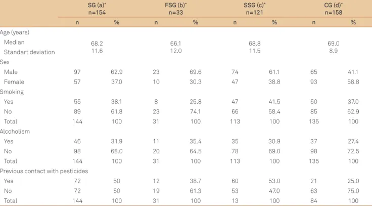
Table 2 shows socio-demographic data such as age, sex and lifestyle (tobacco use, alcoholism and previous contact with pesticides). Age was similar among groups (p>0.05), whereas male prevailed in SG (62.9%), FSG (69.6%) and SSG
(61.1%), compared with CG (41.1%; p=0.002; p=0.005; p=0.001,
respectively). Tobacco use and alcoholism had low frequency and were similar among the groups (p>0.05). On the other hand, patients were more exposed to pesticides (50%) than
controls (25%; p=0.0004), as well as SSG (53.0%) compared with CG (25%; p=0.0001), meanwhile FSG (38.7%) showed no signiicant diference from CG (25%; p=0.226).
Tables 3 to 5 present the groups distribution according to genetic variants and environmental factors. SG, SSG and CG were similar concerning tobacco use (Table 3) and alco-holism (Table 4). However, in FSG and smoker patients there
Figure.GSTP1-Alw 26I electrophoresis.
GSTP1- Alw26I FSG (a)*
n=33
SSG (b)*
n=121
SG (c)*
n=154
CG (d)*
n=158
Genotype n % n % n % n %
I/I 21 63.6 46 38.0 67 43.5 53 33.5
I/V 10 30.3 68 56.1 78 50.6 98 62.0
V/V 2 6.0 7 5.7 9 5.8 7 4.4
Total 33 100 121 100 154 100 158 100
Allele n AF n AF n AF n AF
I 52 0.78 160 0.66 212 0.68 204 0.64
V 14 0.21 82 0.33 96 0.31 112 0.35
Total 66 1.00 242 1.00 308 1.00 316 1.00
Table 1. Genotypic and allelic frequencies for GSTP1-Alw26I in patients with Parkinson’s disease, grouped in study group, familial or sporadic, and individuals without the disease, or controls.
Genotype
SG (a) FSG (b) SSG (c) CG (d)
p-value
T nT T nT T GEE
nT T nT
n % n % n % n % n % n % n % n % axd bxd cxd bxc
I/I 24 43.6 40 7.8 6 75 13 56.5 18 38.9 26 39.3 16 32.0 26 30.5 0.950 0.839 0.963 0.676 I/V 29 52.7 42 47.1 2 25 8 34.7 27 57.4 35 53.0 34 68.0 53 62.3 0.950 0.313 0.705 0.297
V/V 2 3.6 7 44.9 0 0 2 8.6 2 4.2 5 7.5 0 0 6 7.0 0.485 0.461 1.000
Total 55 100 89 100 8 100 23 100 47 100 66 100 50 100 85 100
FSG: familial Parkinson’s disease study group; SSG: sporadic Parkinson’s disease study group; SG: study group; CG: control group; T: smokers; nT: no smokers; Smokers intra-group analysis (II x -/V) – axd: SGxCG=0.305; bxd: FSGxCG=0.043; cxd: SSGxCG=0.662; bxc: FSGxSSG=0.067.
p-value (χ2 or Fisher): significance for p<0.05.
Table 3. Distribution of Parkinson’s disease patients, grouped in study group, familial or sporadic, and individuals without the disease, smokers and no smokers, according to their genotypes for GSTP1-Alw26I.
Genotype
SG (a) FSG (b) SSG (c) CG (d)
p-value
A nA A nA A nA A nA
n % n % n % n % n % n % n % n % axd bxd cxd bxc
I/I 22 47.8 42 42.8 8 72.7 11 55 14 40 30 38.4 12 32.4 30 30.6 0.679 0.454 0.926 0.618 I/V 20 43.4 51 52.4 2 18.1 8 40 18 51.4 44 54.4 25 67.5 62 63.2 0.937 0.721 0.968 0.715 V/V 4 8.6 5 5.1 1 9.0 1 5 3 8.5 4 5.1 0 0 6 6.1 0.103 0.250 0.192 1.000 Total 46 100 98 100 11 100 20 100 35 100 78 100 37 100 98 100
Table 4. Distribution of Parkinson’s disease patients, grouped in study group, familial or sporadic, and individuals without the disease, alcoholics and no alcoholics, according to their genotypes for GSTP1-Alw26I.
FSG: familial Parkinson’s disease study group; SSG: sporadic Parkinson’s disease study group; SG: study group; CG: control group; A: alcoholics; nA: no alcoholics; Alcoholics intra-group analysis (II x -/V) – axd: SGxCG=0.232; bxd: FSGxCG=0.033; cxd: SSGxCG=0.672; bxc: FSGxSSG=0.082. p-value (χ2 or Fisher): significance
for p<0.05.
SG (a)*
n=154
FSG (b)*
n=33
SSG (c)*
n=121
CG (d)*
n=158
n % n % n % n %
Age (years)
Median 68.2
11.6 66.112.0 68.811.5 69.08.9
Standart deviation Sex
Male 97 62.9 23 69.6 74 61.1 65 41.1
Female 57 37.0 10 30.3 47 38.8 93 58.8
Smoking
Yes 55 38.1 8 25.8 47 41.5 50 37.0
No 89 61.8 23 74.1 66 58.4 85 62.9
Total 144 100 31 100 113 100 135 100
Alcoholism
Yes 46 31.9 11 35.4 35 30.9 37 27.4
No 98 68.0 20 64.5 78 69.0 98 72.5
Total 144 100 31 100 113 100 135 100
Previous contact with pesticides
Yes 72 50 12 38.7 60 53.0 21 25.0
No 72 50 19 61.3 53 47.0 63 75.0
Total 144 100 31 100 13 100 84 100
FSG: familial Parkinson’s disease study group; SSG: sporadic Parkinson’s disease study group; SG: study group; CG: control group; axd: SGxCG; bxc: FSGxSSG; bxd: FSGxCG; cxd: SSGxCG; p-value: Age – axd=0.479; bxc=0.54; bxd=0.196; cxd=0.830; Sex – axd=0.0002; bxc=0.485; bxd=0.005; cxd=0.001; Smoking – axd=0.939; bxc=0.163; bxd=0.330; cxd=0.547; Alcoholism – axd=0.485; bxc=0.795; bxd=0.499; cxd=0.634; Pesticides – axd=0.0004; bxc=0.223; bxd=0.226; cxd=0.0001. χ2: or Fisher’s tests, p<0.05 significance.
was a 75% prevalence of wild homozygote genotype (I/I),
compared with GC (32%; p=0.043). I/I genotype also pre
-vailed in FSG and alcoholic patients (FSG=72.7%; CG= 27.4%; p=0.033). Heterozygote genotype (I/V) combined with pesti
-cides prevailed in SG (60.5%=43 exposed patients/71 hetero -zygote patients, versus CG=24.0%=13 exposed individuals/54 heterozygote individuals; p= 0.0001). Likewise, I/V presented
higher frequency combined with exposure to pesticides in
SSG (59.6%=37 exposed patients/62 heterozygote patients); and FSG (60%=6 exposed patients/10 heterozygote patients), compared with GC (24.0%=13 exposed individuals/54 het
-erozygote individuals; p=0. 0002 and p=0.053, respectively).
Earlier PD or late PD patients presented similar distri-bution for GSTP1-Alw26I genotype (p>0.05), also when com-bined with tobacco use and alcoholism (p>0.05). On the oth-er hand, hetoth-erozygote genotype (I/V) prevailed in patients with earlier PD and previous exposure to pesticides (90%), compared with late PD, also exposed (63.9%), with no
signif-icant diference between groups, though (p=0.092). Results
were also similar considering genotype distribution between
≤68 and >68 years patients (p>0.05).
he logistic regression analysis for sex, age, tobacco use, alcoholism, exposure to pesticides and GSTP1-Alw26I
genotypes (Logit Y= -0.397997 +1.031093 sex +1.808364 age -0.562085 smoking -0.130537 alcoholism +1.109846 pes
-ticide -0.654763 genotype) pointed male sex (p=0.0034), age >68 years (p<0.0001) and exposure to pesticides (p=0.001) as
risk factors for PD.
DISCUSSION
In this study, with mixed ethnic Brazilian casuistics, the
GSTP1-Alw26I polymorphism genotypic distribution is sim-ilar to that in the general population, considering distinct racial groups8,13,14. Heterozygote genotype (I/V) prevailed
among familial or sporadic PD patients, and also in the con-trol group. his distribution, although supported by some au-thors8, is diferent from other studies4,14,22. he V/V
homozy-gote did not show any association with PD, which was also reported by other authors8,14. he presence of the Val105 (V)
allelic variant of GSTP1 is associated with the decrease of the
enzyme activity, which would favor dopaminergic neurons degeneration in PD4,15. However, in this study, the wild
geno-type (I/I) presented association with familial PD. Studies in-volving GSTP1 polymorphisms and familial PD are scarce9,
making population comparisons diicult.
In this study, the familial PD group exhibited the pat-tern predicted by Hardy–Weinberg equilibrium, different from the pattern of other groups studied, which is also ob-served in other case-control studies of different genetic polymorphisms23,24. The selection criteria adopted in this
study was to form groups of older individuals, since PD mainly affects elderly patients. Furthermore, the disease is associated with occupational exposure to pesticides, which is more common in males. This was confirmed by the logistic regression analysis that demonstrated male sex, age >68 years and exposition to pesticides as risk fac-tors for PD. Therefore, the profiles of patient and control groups do not represent the general population concern-ing sex and age, influencconcern-ing the distribution of genotypes. FSG also included younger patients, suggesting better representation of the general population. Additionally, ab-sence of Hardy-Weinberg equilibrium would be expected for a wide group of genetic diseases, considering the gene contribution — although modest — for complex diseas-es. However, given the numerous candidate gene stud-ies in different cases, genetic markers showing disequi-librium are scarce, which allows investigators to ignore the distribution of genotypes suggesting disequilibrium, therefore ignoring valuable information to identify casual polymorphisms23.
he polymorphism GSTP1-Alw26I, when related to to-bacco use and alcoholism, was the same in the SG, SSG and CG groups, as well as in other studies involving smok-ing and PD10,22,25. On the other hand, there are references of
tobacco protection in PD, including haplotypes of GSTP110.
he catalytic eiciency of the GSTP1 variants difers from that of the wild type, and it varies according to the charac-teristics of the substrates, manly distinct26. his explains the
protective efect of the mutant allele regarding diol epoxides found in tobacco products10, in contrast with its reduced
ef-fects upon detoxiication of pesticides7. In this study, there
was an association between smoking and wild homozygote
Genotype
SG (a) FSG (b) SSG (c) CG (d)
p-value
P nP P nP P nP P nP
n % n % n % n % n % n % n % n % axd bxd cxd bxc
I/I 23 31.9 41 56.9 5 41.6 14 73.6 18 30.0 26 49.0 7 33.3 18 28.5 0.643 0.901 0.416 0.393 I/V 43 59.7 28 38.8 6 50 4 21.0 37 61.6 25 47.1 13 61.9 41 65.0 0.0001 0.053 0.0002 1.000 V/V 6 8.3 3 4.1 1 8.3 1 5.2 5 8.3 2 3.77 1 4.7 4 6.3 0.265 1.000 0.242 1.000 Total 72 100 72 100 12 100 19 100 60 100 53 100 21 100 63 100
Table 5. Distribution of Parkinson’s disease patients, grouped in study group, familial or sporadic, and individuals without the disease, with or without previous contact with pesticides, according to their genotypes for GSTP1-Alw26I.
genotype in familial PD patients, conirming the studies by Pal et al.26 and De Palma et al.10.
Likewise, association between I/I genotype and alcohol-ism was found in familial PD patients. Admittedly, alcohol abuse damages brain structures and their functions, which leads to neurodegeneration27. he efects of alcohol in the
brain are not uniform, afecting mainly the pre- frontal cor-tex, the hippocampus, the cerebellum, the substantianigra
and the glia27. Dopaminergic neurons in the substantia
nig-ra are believed to be damaged by alcohol during intnig-rauterine development27,28. he efect of alcohol in this region is still
unclear concerning brains are already developed28. Studies
involving GSTs, alcohol, substantia nigra and PD are rare. On the other hand, GST activity is known to be reduced in hepatocytes due to alcohol exposure29. Retinoid X receptor
α-deicient (RXRα KO) mice, which are more susceptible to ethanol-induced hepatotoxicity, showed a 56% decrease in
GSTP1 activity, which demonstrates its role in the detoxii-cation of alcohol in the liver29. herefore, GSTP1
polymor-phisms seem to intensify the damage caused by ethanol in hepatocytes, leading to alcoholic cirrhosis and pancreatitis19.
he present study revealed the combination of the wild geno-type (I/I) with alcoholism in familial PD patients. he small casuistic of familial patients analyzed in this study speaks in favor of detailed studies involving familial PD patients, GSTP1
polymorphisms and lifestyle.
PD patients group showed more exposure to pesticides, especially concerning the combination of such exposure and the heterozygote genotype (I/V). he GST enzymes, mainly
GSTP14,7, are involved in metabolizing pesticides8. Studies
demonstrated that some kinds of pesticides, like rotenone, can cause PD-like symptoms30, acting as inhibitors of
mito-chondrial I complex. In this case, there is some evidence that dopaminergic neurons are particularly vulnerable to mito-chondrial dysfunction30. he toxin is captured by dopamine
and noradrenalin transporters and is accumulated in the cy-tosol, causing cellular death induced by oxidative stress and deiciency of the breathing mitochondrial chain30. Shi et al.15
demonstrated protection of the neuronal dopaminergic cells of mice exposed to rotenone by the expression of the wild
GSTP1 (I allele), and reduced protection by its variants ex-pression (V allele). hese data were conirmed in the present study, by means of the association of GSTP1-Alw26I heterozy-gote, exposure to pesticides and PD.
he V allele have been associated to late onset PD, given its prevalence in patients with more than 68 years of age4,15,
de-spite the lack of association between GSTP1 polymorphisms and PD onset age in North American casuistics14. In this study,
there was no association of GSTP1-Alw26I genotypes, current age and PD onset age. However, considering onset age, previous exposure to pesticides and the presence of the mutant allele of
GSTP1-Alw26I, the group with earlier PD (PD onset before than 50 years) was signiicant, if compared with those with late PD onset age, which requires conirmation in wide casuistics.
The regulation of the apoptotic kinase c–jun termi-nal ( JNK) by protein–protein direct binding and the de-toxification of electrophilic compounds by conjugation with reduced glutathione have been the mainly reported
GSTP1 functions15. The former mechanism could be
asso-ciated to late onset PD in GSTP1 heterozygote patients. The GSTP1 enzyme would play an important role in the thermal shock protein system — also responsible for regu-lating JNK apoptosis4 failed to act due to aging. Shi et al.15
demonstrated that in the brains of mice treated with rote-none there was no activation of the JNK pathway of apop-tosis. High level of oxidative stress was detected though, and GSTP1 detoxification with reduced glutathione was essential for neuron protection in this case15. That would
explain the possible relation suggested in this study, be-tween heterozygote genotype for GSTP1 in patients with previous exposure to pesticides, and the earlier PD, to be confirmed in further studies.
In conclusion, this study conirms the association among PD, male sex, ageing and exposure to pesticides, whose ef-fects can be enhanced in combination with I/V genotype of
GSTP1-Alw26I, reinforcing the relation between genetic poly-morphisms involved in xenobiotics metabolism and environ-mental factors in PD. his is conirmed by the prevalence of the I/I genotype and tobacco or alcohol use only in the FSG, suggesting that diferent efects of GSTP1-Alw26I variants, due to lifestyle, determine speciic subgroups of patients, which may be conirmed in other casuistics.
ACKNOWLEDGMENTS
he authors would like to thank Moacir Fernandes de Godoy for assistance with the statistical analysis.
1. Banco de Dados do Sistema Único de Saúde (DATASUS). Brazilian Health System Official Data 2010. [cited 2013 Apr]. Available at: http:// portal.saude.gov.br/portal/arquivos/pdf/pcdt_doenca_parkinson_ livro_2010.pdf
2. Ischiropoulos H, Beckman JS. Oxidative stress and nitration in neurodegeneration: cause, effect, or association? J Clin Invest 2003;111:163-169.
3. Wood-Kaczmar A, Gandhi S, Wood NW. Understanding the molecular causes of Parkinson’s disease. Trends Mol Med 2006;12:521-528.
4. Vilar R, Coelho H, Rodrigues E, et al. Association of A313 G polymorphism (GSTP1*B) in the glutathione–S-transferase P1 gene with sporadic Parkinson’s disease. Eur J Neurol 2007; 14:156-161.
5. Taningher M, Malacarne D, Izzotti A, Ugolini D, Parodi S. Drug metabolism polymorphisms as modulators of cancer susceptibility. Mutat Res 1999;436:227-261.
6. Board P, Harris M, Flanagan J, Langton L, Coggan M. Genetic heterogeneity of the structure and function of GSTT2 and GSTP1. Chem Biol Interact 1998;111-112:83-89.
7. Menegon A, Board PG, Blackburn AC, Mellick GD, Le Couteur DG. Parkinson’s disease, pesticides, and glutathione transferase polymorphisms. Lancet 1998;352:1344-1346.
8. Kiyohara C, Miyake Y, Koyanagi M, et al. GST polymorphisms, interaction with smoking and pesticide use, and risk for Parkinson’s disease in a Japanese population. Parkinsonism Relat Disord 2010;16:447-452.
9. Wilk JB, Tobin JE, Suchowersky O, et al. Herbicide exposure modifies GSTP1 haplotype association to Parkinson onset age: the GenePD Study. Neurology 2006;67: 2206-2210.
10. De Palma GD, Dick FD, Calzetti S, et al. Geoparkinson Study Group. A casecontrol study of Parkinson’s disease and tobacco use: gene-tobacco interactions. Mov Disord 2010;25:912-919.
11. Board PG, Webb GC, Coggan M. Isolation of a cDNA clone and localization of the human glutathione S-transferase 3 genes to chromosome bands 11q13 and 12q13–14. Ann Hum Genet 1989;53:205-213.
12. Ali-Osman F, Akande O, Antoun G, Mao JX, Buolamwini J. Molecular cloning, characterization, and expression in Escherichia coli of full-length cDNAs of three human glutathione S-transferase Pi gene variants. Evidence for differential catalytic activity of the encoded proteins. J Biol Chem 1997;272:10004-10012.
13. Chan QKY, Khoo U, Ngan HYS, et al. Single nucleotide polymorphism of Pi-class glutathione S-transferase and susceptibility to endometrial carcinoma. Clin Cancer Res 2005;11:2981-2985.
14. Kelada SN, Stapleton PL, Farin FM, et al. Glutathione S-transferase M1, T1, and P1 polymorphisms and Parkinson’s disease. Neurosci Lett 2003;337:5-8.
15. Shi M, Bradner J, Bammler TK, et al. Identification of glutathione S-transferase pi as a protein involved in Parkinson disease progression. Am J Pathol 2009;175:54-65.
16. Parra FC, Amado RC, Lambertucci JR, Rocha J, Antunes CM, Pena SD. Color and genomic ancestry in Brazilians. Proc Natl Acad Sci USA 2003;100:177-182.
17. Pankratz ND, Wojcieszek J, Foroud T. Parkinson Disease Overview. 2009. [cited 2011 Dec]. Available at: http://www.ncbi.nlm.nih.gov/ books/NBK1223/
18. Jankovic J. Parkinson’s disease: clinical features and diagnosis. J Neurol Neurosurg Psychiatry 2008;79:368-376.
19. Burim RV, Canalle R, Martinelli Ade L, Takahashi CS. Polymorphisms in glutathione S-transferases GSTM1, GSTT1 and GSTP1 and cytochromes P450 CYP2E1 and CYP1A1 and susceptibility to cirrhosis or pancreatitis in alcoholics. Mutagenesis 2004; 19:291-298.
20. Salazar LA, Hirata MH, Cavalli SA, Machado MO, Hirata RD. Optimized procedure for DNA isolation from fresh and cryopreserved clotted human blood useful in clinical molecular testing. Clin Chem 1998;44:1748-1750.
21. Ishii T, Matsuse T, Teramoto S, et al. Glutathione S-transferase P1 (GSTP1) polymorphism in patients with chronic obstructive pulmonary disease. Thorax 1999;54:693-696.
22. Wahner AD, Glatt CE, Bronstein JM, Ritz B. Glutathione S-transferase mu, omega, pi, and theta class variants and smoking in Parkinson’s disease. Neurosci Lett 2007;413:274- 278.
23. Xu J, Turner A, Little J, Bleecker ER, Meyers DA. Positive results in association studies are associated with departure from Hardy-Weinberg equilibrium: hint for genotyping error? Hum Genet 2002;111:573-574.
24. Wittke-Thompson JK, Pluzhnikov A, Cox NJ. Rational inferences about departures from Hardy-Weinberg equilibrium. Am J Hum Genet 2005;76:967-986.
25. Sugita M, Izuno T, Tatemichi M, Otahara Y. Meta-analysis for epidemiologic studies on the relationship between smoking and Parkinson’s disease. J Epidemiol 2001;11:87-94.
26. Pal A, Desai DH, Amin S, et al. Location of the epoxide function determines specificity of the allelic variants of human glutathione transferase Pi toward benzo[c]chrysene diol epoxide isomers. FEBS Lett 2000;486:163-166.
27. Alfonso-Loeches S, Guerri C. Molecular and behavioral aspects of the actions of alcohol on the adult and developing brain. Crit Rev Clin Lab Sci 2011;48:19-47.
28. Grinfeld H. What effects can be expected of prenatal ethanol exposure in pregnant mice and their offspring? Einstein 2004;2:187-192.
29. Gyamfi MA, Wan YJ. The effect of ethanol, ethanol metabolizing enzyme inhibitors, and Vitamin E on regulating glutathione, glutathione S-transferase, and S-adenosylmethionine in mouse primary hepatocyte. Hepatol Res 2006;35:53-61.