To tal and se gm e ntal co lo nic transit tim e
in co nstipate d patie nts with Chagas’
dise ase witho ut m e gae so phagus o r
m e gaco lo n
1Disciplina de Gastroenterologia, Departamento de Clínica Médica and 2Departamento de Radiologia, Faculdade de Ciências Médicas,
Universidade Estadual de Campinas, Campinas, SP, Brasil S.L. Santos1,
I.K. Barcelos2 and
M.A. Mesquita1
Abstract
Manometric and pharmacological tests have shown that motor abnor-malities may occur in the non-dilated colons of chagasic patients. In order to investigate the presence of abnormalities of colonic function in constipated patients with Chagas disease (ChC) without megae-sophagus or megacolon, studies of total and segmental colonic transit time with radiopaque markers were performed on 15 ChC patients, 27 healthy volunteers and 17 patients with idiopathic constipation (IC). The values obtained for the control group were similar to those reported in the literature (total colonic time: 34.1 ± 15.6 h; right colon: 9.9 ± 7.3 h; left colon: 10.8 ± 10 h, and rectosigmoid: 12.6 ± 9.9 h). Colonic transit time data permitted us to divide both IC and ChC patients into groups with normal transit and those with slow colonic transit. Colonic inertia was detected in 41% of IC patients and in 13% of ChC patients; left colon isolated stasis (hindgut dysfunction) was detected in 12% of IC patients and 7% of ChC patients, and outlet obstruction was detected in 6% of IC patients and 7% of ChC patients. There were no significant differences in total or segmental colonic transit times between slow transit IC and slow transit ChC patients. In conclusion, an impairment of colonic motility was detected in about 30% of constipated patients with Chagas disease without megae-sophagus or megacolon. This subgroup of patients presented no distinctive clinical feature or pattern of colonic dysmotility when compared to patients with slow transit idiopathic constipation.
Co rre spo nde nce
M.A. Mesquita
Disciplina de Gastroenterologia Departamento de Clínica Médica FCM, UNICAMP
Caixa Postal 6111 13081-970 Campinas, SP Brasil
Fax: + 55-19-289-1682 Publication supported by FAPESP.
Received September 28, 1998 Accepted September 27, 1999
Ke y wo rds
·Colonic transit
·Chagas’ disease
·Constipation
·Radiopaque markers
·American trypanosomiasis
Intro ductio n
Chronic intestinal constipation is one of the most frequent complaints in clinical prac-tice (1). In countries like Brazil, where Chagas disease is endemic, constipation may reflect disorders of colorectal motility in-duced by the intrinsic denervation seen in
esophagus (8), the study of colonic motility may identify functional abnormalities in a stage preceding colon dilation.
The assessment of total and segmental colonic transit time (CTT) with ingested ra-diopaque markers (9,10) has been widely employed to evaluate colorectal function in constipated patients. It is a simple noninva-sive technique, readily applicable within the clinical context, which allows patients to be classified into four groups: intestinal consti-pation with normal colonic transit time, co-lonic inertia (prolonged transit time through-out the colons, especially the right colon), hindgut dysfunction (prolonged transit time in the left colon, but normal in the right one), and outlet obstruction (prolonged transit time in the rectosigmoid but normal in the re-maining areas).
In the present study, this technique was used to evaluate constipated patients with Chagas disease (ChC) without megaesopha-gus or megacolon in order to investigate: 1) whether abnormalities of colonic function could be demonstrated at this stage of the disease and 2) to compare these abnormali-ties with those found in a group of patients with idiopathic intestinal constipation (IC).
Mate rial and Me tho ds
Study po pulatio n
Table 1 shows the clinical features of the study population. Informed written consent was obtained from all subjects involved in the study. The protocol was approved by the Ethics Committee of the Hospital das Clínicas, UNICAMP.
Control group. Twenty-seven healthy vol-unteers (14 males and 13 females) were recruited from the medical and paramedical staff. The selection criteria included a stool frequency of 2 stools/day to 3 stools/week, no difficulty in passing stools and normal stool consistency. No subject was taking any medication known to affect gastrointestinal
motility or reported changes in bowel move-ments induced by stress. No volunteer had an epidemiological background for Chagas disease.
Patients with IC. This group consisted of 17 patients (16 females and 1 male) who were referred to our outpatient clinic for evaluation of chronic constipation, defined as less than 3 stools/week and passing of hard stools for at least one year. All patients had barium enema to document the absence of any organic colonic lesions and serologic tests to exclude Chagas disease. There was no clinical evidence of diabetes mellitus, collagen disease or neurological disorders.
Patients with ChC. These patients were selected from a group regularly followed up at GEDOCH (Chagas disease study group) with positive serologic reactions (comple-ment fixation test and/or immunofluores-cence reaction) for Chagas disease, with no evidence of heart failure, megaesophagus or megacolon, with the latter two being con-firmed by esophagogram and barium enema. Fifteen consecutive patients (10 females and 5 males) who also fulfilled the criteria for a diagnosis of chronic constipation in a sepa-rate interview assessing specifically their bowel habits were enrolled in the study. There was no clinical evidence of diabetes mellitus or collagen disease in this group. The symptoms of constipation were similar to those reported by IC, but fewer patients reported the use of laxatives to promote evacuation (Table 1).
Co lo nic transit studie s
Total and segmental CTT was assessed as previously described by Metcalf et al. (10). Patients were asked to maintain their usual diet and received a fiber supplement of 20 g/day (Metamucil®
partici-pants ingested 24 radiopaque markers con-tained within a gelatin capsule (SITZMARKS®
, Konsyl Pharmaceuticals Inc., Fort Worth, TX, USA) at the same time, after breakfast, on three consecutive days. An abdominal X-ray was taken on days 4 and 7 at the time of marker ingestion. Markers were identified and counted on the abdominal films. The right colon, left colon and rectosigmoid were recognized by bony landmarks and/or by their clear identification due to gas shadows as described by Arhan et al. (9). The spinal processes and imaginary lines from the fifth lumbar vertebra to the pelvic outlet served as landmarks.
Total and segmental CTT were then cal-culated using the following formula:
where Dt = mean transit time, T = time interval between X-rays, N = number of ingested markers, j = number of X-rays taken, and ni = total number of markers present on a given film sector at time ti. Since the ab-dominal X-rays were always taken at 72-h intervals and the total number of markers was kept constant at 72, the above formula can be simplified to:
Statistical analysis
Results are reported as mean ± SD. The upper limits of normality of colonic transit times were considered as the mean + 2 SD. The results were analyzed by the Mann-Whitney test for comparison of quantitative data and by the chi-square test or Fishers exact test for comparison of qualitative data. Differences were considered statistically sig-nificant when P<0.05.
Re sults
Table 2 shows the mean values of total
and segmental CTT for controls and pa-tients.
Co ntro l gro up
The upper limits of normality for total and segmental CTT were: total colonic tran-sit time = 65 h; right colonic trantran-sit time = 25 h; left colonic transit time = 31 h, and rec-tosigmoid transit time = 32 h. No statistically
Table 1 - Clinical features of the study population.
IC, Idiopathic constipation; ChC, Chagas’ disease constipation. * P<0.05 vs controls and * * P<0.05 vs ChC (c2 and Fisher’s exact tests).
Variables IC (N = 17) ChC (N = 15) Control (N = 27)
M ean age ± SD (years) 35 ± 11 48 ± 11* 41 ± 12
Gender (% females) 94.2* 66.6* 48.1
Symptoms duration (years) >7 >10
-Bow el movements/w eek 1 1-2 3-14
Straining (% ) 71 80
-Hard stools (% ) 94 93
-Laxative use (% ) 100* * 66.6
-Table 2 - Total and segmental colonic transit time (CTT) in patients w ith idiopathic constipation (IC) and constipated patients w ith Chagas’ disease (ChC).
Data are reported as means ± SD in hours. In patients w ith slow transit constipation the values of total CTT are above the upper limits of normality. TCTT: Total CTT; RCTT: right CTT; LCTT: left CTT, and RSTT: rectosigmoid transit time.
TCTT RCTT LCTT RSTT
Controls 34.1 ± 15.6 9.9 ± 7.4 10.8 ± 10 12.6 ± 10
Upper normal limits 65 25 31 32
Normal transit IC 39.2 ± 17 18.1 ± 15 12.2 ± 13.1 9 ± 6
Normal transit ChC 42.5 ± 15.1 11.0 ± 10 12.8 ± 10 17.9 ± 13
Slow transit IC 110.4 ± 30.4 36.4 ± 27.3 46.0 ± 29 28 ± 24.6
Slow transit ChC 100.2 ± 25.6 25.7 ± 17.3 45.2 ± 41.7 29.2 ± 31.2
Table 3 - Effect of age and gender on total and segmental colonic transit times (CTT) in 27 healthy volunteers.
Results are reported as mean ± SD hours. For abbreviations see legend to Table 2.
TCTT RCTT LCTT RSTT
M ale (N = 14) 30.7 ± 17.1 9.4 ± 6.9 8.7 ± 11.4 12.1 ± 9.9
Female (N = 13) 37.7 ± 13.5 7.0 ± 8.1 13.5 ± 8.1 14.5 ± 10.2
Age >40 years (N = 8) 32.3 ± 11 13.2 ± 6.5 10.4 ± 9.7 11.5 ± 12.2
when a decrease of markers was observed on the X-ray. IC and ChC patients were divided according to total CTT into those with nor-mal transit and those with slow transit, the latter when total CTT was longer than 65 h (Figure 1). Slow colonic transit was shown in 65% of IC (11 out of 17, 10 females) and 27% of ChC (4 out of 15, 3 females; P = 0.03). There was no difference between slow transit IC and slow transit ChC patients in total CTT (Table 2). In addition, all patients with prolonged transit reported the chronic use of laxatives.
Segmental CTT. The study of the seg-mental CTT classified slow colonic transit patients into the subgroups previously de-fined (Figures 2-4). Among IC patients, 41% (7 of 17) had a pattern of colonic inertia, 12% (2) had hindgut dysfunction, 6% (1) had outlet obstruction, and 6% (1) showed a prolonged total CTT with normal segmental transit time. Among ChC patients, 13% (2) showed a pattern of colonic inertia, 7% (1) had hindgut dysfunction and 7% (1) had outlet obstruction.
D iscussio n
The study of total and segmental colonic transit times with radiopaque markers proved to be a simple, practical and reliable method. The results obtained for our control group are comparable to those described in the literature, including a Brazilian study (11). No significant difference in colonic transit times was found between our male and fe-male controls. Gender effect on colonic tran-sit time has been controversial. Many stud-ies have reported a more prolonged colonic transit in women (10,11). However, these differences have been recently attributed to subject selection (12,13). It has been sug-gested that many women recruited as healthy controls may suffer from irritable bowel syn-drome. It is possible that our routine of ask-ing subjects about changes in bowel habits induced by stress contributed to our findings
Figure 1 - Total colonic tran-sit time (TCTT) in patients w ith idiopathic constipation (IC) and constipated pa-tients w ith Chagas’ disease (ChC) w ithout megacolon. The horizontal dotted line indicates the upper limits of normality of the test. Both IC and ChC have been di-vided into those w ith nor-mal transit and those w ith slow transit on the basis of the dotted line.
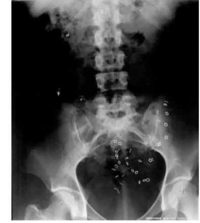
Figure 2 - Colonic inertia in a constipated patient w ith Chagas’ disease (ChC). An X-ray obtained on day 7 show s stasis of the radiopaque markers in the ascending colon.
T
C
T
T
(
h
)
150
120
90
60
30
0
Controls IC ChC
significant effect of age or gender on colonic transit times was demonstrable (Table 3), although there was a trend for women to present a slower left colon transit than men.
Patie nt gro ups
Figure 4 - Outlet obstruction in a patient w ith idiopathic constipation (IC). An X-ray taken on day 4 show s aggregation of markers in the rectosigmoid. The same pattern w as observed on day 7.
of similar results for men and women. Age also did not influence colonic transit time, confirming previous reports (10).
Colonic transit of radiopaque markers divided both IC and ChC patients into those with normal CTT and those with prolonged CTT. The complaints of constipation in pa-tients with normal transit time have been associated either with a low fiber diet or with psychological disorders (14). Since the in-gestion of fibers varied among our patients, and considering that a fiber supplement may be helpful to exclude a low fiber intake as a cause of constipation (12), the diets of our patients were enriched with 20 g of fiber. Although it seems reasonable to assume that most of our patients with normal transit were constipated for dietary reasons, it is difficult to determine it from our data, since previous CTT studies without fiber supplement had
not been performed on these patients. Like-wise, our study was not designed to assess the role of psychological factors in these patients.
Although the symptoms of constipation were similar for all patients of both groups, slow colonic transit was observed only in 27% of ChC patients and 65% of IC patients. These findings agree with several studies which have demonstrated the limited useful-ness of clinical symptoms and physical ex-amination in distinguishing constipated pa-tients with normal or delayed colonic transit (15). Therefore, measurements of colonic transit time provide an objective assessment for this heterogeneous condition.
The proportion of slow colonic transit observed among our ChC patients was simi-lar to that reported by Pemberton et al. (16) who found a demonstrable abnormality in
29% of 277 patients with constipation seen at the Mayo Clinic. However, the frequency of colonic transit dysfunction reported in the literature varies widely (30-80%) (17). It has been suggested that these abnormalities may be more frequent among constipated patients referred to tertiary gastroenterology prac-tices. This might be one explanation for the higher frequency of slow transit found a-mong our IC patients, all of them referred to our clinic for the evaluation of constipation. One possible pathogenic mechanism for prolonged colonic transit in ChC patients may be the colonic denervation due to Chagas disease. Alternatively, slow transit in these patients may be associated with the same pathogenic factors related to idiopathic constipation. Abnormalities of the myen-teric and submucosal plexuses have been shown in colonic tissue of patients with idio-pathic constipation submitted to colectomy (18). One of the factors implicated in the enteric neuropathy shown by those patients is the long-term use of laxatives (19). It is noteworthy that the chronic use of laxatives was reported by all IC and ChC patients with slow colonic transit.
Moreover, slow transit patients of both groups also had in common the female pre-ponderance and the low rate of delayed rec-tosigmoid transit. Our data agree with those of studies of idiopathic constipation which have shown that slow colonic transit is al-most exclusively confined to women (20,21). On the other hand, in contrast to the higher proportion of females found in our ChC group, the proportion of males and females is almost the same among patients with cha-gasic megacolon (22).
Colonic inertia was the main pattern of colonic dysfunction, observed in 43% and 13% of IC and ChC patients, respectively, followed by hindgut dysfunction and outlet
obstruction. Based on the knowledge of the colonic involvement in Chagas disease, which shows a preferential dilation of rec-tosigmoid (23), a delayed recrec-tosigmoid tran-sit might be expected as the main dysfunc-tion in our ChC patients. However, despite the small number of ChC patients with slow transit, the abnormalities detected apparently followed the same distribution as seen for IC patients: colonic inertia, hindgut dysfunc-tion and outlet obstrucdysfunc-tion, the latter present in only one chagasic patient.
It is tempting to assume from the data discussed above that the colonic dysfunction found in this particular group of patients with Chagas disease may be related to the same underlying factors responsible for id-iopathic constipation, such as the denerva-tion induced by laxative abuse. Our selec-tion of chagasic patients excluded those with esophagopathy, a fact that may have ac-counted for the low rate of rectosigmoid transit abnormalities in this group. It is known from Chagas disease natural history that megacolon is recognized later than megae-sophagus, due to either a slower evolution or to a delay in seeking medical care (22). However, previous pharmacological studies investigating denervation (4) have reported that 20% of cases of nondilated sigmoid colon in patients with megaesophagus showed hyper-reactivity to metacholine. Therefore, additional studies of colonic tran-sit time in patients with megaesophagus are indicated.
Ackno wle dgm e nts
Re fe re nce s
1. Drossman DA, Sandler RS, M cKee DC & Lovitz AJ (1982). Bow el patterns among subjects not seeking health care. Use of a questionnaire to identify a population w ith bow el dysfunction. Gastroenterology, 83: 629-634.
2. M eneghelli UG (1985). Chagas’ disease: a model of denervation in the study of di-gestive tract motility. Brazilian Journal of M edical and Biological Research, 18: 255-264.
3. M acedo JFS, M eneghelli UG, Oliveira RB, Godoy RA, Troncon LEA & Dantas RO (1986). Effect of CCK-OP and intraduode-nal administration of essential amino ac-ids on intraluminal pressures of sigmoid and rectum in patients w ith chagasic megacolon. Digestive Diseases and Sci-ences, 31: 145-150.
4. Vieira CB, Godoy RA, M eneghelli UG & Carril CF (1966). Resposta do cólon sig-móide não ectásico à metacolina na forma crônica da moléstia de Chagas. Arquivos de Gastroenterologia, 3: 21-26.
5. M eneghelli UG, Godoy RA, Oliveira RB, Santos JCM , Dantas RO & Troncon LEA (1983). Effect of pentagastrin on the mo-tor activity of the dilated and nondilated sigmoid and rectum in Chagas’ disease. Digestion, 27: 152-158.
6. Habr-Gama A, Raia A & Correa Netto A (1970). M otility of sigmoid colon and rec-tum: Contribution to the pathophysiology of megacolon in Chagas’ disease. Dis-eases of the Colon and Rectum, 14: 291-304.
7. M eneghelli UG, Godoy RA, M acedo JFS, Oliveira RB, Troncon LEA & Dantas RO (1982). Basal motility of dilated and non-dilat ed sigm oid colon and rect um in Chagas’ disease. Arquivos de
Gastroente-rologia, 19: 127-132.
8. Oliveira RB, Rezende Filho J, Dantas RO & Iazigi N (1995). The spectrum of esoph-ageal motor disorders in Chagas’ disease. American Journal of Gastroenterology, 90: 1119-1124.
9. Arhan P, Devroede G, Jehannin B, Lanza M , Faverdin C, Dornic C, Persoz B, Tétreault L, Perey B & Pellerin D (1981). Segmental colonic transit time. Diseases of the Colon and Rectum, 24: 625-629. 10. M etcalf AM , Phillips SF, Zinsmeister AR,
M acCarty RL, Beart RW & Wolff BC (1987). Simplified assessment of segmen-tal colonic transit time. Gastroenterology, 92: 40-47.
11. Jorge JM N & Habr-Gama A (1991). Tempo de trânsito colônico total e segmentar: análise crítica dos métodos e estudo em indivíduos normais com marcadores ra-diopacos. Revista Brasileira de Coloproc-tologia, 11: 55-60.
12. Chaussade S, Roche H, Khyara A, Coutu-rier D & Guerre J (1986). M esure du temps de transit colique (TTC): descrip-tion et validadescrip-tion d’une nouvelle tech-nique. Gast roent érologie Clinique et Biologique, 10: 385-389.
13. Bouchoucha M , Devroede G, Arhan P, Strom B, Weber J, Cugnenc PH, Denis P & Barbier JP (1992). What is the meaning of colorectal transit time measurement? Diseases of the Colon and Rectum, 35: 773-782.
14. Wald A (1986). Colonic transit time and anorectal manometry in chronic idiopathic constipation. Archives of Internal M edi-cine, 14: 1713-1716.
15. Chaussade S, Khyari A, Roche H, Garret M , Gaudric M , Couturier D & Guerre J (1989). Determination of total and
seg-mental colonic transit time in constipated patients. Results in 91 patients w ith a new simplified method. Digestive Dis-eases and Sciences, 34: 1168-1172. 16. Pemberton JH, Rath DM & Ilstrup DM
(1991). Evaluation and surgical treatment of severe chronic constipation. Annals of Surgery, 214: 403-411.
17. Surrenti E, Rath DM , Pemberton JH & Camilleri M (1995). Audit of constipation in a tertiary referral gastroenterology prac-tice. American Journal of Gastroenterol-ogy, 90: 1471-1475.
18. Kamm M A (1991). Constipation. In: Kamm M A & Lennard-Jones JE (Editors), Gas-trointestinal Transit. Pathophysiology and Pharm acology. W right son Biom edical Publishing Ltd., Petersfield, UK, 133-140. 19. Riemann JF & Schmidt H (1982). Ultra-structural changes in the gut autonomic nervous system follow ing laxative abuse and in other conditions. Scandinavian Journal of Gastroenterology, 71: 111-124. 20. Preston DM & Lennard-Jones JE (1986). Severe chronic constipation of young w omen: idiopathic slow transit constipa-tion. Gut, 27: 41-48.
21. Johanson JF, Sonnenberg A & Koch TR (1989). Clinical epidemiology of chronic constipation. Journal of Clinical Gastroen-terology, 11: 525-536.
22. Rezende JM (1993). M anifestações diges-tivas da doença de Chagas. In: Dani R & Castro LP (Editors), Gastroenterologia Clínica. Guanabara Koogan, Rio de Janeiro, 1729-1755.