NAIARA VIANA CAMPOS
ARSENIC HYPERACCUMULATION IN Pityrogramma calomelanos (L.) LINK
(PTERIDACEAE): MORPHOPHYSIOLOGICAL MECHANISMS OF
TOLERANCE
Thesis presented to the Universidade Federal de Viçosa as part of the requirement of the Post-Graduate Program in Botany for obtention of the degree of Doctor Scientiae.
VIÇOSA
MINAS GERAIS – BRASIL
Fichi citilográfici prepiridi peli Biblioteci Centril di Universidide Federil de Viçosi - Câmpus Viçosi
T
Campos, Naiara Viana, 1985-C198a
2014 calomelanosArsenic hyperaccumulation in (L.) Link (Pteridaceae) : morphophysiologicalPityrogramma mechanisms of tolerance / Naiara Viana Campos. - Viçosa, MG, 2014.
xiv, 117f. : il. (algumas color.) ; 29 cm.
Orientador : Aristéa Alves Azevedo.
Tese (doutorado) - Universidade Federal de Viçosa. Inclui bibliografia.
1. Samambaias. 2. Pityrogramma calomelanos. 3. Pityrogramma calomelanos - Tolerância - Arsênio. 4. Fitorremediação. I. Universidade Federal de Viçosa. Departamento de Biologia Vegetal. Programa de Pós-graduação em Botânica. II. Título.
CDD 22. ed. 628.55
FichaCatalografica :: Fichacatalografica https://www3.dti.ufv.br/bbt/ficha/cadastrarficha/visua...
NAIARA VIANA CAMPOS
ARSENIC HYPERACCUMULATION IN Pityrogramma calomelanos (L.) LINK
(PTERIDACEAE): MORPHOPHYSIOLOGICAL MECHANISMS OF
TOLERANCE
Thesis presented to the Universidade Federal de Viçosa as part of the requirement of the Post-Graduate Program in Botany for obtention of the degree of Doctor Scientiae.
APPROVED: December 12th of 2014.
Adriano Nunes Nesi Cléberson Ribeiro
Marcelo Braga Bueno Guerra Wagner Campos Otoni
ii
This thesis is dedicated...
To my great love, Vitor,
To my parents, Lídia and Oldac,
iii
“Let nothing disturb you,
Let nothing frighten you,
All things are passing away:
God never changes.
Patience obtains all things
Whoever has God lacks nothing;
God alone suffices”.
iv
ACKNOWLEDGEMENTS
I would like to thank to Universidade Federal de Viçosa and Department of Plant Biology, for the support and the opportunity to conduct my thesis.
To CNPq and FAPEMIG, for the financial support to this research.
To my adviser, Dra. Aristéa Alves Azevedo, I sincerely thank you for allowed me to develop this project, advising and encouraging me, being patient and friendly along these almost 10 years. Your passion and dedication are source of inspiration for me. I feel grateful to be your ‘scientific daughter’.
To my co-advisers Dr. Marcelo Ehlers (DBV/UFV) and Dr. Cléberson Ribeiro (DBG/UFV) who provided invaluable support, criticisms and guidance when I needed.
To Dr. Bruno Lemos (UFABC-SP), co-adviser, for availability and assistance in the arsenic speciation analysis. And also to Dr. Fernando Barbosa Jr. who allowed me to work in the Laboratory of Toxicology and Essentiality of Metals (USP-Ribeirão Preto).
To Dr. Jaime Wargas (DPS/UFV), co-adviser, for the attention and support in the ICP OES analysis. Also to Mario, for the technical assistance with the ICP OES.
To Dr. Adriano Nunes Nesi, for all your attention, advice, and important teachings, especially in the metabolic analyses.
To Nívea Vieira who do not measure efforts to assist me with the GC-MS analysis of metabolites. You are a very special person and friend!
To all team of the Nucleus of Biomolecules Analysis (UFV) that supported the GC-MS analysis, and to Denise Fernandes who help me in several steps.
v
a good friend! I also thank to Dr. Carlos Schaefer for the partnership, and Pablo and Elton for the help in the micro-EDXRF analysis.
To Dr. Francisco Krug who allowed me to work in the Analytical Chemical Laboratory (CENA-Piracicaba) and to Tata for the technical assistance in the sample preparation.
To Karla Ribeiro and Gilmar Valente (Nucleus of Microscopy and Microanalysis/UFV) for the technical assistance, attention and friendship.
To Dra. Catarina Megumi for the permission to use the cryomicrotome and to Luana De Jesus for the friendship and assistance in this analysis.
To all teachers of the Plant Biology Department for sharing knowledge and experiences. To Dra. Luzimar Campos and Dra. Renata Meira for the encouragement and friendship. To Wagner Otoni, for your attention and help with the fern propagation.
To Dr. Fábio Da Matta and Dr. Wagner Araújo for allowed me to work in the Plant Physiology Laboratories and in the Plant Growth Unit (UCP).
To all my colleagues and friends of the Laboratory of Plant Anatomy for friendship, for the attention, friendship, for the good moments and coffee breaks! Special thanks to Samara Arcanjo, Ivan Becari, Larisse Freitas and Talita de Oliveira who have supported me in all stages, working so hard and tirelessly during these years. You are very special friends, who I known that I can consider forever. To Danilo, Alexandre, Priscila and Daniela for the valorous help in the experiments and analyses.
To Aurora and Patricia also for the technical assistance.
To my colleagues and friends of the Plant Physiology Laboratories. Especially to Alice Pitta, Ana Carla, Camilo, Danielle Brito, Franklin, Giuliana, Ignacio, Lilian and Sabrina for the assistance in the analysis, teachings and friendship.
To Merces, Antonio Cordeiro and Rogerio Gomide for the technical assistance.
To my special friends Nínive Almeida, Mariana Fonseca, Patricia Feliciano who share with me many histories, dreams, works and delicious food!
vi
To the priceless friends and brothers of the GOU Cenáculo, MUR (Viçosa/ BH/ MG), and RCC, thanks for the compression, teachings, prayers and friendship. And to all friends around the world!
A special thank to Fernanda Fontes who have been always with me. Thanks for attention, your care and important prayers. Friends forever S2!
I also thank, especially, to Samara Arcanjo and Sanzio Dias, my brothers and ‘godchildren’, for all attention, host and affection. You are very special to me!
To all my brothers and friends of the Fraternidade Pequena Via (Viçosa/ Campos dos Goytacazes) who have been tough me with their lives! Especially to my friends Maria Tereza and Naiara Barbosa who I love so much.
To my new family in Macaé, my colleagues and friends of the Nupem (UFRJ), especially to Tatiana Konno, Lísia and Raquel Gestinary, Ana Petry, Aldo Caccavo, Bruna Pagliani, Paula Catelani, Ana Paula, Ariela, Erline, Mirna and Uliana. To my friends of Petrobrás, and to my brothers of the Toca de Assis and Paróquia de Fátima.
I would like to convey my deepest love and heartfelt thanks to my parents, Lidia and Oldac, who has provided me with great strength and has sustained me throughout these challenging times. Thank for the love and prayers. Also thank to my brothers, brother- and parents-in-law, and nephews, who always give me attention, love and protection.
To my husband, Vitor, for the complicity, compression, care, friendship, help, encouragement and eterne love. You have been truly always with me…“in joy and in sorrow, in plenty and in want, in sickness and in health”….You are my best God’s gift!
vii BIOGRAPHY
NAIARA VIANA CAMPOS, Brazilian, married, daughter of Oldac Campos and Ilídia Vieira Viana Campos, was born in Sete Lagoas-MG, on October 16, 1985.
In March of 2004 initiated an undergraduate course in Biological Sciences at the Universidade Federal de Viçosa. In April of 2006 began the scientific initiation, working for two years in the project “Evaluation of the phytotoxic effects of fluoride in Spondias dulcis Forst F. (Anacardiaceae), a tropical sensitive species”. In the third year of scientific initiation developed the project of monograph “Accumulation and phytotoxic effects of fluoride in boldo-gambá and capim-cidreira used for tea”. In January of 2009 concluded the Bachelor's and Licentiate's degree in Biological Sciences receiving praise votes for the Biological and Health Sciences Center.
In March of 2009 began the MSc studies in Botany at Plant Biology Department (DBV/UFV). In June of 2010 participated in the “IX Programa de Jóvenes Liberoamericanos – Una inmersión en la realidade política, social y económica de Espanã y la Unión Européa”, organized by the Fundación Carolina (Madri/ES). In February of 2011 concluded the dissertation entitled “Effects of arsenic and phosphorus interaction on the arsenic tolerance of two populations of Borreria verticillata (Rubiaceae)”.
viii
TABLE OF CONTENTS
RESUMO ... xi
ABSTRACT ... xiii
INTRODUCTION ... 1
CAPÍTULO 1: Alterações fisiológicas em pina e raiz de Pityrogramma calomelanos (L.) Link para minimizar efeitos deletérios do arsênio ... 13
RESUMO ... 13
ABSTRACT ... 14
1. Introduction ... 15
2. Materials and methods ... 16
2.1. Site characterization and sampling ... 16
2.2. In vitro development of gametophytes and sporophytes ... 17
2.3. Experimental design ... 17
2.4. Growth parameters and chlorophyll content ... 18
2.5. Total arsenic and nutrient determination ... 19
2.6. Arsenic species determination ... 19
2.7. Enzyme extraction and activity assays ... 19
2.8. Protein assay ... 20
2.9. Determination of total and non-protein thiols ... 20
2.10. Lipid peroxidation assay ... 21
2.11. Statistical analysis ... 21
3. Results ... 22
3.1. Arsenic accumulation and speciation and its effects on fern nutritional homeostasis ... 22
3.2. Arsenic effects on growth parameters and chlorophyll content ... 25
3.3. Oxidative damage and antioxidant responses induced by arsenic ... 25
4. Discussion ... 27
4.1. Arsenic reduction and translocation leads to arsenic hyperaccumulation ... 27
4.2. Arsenic affects nutrient concentrations in different organs ... 29
4.3. Roots and pinnae differ in enzymatic and non-enzymatic antioxidant responses to arsenic toxicity... 30
5. Conclusion ... 32
6. Acknowledgments ... 32
ix
CAPÍTULO 2: Acúmulo e distribuição espacial de arsênio e fósforo em Pityrogramma
calomelanos usando fluorescência de raios-X com energia dispersiva ... 38
RESUMO ... 38
ABSTRACT ... 39
1. Introduction ... 41
2. Material and methods ... 42
2.1. Experimental conditions and sampling ... 42
2.2. Determination of arsenic and phosphorus by ICP OES ... 43
2.3. Micro-EDXRF analysis ... 43
2.4. Statistical analysis ... 46
3. Results ... 47
3.1. Determination of As and P by ICP OES ... 47
3.2. Micro-EDXRF analysis of pelletized samples ... 47
3.3. Microchemical As and P mapping ... 50
4. Discussion ... 52
4.1. Micro-EDXRF is an appropriate analytical tool for As and P determination in pelletized fern samples ... 52
4.2. Advantages and limitations of As and P localization by micro-EDXRF analysis in plants ... 53
5. Conclusions and outlook ... 55
6. Acknowledgements ... 55
7. References ... 56
CAPÍTULO 3. Hiperacumulação de arsênio em Pityrogramma calomelanos (L.) Link: características adaptativas para lidar com elevadas concentrações do metaloide ... 59
RESUMO ... 59
ABSTRACT ... 60
1. Introduction ... 61
2. Material and methods ... 62
2.1. Plant material and growth conditions ... 62
2.2. Dry weight, arsenic and phosphorus determination in fern tissues ... 63
2.3. Visual and anatomical characterization ... 63
2.4. Chlorophyll fluorescence imaging ... 64
2.5. Statistical analysis ... 65
3. Results ... 65
x
3.2. Structural characterization in light microscopy ... 69
3.3. Chlorophyll a imaging fluorescence analysis ... 73
4. Discussion ... 75
5. Conclusion ... 79
6. Acknowledgments ... 79
7. References ... 80
CAPÍTULO 4: Hiperacumulação de arsênio induz reprogramação metabólica em Pityrogramma calomelanos (L.) Link para evitar o estresse oxidativo ... 86
RESUMO ... 86
ABSTRACT ... 87
1. Introduction ... 88
2. Material and methods ... 90
2.1. Reagents ... 90
2.2. Experimental design and sampling ... 90
2.3. Determination of the maximum quantum yield of photosystem II (PSII) and photosynthetic pigments concentrations ... 91
2.4. Determination of arsenic and phosphorus ... 91
2.5. Biochemical analysis of metabolites ... 91
2.6. GC-MS metabolite profiling ... 92
2.7. Antioxidant activity and cellular damage ... 93
2.8. Statistical analysis ... 94
3. Results ... 94
4. Discussion ... 99
5. Acknowledgments ... 103
xi RESUMO
CAMPOS, Naiara Viana, D.Sc., Universidade Federal de Viçosa, dezembro de 2014. Hiperacumulação de arsênio em Pityrogramma calomelanos (L.) Link (Pteridaceae): mecanismos morfofisiológicos de tolerância. Orientadora: Aristéa Alves Azevedo. Coorientador: Bruno Lemos Batista.
xii
xiii ABSTRACT
CAMPOS, Naiara Viana, D.Sc., Universidade Federal de Viçosa, December, 2014. Arsenic hyperaccumulation in Pityrogramma calomelanos (L.) Link (Pteridaceae): morphophysiological mechanisms of tolerance. Adviser: Aristéa Alves Azevedo. Co-adviser: Bruno Lemos Batista.
xiv
1 INTRODUCTION
Arsenic (As) is a metalloid that belongs to the group 15 of periodic table of elements, along with nitrogen, phosphorus, antimony, and bismuth. The most common valence states of arsenic are −γ, 0, +γ and +5 (Henke, β009). In its elemental form As is a brittle, grayish crystal that becomes darker (yellow) when exposed to air (Krebs, β006). According to Krebs (β006) the term ‘arsenic’ is probably derived either from the Latin word ‘arsenicum’ or the Greek word ‘arsenikon’, both meaning a yellow pigment.
Arsenic is widely distributed in the Earth's crust, which contains about 3.4 mg kg-1 As (Wedepohl, 1991). It is mostly found in nature in minerals, such as realgar (arsenic monosulfide, AsS), orpiment (arsenic trisulfide, As2S3), and arsenopyrite (iron
arsenosulfide, FeAsS); and it is also present in the most sulfide ores of other metals (Krebs, 2006; Zhao et al., 2010). Arsenic is naturally released into the environment through activities such as volcanic action, low temperature volatilization, erosion of rocks and forest fires (Tripathi et al., 2007). Anthropogenic sources of arsenic include metal mining and smelting (e.g. copper and gold), pesticide application, coal combustion, wood combustion, and waste incineration (Zhao et al., 2010). The oxidation of arsenosulfides in natural rock formations or mining wastes can release As into ground- and surface-waters and represents a serious risk to human health and the environment (Henke, 2009).
Arsenic is currently the most hazardous substances to human health, according to the Agency for Toxic Substances and Disease Registry (ATSDR, 2013). It is a carcinogen element and the chronic or long-term human intake of toxic inorganic As from drinking water and food may result in arsenicosis (Hughes, β011; Shankar et al., 2014). Arsenicosis is a common name generally used for As related health problems including skin disorders, skin cancers, internal cancers (bladder, kidney, and lung), cardiovascular and peripheral vascular diseases and possibly diabetes (Sun et al., 2014 for review). Studies suggest that As inhibits cell cycle check point proteins, DNA repair system and DNA methylation, which ultimately lead to the tumor development (Reichard and Puga, 2010; Sinha et al., 2013). A synthetic view of the deleterious As effects in animal cells is shown in the Figure 1.
2
are estimated to be affected and new affected areas are continuously discovered (Ravenscroft et al., 2009). Bengal (Bangladesh) and West Bengal (India) are the two major impacted areas, which are dependent on As-contaminated groundwater for drinking purposes (Henke, 2009; Shankar et al., 2014). A global overview of the As-contaminated areas is shown in the Figure 2.
Figure 1: Effects of arsenic toxicity in humans and rats (Sun et al., 2014).
3
4
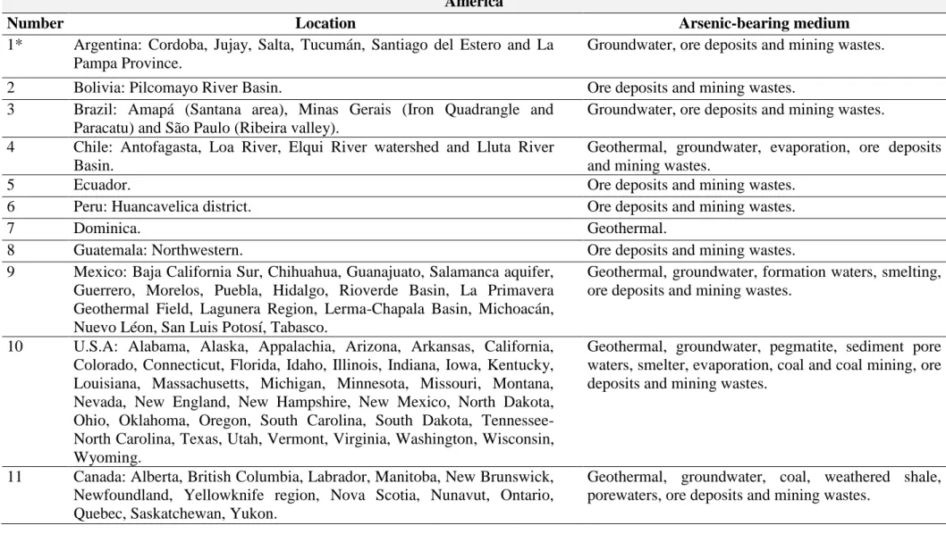
Table 1. List of the global arsenic As-contaminated areas with the correspondent local sources (adapted from Henke, 2009). America
Number Location Arsenic-bearing medium
1* Argentina: Cordoba, Jujay, Salta, Tucumán, Santiago del Estero and La Pampa Province.
Groundwater, ore deposits and mining wastes.
2 Bolivia: Pilcomayo River Basin. Ore deposits and mining wastes.
3 Brazil: Amapá (Santana area), Minas Gerais (Iron Quadrangle and Paracatu) and São Paulo (Ribeira valley).
Groundwater, ore deposits and mining wastes. 4 Chile: Antofagasta, Loa River, Elqui River watershed and Lluta River
Basin.
Geothermal, groundwater, evaporation, ore deposits and mining wastes.
5 Ecuador. Ore deposits and mining wastes.
6 Peru: Huancavelica district. Ore deposits and mining wastes.
7 Dominica. Geothermal.
8 Guatemala: Northwestern. Ore deposits and mining wastes.
9 Mexico: Baja California Sur, Chihuahua, Guanajuato, Salamanca aquifer, Guerrero, Morelos, Puebla, Hidalgo, Rioverde Basin, La Primavera Geothermal Field, Lagunera Region, Lerma-Chapala Basin, Michoacán, Nuevo Léon, San Luis Potosí, Tabasco.
Geothermal, groundwater, formation waters, smelting, ore deposits and mining wastes.
10 U.S.A: Alabama, Alaska, Appalachia, Arizona, Arkansas, California, Colorado, Connecticut, Florida, Idaho, Illinois, Indiana, Iowa, Kentucky, Louisiana, Massachusetts, Michigan, Minnesota, Missouri, Montana, Nevada, New England, New Hampshire, New Mexico, North Dakota, Ohio, Oklahoma, Oregon, South Carolina, South Dakota, Tennessee-North Carolina, Texas, Utah, Vermont, Virginia, Washington, Wisconsin, Wyoming.
Geothermal, groundwater, pegmatite, sediment pore waters, smelter, evaporation, coal and coal mining, ore deposits and mining wastes.
11 Canada: Alberta, British Columbia, Labrador, Manitoba, New Brunswick, Newfoundland, Yellowknife region, Nova Scotia, Nunavut, Ontario, Quebec, Saskatchewan, Yukon.
5 Africa
12 Botswana. Groundwater.
13 Ghana. Groundwater, ore deposits and mining wastes.
14 Nigeria. Groundwater.
15 South Africa. Ore deposits and mining wastes.
16 Tanzania: Serengeti NP (Orangi River). Ore deposits and mining wastes.
17 Tunisia. Ore deposits and mining wastes.
18 Zimbabwe. Ore deposits and mining wastes.
Europe
19 Austria: East Alps. Ore deposits and mining wastes.
20 Belgium and Netherlands. Groundwater.
21 Bulgaria: Plovdiv-Assenovgrad area, Southwest. Geothermal, coal, ore deposits, smelting and mining.
22 Cyprus: Mathiatis Mine. Ore deposits and mining wastes.
23 Czech Republic: Bohemian Massif, Krusné hory Mountains, Sokolov Basin, Ostrava-Karvina Basin.
Mineralization, coal and coal mining wastes, ore deposits and mining wastes.
24 Finland: Ilomantsi, Kittila, Pirkanmaa, Seinajoki district and Ylojarvi. Tills, groundwater, ore deposits and mining wastes. 25 France: Corsica, Douai area, Massif Central, Rhône River. Smelter, geothermal, ore deposits and mining wastes. 26 Germany: Bavaria, Mulde River, Dessau area, Freiberg, Koenigstein
Mine, Mansfeld region, Southeast Harz Forelands and Ebersdorf Coal Deposit.
Groundwater, chemical industry, coal deposits and mining wastes, ore deposits and mining wastes.
27 Greece: Central Macedonia, Crete, Elassona Basin, Northwestern, Mygdonia region, Eastern Attica and Santorini.
Groundwater, geothermal, coal and coal mining wastes, ore deposits and mining wastes.
28 Hungary, Romania and Slovakia: Great Hungarian Plain, Mátra Mountains, Pannonian Basin, Danube Basin, Baia Mare Region and Cierna Lehota.
Groundwater, Mining wastes, Mineralized Shales, ore deposits and mining wastes.
6
30 Poland: Sudety Mountains and Lyublin Basin. Mineralization, coal deposits and mining wastes, ore deposits and mining wastes.
31 Portugal: Castromil gold deposit. Ore deposits and mining wastes.
32 Russia: Kola Peninsula. Smelter.
33 Spain: Anllóns River, Asturias and León, Aznalcóllar, Central, Eastern Pyrenees, Madrid Aquifer and Puertollano Basin.
Groundwater, coal and coal mining wastes, ore deposits and mining wastes.
34 Sweden: Kalix River and Bothnian Bay, Kristineberg mining site, Ronnskar Smelter, Adak Mine and Vormbacken River.
Smelter, ore deposits and mining wastes.
35 Switzerland: Alps, Camignolo area and Malcantone watershed. Geothermal, groundwater, hydrothermal deposits, ore deposits and mining wastes.
36 Turkey: Central Anatolia, Emet, Gediz and Kutahya. Geothermal, coal, groundwater, volcanics, ore deposits and mining wastes.
37 Ukraine-Russian: Donbas Basin, L´vov-Volynsk Basin. Groundwater, coal deposits and mining wastes.
38 United Kingdom: Cornwall, Scotland and South Wales. Groundwater, intrusive igneous rocks, coal, ore deposits, smelting, and mining wastes.
Asia
39 Bangladesh. Groundwater.
40 Cambodia. Groundwater.
41 Caspian Sea. Mine runoff and other anthropogenic sources
42 China: Anhui, Beijing, Guangdong, Guizhou, Henan, Hunan, Inner Mongolia, Jilin, Liaoning, Ningxia, Northeast, Qinghai, Shandong, Shanxi, Sichuan, Xinjiang, Yunnan.
Groundwater, coal and coal mining, ore deposits and mining wastes.
43 India: Andhra Pradesh, Assam, Bihar and Jharkhand, Chhattisgarh, Karnataka, Punjab, Haryana, Himachal Pradesh, Rajasthan, Tamil Nadu, Uttar Pradesh and West Bengal.
Groundwater, coal, schist belt, ore deposits and mining wastes.
44 Indonesia: Ngada district (Bejawa geothermal area), North Sulawesi and West Java.
Geothermal, volcanic deposits, ore deposits and mining wastes.
7
46 Japan: Ichinokawa Mine, Honshu, Kyushu and Osaka. Geothermal, groundwater, ore deposits and mining wastes.
47 Korea, South. Ore deposits and mining wastes.
48 Laos. Groundwater.
49 Malaysia: Peninsula, Sabah and Sarawak. Ore deposits and mining wastes.
50 Myanmar: Ayeyarwaddy Division, Bago and Rakhine state. Groundwater.
51 Nepal: Southern. Groundwater.
52 Pakistan: Punjab and Sindh. Groundwater and drinking water.
53 Philippines: Luzon, Mindanao and Palawan. Geothermal, ore deposits and mining wastes. 54 Russia: Altai Mountains, Kamchatka Peninsula, Kemerovo region and
Ural Mountains.
Geothermal, smelting, coal and coal mining, ore deposits and mining wastes.
55 Sri Lanka. Groundwater.
56 Taiwan. Groundwater and smelter
57 Thailand: Mae Moh and Nakhon Si Thammarat Province. Groundwater, coal mining and combustion, ore deposits and mining wastes.
58 Vietnam: Red River region and Mekong River region. Groundwater. Oceania
59 Australia: New South Wales, Queensland, Western Australia. Groundwater, smelter and industrial complex, ore deposits and mining wastes.
60 New Zealand: North and South Island. Geothermal, coal and coal mining, ore deposits and mining wastes.
61 Papua New Guinea: Ambitle Island (Tatum Bay). Geothermal.
8
In response to the widespread As contamination, many governments have instituted regulations on the disposal of arsenic bearing wastes and As emission from ore smelters and coal-combustion. Remediation of As-contaminated soils and waters is needed to protect the biosphere. Conventional physicochemical technologies have been applied to remediate As such as vitrification, soil washing/flushing and precipitation, however, without success (Mondal et al., 2006; Mirza et al., 2014). Most of these methods are found very costly (75 – 500 dollars per ton of soil), and thus low-cost technologies are needed to effectively treat As-contaminated sites (Mirza et al., 2014). Phytoremediation has been suggested as the most cost effective and efficient method for removal or minimization of metal contamination both in soil and water.
Phytoremediation involves the use of plants to remove, transfer, stabilize, and/or degrade contaminants in soil, sediment, and water (Hughes et al., 1997). Different processes can be involved in the phytoremediation of As-contaminated sites: phytostabilization, which refers to the use of plants to minimize As dispersion from soil to water or air; phytoextraction (or phytofiltration), which refers to the use of plants to remove As from soil or water; and phytovolatilization that involves the transformation of As to volatile compounds and their emission from plants (Zhu and Rosen, 2009).
Plants exhibit a wide variation in their response to As. Not considering the As-sensitive, plants can be classified as excluders, accumulators and hyperaccumulators according to their physiological mechanisms of resistance/tolerance (Table 2). Excluders are plants adapted to grow in soils that have high As concentrations, without accumulating it (Moreno-Jiménez et al., 2012). The concentration of As in non-accumulator plants rarely exceeds 2 mg As per kg in aerial parts (Horswell and Speir, 2006). Plants that can accumulate high concentrations of metals in the aboveground biomass (higher than 1000 mg As per kg), without suffering growth constraints, have been referred as hyperaccumulators (Reeves and Baker, 2000; Kramer, 2010). Excepting hyperaccumulators, most plants accumulate As in roots (accumulators).
9
vacuole of leaf cells combined with a higher antioxidant capacity (Lombi et al., 2002; Srivastava et al., 2005; Singh et al., 2006; Zhao et al., 2009).
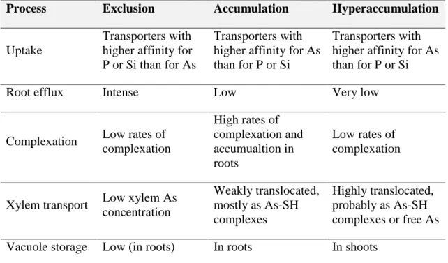
Table 2. Physiological mechanisms involved in As exclusion, accumulation and hyperaccumulation by plants. Adapted from Moreno-Jiménez et al. (2012).
Process Exclusion Accumulation Hyperaccumulation
Uptake
Transporters with higher affinity for P or Si than for As
Transporters with higher affinity for As than for P or Si
Transporters with higher affinity for As than for P or Si
Root efflux Intense Low Very low
Complexation Low rates of complexation
High rates of complexation and accumualtion in roots
Low rates of complexation
Xylem transport Low xylem As concentration
Weakly translocated, mostly as As-SH complexes
Highly translocated, probably as As-SH complexes or free As Vacuole storage Low (in roots) In roots In shoots
Pityrogramma calomelanos (L.) Link is considered to be of American origin (Wardlaw, 1962) and is now virtually pan-tropical in its distribution. This species is known to accumulate unusually high levels of As, and is commonly found associated with mine sites in Australia, Brazil and Thailand (Visoottiviseth et al., 2002; Melendez et al., 2011; Niazi et al., 2012). Pityrogramma calomelanos has been pointed as a suitable species for phytoextraction/phytoremediation (Jankong et al., 2007), however, the mechanisms responsible for its tolerance remain unknown, as well the As effects on its development.
This study aimed to characterize the morphoanatomical and physiological responses of Pityrogramma calomelanos (L.) Link to arsenic exposure in order to understanding its mechanisms of As-accumulation and tolerance and provide data to improve its features for phytoremediation approaches.
10 REFERENCES
ATSDR, 2013. The ATSDR 2013 substance priority list. Agency for Toxic Substances and Disease Registry. Available in http://www.atsdr.cdc.gov/SPL/index.html (accessed on November 26, 2014).
Borba RP, Figueiredo BR, Matschullat J, 2003. Geochemical distribution of arsenic in waters, sediments and weathered gold mineralized rocks from Iron Quadrangle, Brazil. Environ Geol 4:39–52.
Deschamps ME, Ciminelli VST, Weidler PG, Ramos AY, 2003. Arsenic sorption onto soils enriched with manganese and iron minerals. Clays Clay Miner 51:197.
Henke KR, 2009. Environment chemistry, health threats and waste treatment. John Wiley & Sons Ltd, West Sussex, UK. 567p.
Horswell J, Speir T, 2006. Arsenic phytotoxicity: effect on crop yield and crop quality. In: P Bhattacharya, E Smith, R Naidu, P Nadebaum, G Owens (eds), Managing Arsenic in the Environment. CSIRO Publishing, Melbourne, Australia, pp 183–207. Hughes JB, Shanks J, Vanderford M, Lauritzen J, Bhadra R, 1997. Transformation TNT
by aquatic plants and plant tissue cultures. Environ Sci Technol 31: 266–271.
Hughes MF, Beck B, Chen Y, Lewis AS, Thomas DJ, 2011. Arsenic exposure and toxicology: a historical perspective. Toxicol Sci 123:305–332.
Jankong P, Visoottiviseth P, Khokiattiwong S, 2007. Enhanced phytoremediation of arsenic contaminated land. Chemosphere 68:1906–1912.
Kramer U, 2010. Metal hyperaccumulation in plants. Annu Rev Plant Biol 61:517-534. Krebs RE, 2006. The history and use of our earth’s chemical elements: a reference
guide. Greenwood Press, London. 422p.
Lombi E, Zhao FJ, Fuhrmann M, Ma LQ, McGrath SP, 2002. Arsenic distribution and speciation in the fronds of the hyperaccumulator Pteris vittata. New Phytol 156:195– 203.
Ma LQ, Komar KM, Tu C, Zhang W, Cai Y, Kennelley ED, 2001. A fern that hyperaccumulates arsenic. Nature 409:579.
Matschullat J, Borba RP, Deschamps E, Figueiredo BR, Gabrio T, Schwenk M, 2000. Human and environmental contamination in the Iron Quadrangle, Brazil. Appl Geochem 15:181–190.
11
Mondal P, Majumder CB, Mohanty B, 2006. Laboratory-based approaches for arsenic remediation from contaminated water: recent developments. J Hazard Mater 137:464–479.
Moreno-Jiménez E, Esteban E, Peñalosa JM, 2012. The fate of arsenic in soil-plant systems. Rev Environ Contam Toxicol 215:1–37.
Murcott S, 2012. Arsenic contamination in the world: an international sourcebook. IWA Publishing. London.
Niazi NK, Singh B, Zwieten LV, Kachenko AG, 2012. Phytoremediation of an arsenic-contaminated site using Pteris vittata L. and Pityrogramma calomelanos var. austroamericana: a long-term study. Environ Sci Pollut Res 19:3506–3515.
Ravenscroft P, Brammer H, Richards K, 2009. Arsenic Pollution: A Global Synthesis. John Wiley & Sons, West Sussex, UK. 618p.
Reeves RD, Baker AJM, 2000. Metal-accumulating plants. In: I Raskin, BD Ensley, (eds), Phytoremediation of toxic metals: using plants to clean up the environment. John Wiley & Sons Inc, New York, pp 19-229.
Reichard JF, Schnekenburger M, Puga A, 2007. Long term low-dose arsenic exposure induces loss of DNA methylation. Biochem Biophys Res Commun 352:188–192. Shankar S, Shanker U, Shikha, 2014. Arsenic Contamination of Groundwater: A
Review of Sources, Prevalence, Health Risks, and Strategies for Mitigation. Sci World J 2014:1–18.
Singh N, Ma LQ, Srivastava M, Rathinasabapathi B, 2006. Metabolic adaptations to arsenic-induced oxidative stress in Pteris vittata L. and Pteris ensiformis L. Plant Sci 170:274–282.
Sinha D, Biswas J, Bishayee A, 2013. Nrf2-mediated redox signaling in arsenic carcinogenesis: a review. Arch Toxicol 87:383–396.
Smedley PL, Kinniburgh DG, 2002. A review of the source, behaviour and distribution of arsenic in natural waters. Appl Geochem 17:517–568.
Srivastava M, Ma LQ, Singh N, Singh S, 2005. Antioxidant responses of hyperaccumulator and sensitive fern species to arsenic. J Exp Bot 56:1335–1342. Srivastava M, Ma LQ, Santos JAG, 2006. Three new arsenic hyperaccumulating ferns.
Sci Total Environ 364:24–31.
12
Tripathi RD, Srivastava S, Mishra S, Singh N, Tuli R, Gupta DK, Maathuis FJM, 2007. Arsenic hazards: strategies for tolerance and remediation by plants. Trends Biotechnol 25:158–165.
Melendez LB, Silva-Filho EV, Miekeley N, Vieira FA, Sella SM, 2011. Determination of arsenic species in P. calomelanos and N. biserrata. J Braz Chem Soc 22:1961– 1967.
Visoottiviseth P, Francesconi K, Sridokchan W, 2002. The potential of Thai indigenous plant species for the phytoremediation of arsenic contaminated land. Environ Pollut 118:453–461.
Wang X, Ma LQ, Rathinasabapathi B, Liu Y, Zeng G, 2010. Uptake and translocation of arsenite and arsenate by Pteris vittata L. Effects of silicon, boron and mercury. Environ Exp Bot 68:222–229.
Wardlaw CW, 1962. A note on Pityrogramma calomelanos (L.) Link, a fern nuisance in Cameroons plantations. J Ecol 50:129–131.
Wedepohl KH, 1991. The composition of the upper Earth’s crust and the natural cycles of selected metals: Metals in natural raw materials: Natural resources. In: E. Merian (ed), Metals and Their Compounds in the Environment. John Wiley, Hoboken, NJ, pp 3–17.
Zhao FJ, Dunham SJ, McGrath SP, 2002. Arsenic hyperaccumulation by different fern species. New Phytol 156:27–31.
Zhao FJ, Ma JF, Meharg AA, McGrath SP, 2009. Arsenic uptake and metabolism in plants. New Phytol 181:777–794.
Zhao FJ, McGrath SP, Meharg AA, 2010. Arsenic as a food chain contaminant: mechanisms of plant uptake and metabolism and mitigation strategies. Annu Rev Plant Biol 61:535–559.
13
CAPÍTULO 1: Alterações fisiológicas em pina e raiz de Pityrogramma calomelanos
(L.) Link para minimizar efeitos deletérios do arsênio
RESUMO: Hiperacumulação de arsênio (As) tem sido descrita para algumas espécies de Pteridales; entretanto, as bases fisiológicas da hiperacumulação permanecem desconhecidas, especialmente em Pityrogramma calomelanos única hiperacumuladora descrita que não pertence ao gênero Pteris. Com o intuito de aprimorar o entendimento acerca das respostas induzidas por As em diferentes partes da planta, indivíduos de P. calomelanos foram expostos a 1 mM As durante 21 dias e foram subsequentemente comparados com indivíduos do tratamento controle. Plantas tratadas com As apresentaram concentrações médias de 3108, 275, and 283 mg kg-1 na matéria seca de pinas, estipes e raízes, respectivamente. Nas pinas, o As foi encontrado principalmente na forma de arsenito, enquanto o arsenato predominou em estipes e raízes. Aumento na atividade da peroxidase do ascorbato (APX) e da peroxidase (POX) e no conteúdo de tióis não-proteicos foi observado nas pinas, as quais não apresentaram sintoma de toxidez. Apesar do aumento da atividade da superóxido dismutase (SOD) e da catalase (CAT), as raízes de plantas tratadas apresentaram menor massa fresca, menores concentrações de P, K, Fe e Mg, e aumento da peroxidação lipídica. Plantas expostas ao As apresentaram, ainda, redução da translocação de K e S. O elevado fator de translocação de As e potencial antioxidante observado para a pina se mostraram essenciais para evitar o aumento da concentração de espécies reativas de oxigênio permitindo o acúmulo crescente de As nas frondes. A hiperacumulação de As em P. calomelanos requer ainda ajustes na nutrição mineral das plantas, em especial em relação às concentrações de K e o P.
14
CHAPTER 1. Physiological changes in the pinna and root of Pityrogramma calomelanos (L.) Link to alleviate arsenic deleterious effects
Campos, N.V.a; Arcanjo-Silva S.a; Viana, I.B.a; Batista, B.L.b; Loureiro, M.E.a; Ribeiro, C.a; Azevedo, A.A.a∗
a
Departamento de Biologia Vegetal, Universidade Federal de Viçosa, Avenida Peter Henry Rolfs, s/n, 36570-900, Viçosa, MG, Brazil.
b
Departamento de Análises Clínicas, Toxicológicas e Bromatológicas, Faculdade de Ciências Farmacêuticas de Ribeirão Preto – Universidade de São Paulo - Avenida do Café, s/n, Monte Alegre, 14040-903, Ribeirão Preto, SP, Brazil.
ABSTRACT: Arsenic (As) hyperaccumulation has been described in a number of Pteridales. However, the physiological basis of hyperaccumulation remains unclear, especially in non-Pteris species such as Pityrogramma calomelanos. Aiming at a better understanding of As-induced responses in different fern parts, P. calomelanos plants were exposed to 1 mM As for 21 days, and subsequently compared with control plants. Chemical analysis revealed As concentrations in pinnae, stipes, and roots of 3108, 275, and 283 mg kg-1 dry weight, respectively. Arsenic was present mainly as arsenite in pinnae and as arsenate in stipes and roots. Increases in ascorbate peroxidase (APX) and peroxidase (POX) activities and non-protein thiol contents were observed in pinnae, without symptoms of As toxicity. Despite increases in superoxide dismutase (SOD) and catalase (CAT) activities, the roots showed reductions in fresh weight and P, K, Fe, and Mg concentrations and an increase in lipid peroxidation. Plants exposed to As showed a reduction in K and S translocation factors. The higher As translocation and antioxidant capacity of the pinnae are essential for quenching oxygen reactive species at lower concentrations, thus enhancing continuous As accumulation in fronds. Arsenic hyperaccumulation in P. calomelanos also requires adjustments in the mineral nutrition of plants, especially with regard to K and P.
Keywords: As-hyperaccumulator; nutrient balance; reactive oxygen species; thiol
*
15 1. Introduction
Hyperaccumulating plants have the ability to accumulate extremely high concentrations of metals or metalloid within their tissues, wherein the threshold concentration used to define a hyperaccumulator depends on the particular element sequestered (van der Ent et al., 2013). Arsenic (As) hyperaccumulating plants must contain at least 1000 mg As kg-1 dry weight (DW) (Kramer, 2010). Nominal threshold criteria, however, should be complemented with a suite of characteristics that include a bioconcentration factor > 1, a shoot-to-root metal concentration quotient > 1 and extreme metal tolerance due to effective biochemical detoxification (Baker and Whiting, 2002).
Arsenic hyperaccumulation has been described for a number of Pteris spp., most notably Pteris vittata, which accumulates up to 22630 mg As kg-1 DW in the frond (Ma et al., 2001), and other ferns such as Pityrogramma calomelanos, with up to 8350 mg As kg-1 DW (Francesconi et al., 2002). Although P. vittata has been widely studied, P. calomelanos remains a lesser-known As-hyperaccumulating fern. Two varieties of P. calomelanos have been recognized: var. calomelanos (Silver back fern), usually referred to only as P. calomelanos, and var. austromericana (Gold dust fern). The Silver back fern has a white indument and is typical of humid tropics, differing from the Gold dust fern, which exhibits a yellow indument and is common in temperate and subtropical environments (Schelpe, 1975; Moran, 1995). The Gold dust fern has also been recognized as an As hyperaccumulator, with As concentrations reaching 5845 mg kg–1 DW in its young fronds (Kachenko et al., 2010).
16
There is considerable evidence that inorganic As exposure results in the generation of reactive oxygen species (ROS) in plants, including superoxide anion, hydroxyl radicals, and hydrogen peroxide (Hartley-Whitaker et al., 2001). Increased levels of ROS induce lipid peroxidation and cause a decrease in biomass and other physiological disorders in non-hyperaccumulating plants (Hartley-Whitaker et al., 2001; Singh et al., 2006; Garg and Singla, 2011; Campos et al., 2014). Pteris vittata can tolerate high tissue As concentrations due to the enhancement of the antioxidant machinery, leading to As detoxification and hyperaccumulation (Srivastava et al., 2005). Antioxidative defense falls into two general classes: (1) low molecular weight antioxidants, which consist of lipid-soluble membrane-associated antioxidants (e.g., α -tocopherol and -carotene), and the water soluble reductants (e.g., glutathione-GSH and ascorbate); and (2) enzymatic antioxidants (e.g., superoxide dismutase-SOD, catalase-CAT, guaiacol peroxidase-GPX, ascorbate peroxidase-APX) (Cao et al. 2004). The peroxidase POX is another important enzyme induced under stress conditions (Sasaki et al., 2004).
Arsenic hyperaccumulation (absorption, transport, and storage) in P. vittata has been addressed by elsewhere (Tu et al., 2004; Kertulis-Tartar et al., 2005; Hokura et al., 2006; Huang et al., 2007; Wang et al., 2010). However, the physiological bases of As metabolism remain unclear, especially in non-Pteris species, which makes difficult the evolutionary interpretation of the As accumulation and resistance mechanisms in ferns.
In the present research, we evaluated the effects of arsenic on the activities of antioxidant enzymes (APX, CAT, GR, POX, and SOD), acid-soluble thiols, malondialdehyde content and the mineral nutrition of Pityrogramma calomelanos under hydroponic culture. Arsenic speciation analysis was also conducted. The results indicate that As affects nutrient balance in P. calomelanos and that antioxidative systems are differentially involved in fern organs, which can be related to As species partitioning.
2. Materials and methods
2.1. Site characterization and sampling
17
collected in paper bags and placed in a desiccator until spores release (Figure 1 D). Spores were separated from the sporangia and stored at 4 ºC for two months.
2.2. In vitro development of gametophytes and sporophytes
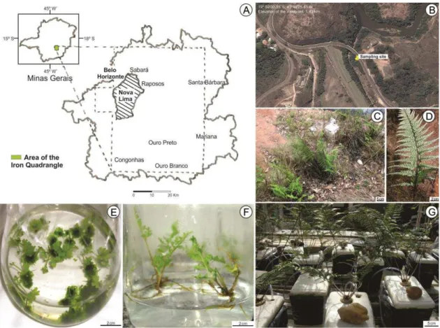
Spores were transferred to 1.5 ml microtubes containing 1.0 ml of 70 % ethanol (EtOH) and centrifuged for 1 min at 13000 g. Then, the supernatant was discarded and the spores were treated with 20 % sodium hypochlorite (NaClO, 2 % of active chlorine) for 15 min and centrifuged for 5 min at 13000 g. The spores were washed and resuspended in autoclaved ultrapure water. Aliquots of the spore suspension were inoculated in Petri dishes (200 μl per dish) containing 20 ml of half-strength Murashige and Skoog (MS) medium (Murashige and Skoog, 1962). The medium was supplemented with Gamborg B5 vitamins (Gamborg et al., 1968; 2.5 ml L-1), sucrose (10 g L-1) and solidified with agar (6.5 g L-1). After spore germination, the gametophytes were transferred to glass jars containing liquid MS medium (1/2 strength) supplemented with Gamborg B5 vitamins (2.5 ml L-1) and sucrose (10 g L-1) (Figure 1 E). The jars were placed on an orbital shaker (110 rpm) in a climate-controlled room with a photoperiod of 16 h of light (100 µmol m-2 s-1). After one month, the sporophytes were transferred to jars containing semi-solid MS (3.0 g L-1 gelrite) (Figure 1 F).
2.3. Experimental design
In vitro produced sporophytes were carefully removed from the media, washed in tap water and transferred to 200-ml pots containing the commercial substrate Plantmax®. Sporophytes were cultivated in a greenhouse with controlled temperature (23 ºC), under 50 %-shading nylon net (Sombrite®), irrigated daily with water and once a week with half-strength Hoagland’s nutrient solution (Hoagland and Arnon, 1950). Ferns at the 4-5 frond stage were transferred to hydroponic system with half-strength Hoagland’s solution, pH 5.5 and continuous aeration. The ferns were cultivated for 40 days before the beginning of the treatments (Figure 1 G). Then, the ferns (n = 5) were treated with 0 (control) and 1 mM As, supplied as sodium arsenate (Na2HAsO4·7H2O),
18
Figure 1. Location map of the sampling area colonized by Pityrogramma calomelanos. Nova Lima district situated in the Iron Quadrangle, Minas Gerais State, Brazil (A). Satellite images of the sampling area by Google Earth (B). Adult individuals of P. calomelanos with fertile fronds (C-D). Gametophyte and sporophyte in vitro development, respectively (E-F). Adult sporophytes in hydroponic system after 40 days of acclimatization (G).
2.4. Growth parameters and chlorophyll content
After 21 days, the ferns were harvested, separated into roots, pinnae and stipe + rachis (referred to here as the stipe), and oven-dried at 60 ºC. The fresh weight (FW) and dry weight (DW) were determined for the pinnae, stipe and roots of each plant. The total FW and DW of the plants were assumed as the sum of the weights of these parts. The moisture content (MC) was given by (FW - DW/ FW × 100%) (Lee and Yu, 2012).
19
root volume was evaluated by measuring the rise in the water level by immersing the organ in a water-filled graduated cylinder.
2.5. Total arsenic and nutrient determination
The dry matter was milled and powdered with a knife mill to evaluate the total concentrations of As, Ca, Cu, Fe, K, Mg, P, and S. Samples (0.1 g) were extracted with nitro-perchloric solution (3:1) in a digestion block as described by Tedesco et al. (1995). The samples were cooled at room temperature and the volume was completed to 10 ml with ultrapure water and then filtered. The samples were analyzed by inductively coupled plasma-optical emission spectrometry (ICP OES) (Perkin Elmer, Shelton, CT, USA).
The translocation factors (TF) of arsenic and nutrients were calculated as the ratio between As concentration in fronds and roots (Huang et al., 2007).
2.6. Arsenic species determination
Arsenic species were evaluated according to Batista et al. (2011). Samples (n = 3, 0.2 g) were accurately weighed into a 50-ml conical tube, added of 10 ml of 2 % (v/v) nitric acid. The tubes were closed and left at room temperature for 48 hours. The mixture was heated in a water bath at 95 ºC for 2 h, cooled at room temperature, filtered with a 0.20-µm cellulose filter and analyzed using high-performance liquid chromatography-inductively coupled plasma mass spectrometry (HPLC-ICP-MS, Perkin Elmer Sciex - Elan DRC II, Framingham, MA, USA). Gallium was used as an internal standard at a final concentration of 5 µg L−1.
2.7. Enzyme extraction and activity assays
20
and 1% PVPP for catalase (CAT, EC 1.11.1.6) and glutathione reductase (GR, EC 1.6.4.2); c) 0.1 M KPB (pH 6.8), 0.1 mM EDTA, 0.1 mM PMSF, and 1 % (w/v) PVPP for ascorbate peroxidase (APX, EC 1.11.1.11). The homogenate was centrifuged at 13000 g for 15 min at 4 ºC and the supernatant was used as the source of crude enzyme. Enzyme activities were determined by adding 0.1 ml of the crude enzyme extract to the following media: a) POX, 1.7 ml of a reaction medium consisting of 50 mM M KPB (pH 6.8), 20 mM pyrogallol, and 20 mM H2O2; b) CAT, 0.9 ml of a reaction
medium consisting of 50 mM KPB (pH 7.0) and 12.5 mM H2O2; c) APX, 1.0 ml of a
reaction medium consisting of 50 mM KPB (pH 6.0), 0.8 mM ascorbic acid, and 1 mM H2O2 (Peixoto et al. 1999); d) GR, 0.9 ml of a reaction medium consisting of 0.1 M
KPB (pH 7.5), 1 mM oxidized glutathione (GSSG), and 0.1 mM NADPH(Carlberg and Mannervik, 1985). In all cases, the mixtures were incubated at 30 ºC and the absorbances were measured during the first minute of the reaction. Enzyme activities were estimated using the following molar extinction coefficients: APX (290 nm, ε: 2.8 mM-1 cm-1), CAT (240 nm, ε: 36 M-1 cm-1), GR (340 nm, ε: 6.22 mM-1 cm-1), and POX (420 nm; ε: 2.47 mM-1 cm-1).
The SOD activity was determined by adding the enzyme extract to a reaction mixture consisting of 50 mM KPB (pH 7.8), 13 mM methionine, 0.1 mM EDTA, 75 µM nitroblue tetrazolium (NBT) and 2 µM riboflavin. The reaction was carried out in a chamber with a 15-W fluorescent lamp at 25 ºC. After 10 min of illumination, the blue formazan formed was measured at 560 nm (Giannopolitis and Ries, 1977). All rates were corrected for non-enzymatic activity. One unit of SOD activity was defined as the amount of enzyme required to cause a 50 % inhibition of the rate of NBT reduction.
2.8. Protein assay
Soluble protein was estimated using the reagent Coomassie Brilliant Blue G-250 and bovine serum albumin as the standard, according to the method of Bradford (1976).
2.9. Determination of total and non-protein thiols
21
8.0), 1 mM Na-EDTA and 1 % (w/v) ascorbic acid. The homogenate was centrifuged for 10 min at 10000 g in a refrigerated centrifuge at 4 °C. For TT analysis, 0.1 ml of the supernatant was mixed with the reaction buffer containing 0.3 ml of KPB (0.2 mM; pH 8.2), 20 μl of DTNB (10 mM) and 1.58 ml of absolute methanol. The samples were incubated for 15 min at 37 °C and the absorbance was measured at 412 nm (Multiskan Spectrum, Spectra 190, Dynex Technologies, USA).
The NPT analysis was carried out using 1.0 ml of the supernatant mixed with 0.2 ml of 50 % trichloroacetic acid (TCA) and 0.8 ml of distilled water. After 1 h in ice bath, the samples were centrifuged for 15 min at 10000 g. Aliquots of 0.4 ml of the supernatant were added to 0.4 M KPB (pH 8.9) and 20 μl of DTNB (10 mM). After 5 min, the absorbance was measured at 412 nm (Multiskan Spectrum, Spectra 190, Dynex Technologies, USA).
The thiol concentration was estimated using the molar extinction coefficient of 13100 mM-1 cm-1. The concentration of protein thiols (PT) was calculated by subtracting NPT from TT.
2.10. Lipid peroxidation assay
Lipid peroxidation in the pinna and root samples was determined as described by Heath and Packer (1968). Freeze-dried samples were mixed with 2 ml of TBA reagent (20 % w/v trichloroacetic acid + 0.5 % w/v thiobarbituric acid (TBA)), heated to 95 °C for 30 min, cooled for 15 min, and centrifuged at 10000 g for 15 min. The amount of malondialdehyde (MDA)-TBA complex was measured by its specific absorbance at 532 nm; the nonspecific absorbance at 600 nm was subtracted from 532 nm.
2.11. Statistical analysis
22 3. Results
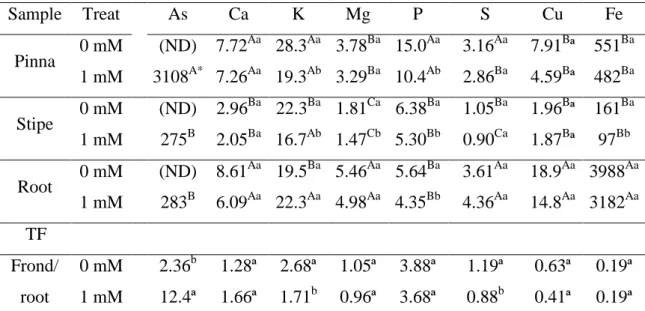
3.1. Arsenic accumulation and speciation and its effects on fern nutritional homeostasis Pityrogramma calomelanos accumulated large amounts of As, with the highest As concentration in pinnae (3108 mg As kg-1 DW). Stipes and roots accumulated similar concentrations of As, 275 and 283 mg As kg-1 DW, respectively. The As translocation factor (TF-As) was 12.0 (Table 1).
Table 1. Concentrations of arsenic (mg kg-1 dry weight, DW), macronutrients (g kg-1 DW) and micronutrients (mg kg-1 DW), and translocation factors (TF) of these elements in plants of Pityrogramma calomelanos of control and 1 mM As treatments (Treat).
Sample Treat As Ca K Mg P S Cu Fe
Pinna 0 mM (ND) 7.72
Aa
28.3Aa 3.78Ba 15.0Aa 3.16Aa 7.91Bª 551Ba 1 mM 3108A* 7.26Aa 19.3Ab 3.29Ba 10.4Ab 2.86Ba 4.59Bª 482Ba Stipe 0 mM (ND) 2.96
Ba
22.3Ba 1.81Ca 6.38Ba 1.05Ba 1.96Bª 161Ba 1 mM 275B 2.05Ba 16.7Ab 1.47Cb 5.30Bb 0.90Ca 1.87Bª 97Bb Root 0 mM (ND) 8.61
Aa
19.5Ba 5.46Aa 5.64Ba 3.61Aa 18.9Aa 3988Aa 1 mM 283B 6.09Aa 22.3Aa 4.98Aa 4.35Bb 4.36Aa 14.8Aa 3182Aa TF
Frond/ root
0 mM 2.36b 1.28ª 2.68ª 1.05ª 3.88ª 1.19ª 0.63ª 0.19ª 1 mM 12.4ª 1.66ª 1.71b 0.96ª 3.68ª 0.88b 0.41ª 0.19ª
*
Means (n = 5) by treatment followed by same letters were not significantly different. Capital letters refer to comparisons between the samples type, for each treatment (Tukey test; p < 0.05), and lowercase letters between the treatments, for each sample type (t test; p < 0.05). (ND) = non-detected.
23
The amounts of nutrients accumulated in pinnae, stipes and roots were compared between the control and As-treated plants. The plants cultivated with As showed 31 %, 17 % and 23 % of reduction in the P concentration of pinnae, stipes and roots, respectively. Arsenic decreased the concentration of K in pinnae and stipes and the concentrations of Fe and Mg in stipes. No changes were observed in the concentrations of Ca, Cu, and S in each fern parts. Plants exposed to As showed reductions of 36 % and 26 % in FT-K and FT-S, respectively (Table 1).
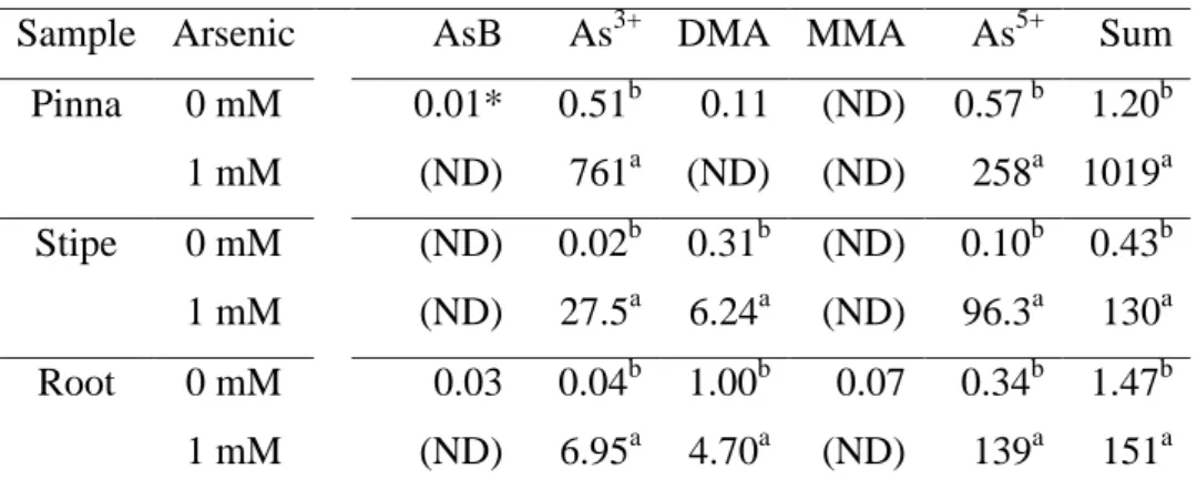
Table 2. Concentration of arsenic species (mg kg-1 dry weight)in the pinna, stipe and root of Pityrogramma calomelanos exposed to 0 and 1 mM As.
Sample Arsenic AsB As3+ DMA MMA As5+ Sum Pinna 0 mM 0.01* 0.51b 0.11 (ND) 0.57 b 1.20b
1 mM (ND) 761a (ND) (ND) 258a 1019a Stipe 0 mM (ND) 0.02b 0.31b (ND) 0.10b 0.43b 1 mM (ND) 27.5a 6.24a (ND) 96.3a 130a Root 0 mM 0.03 0.04b 1.00b 0.07 0.34b 1.47b 1 mM (ND) 6.95a 4.70a (ND) 139a 151a
*Means (n = 3) followed by different letters within the same column, for each sample type, indicate a significant difference (t test; p < 0.05). Legend: (As5+) arsenate, (As3+) arsenite, (AsB) Arsenobetaine/non-retained species on the column, (DMA) dimethylarsinate, (MMA) monomethylarsinate, (ND) non-detected.
24
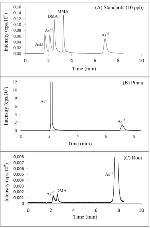
Figure 2. Typical chromatograms obtained for arsenic species in stardard samples (A) in pinna (B), and root (C) samples of Pityrogramma calomelanos after 21 days of exposure to 1 mM As. Legend: arsenobetaine / non-retained species on the column (AsB), arsenite (As3+), dimethylarsinate (DMA), and arsenate (As5+).
(B) Pinna
Time (min)
Inte
nsit
y (c
ps.10
6 )
As+3
As+5
(A) Standards (10 ppb)
Time (min)
Inte
nsit
y (c
ps.10
6 )
As+3
As+5 DMA
MMA
AsB
Time (min)
Inte
nsit
y (c
ps.10
6 )
(C) Root
Time (min)
Inte
nsit
y (c
ps
.10
6 )
As+3
As+5
25
3.2. Arsenic effects on growth parameters and chlorophyll content
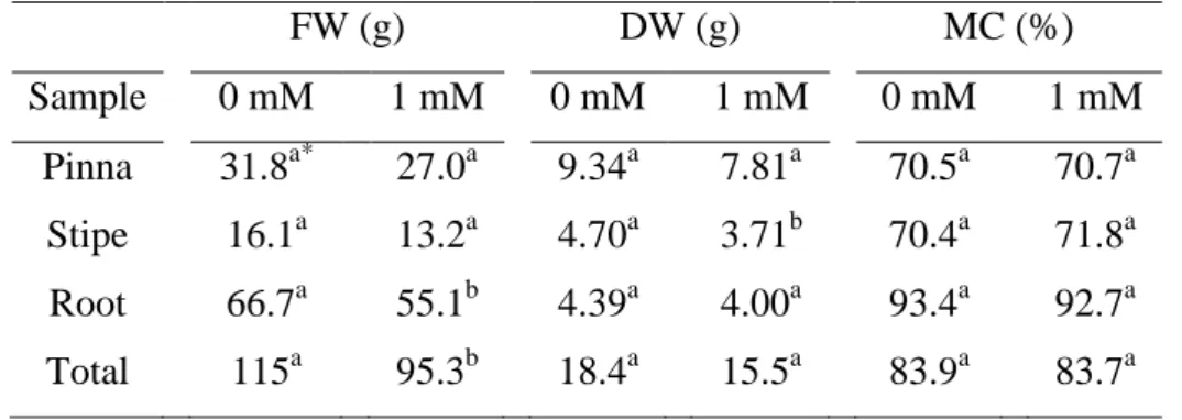
After 21 days of exposure, there were no visual symptoms of As toxicity in the ferns. However, As induced reductions in the root-FW (17 %), total-FW (17 %) and stipe-DW (21 %) (Table 3). No difference was observed between the MC of control and As-treated plants.
Ferns of the control and As treatments showed similar values of chl content (Figure 3 A) and root volume (Figure 3 B) over the time. The chl content did not change with the time in control and As-treated ferns. A significant increase of 29 % (p < 0.05) in the root volume was observed for ferns in both treatments, after three weeks of the beginning of the experiment.
Table 3. Fresh weight (FW), dry weight (DW) and the moisture content (MC) of pinna, stipe, root, and total weight of Pityrogramma calomelanos exposed to 0 and 1 mM As.
FW (g) DW (g) MC (%)
Sample 0 mM 1 mM 0 mM 1 mM 0 mM 1 mM Pinna 31.8a* 27.0a 9.34a 7.81a 70.5a 70.7a
Stipe 16.1a 13.2a 4.70a 3.71b 70.4a 71.8a Root 66.7a 55.1b 4.39a 4.00a 93.4a 92.7a Total 115a 95.3b 18.4a 15.5a 83.9a 83.7a
*Means (n = 5) followed by different letters within the same line, for each parameter, indicate a significant difference (t test; p < 0.05). MC = [(fresh weight - dry weight)/ fresh weight x 100].
3.3. Oxidative damage and antioxidant responses induced by arsenic
26
Days after arsenic exposure
3 6 12 18 21
R o o t v o lu me ( c m 3) 0 40 80 120 160 C h lo ro p h y ll re la ti v e c o n te n t 0 20 40 60 80 Control Arsenic B A
Figure 3. Chlorophyll relative content (A) and root volume (B) of ferns of Pityrogramma calomelanos from control and 1 mM As treatments measured during the exposure time (21 days). Vertical bars represent standart erros (n = 5).
MDA NPT PT TT
m M g F W -1 0 5 10 15 20 25 30 35 40 45 b a
a a a
a
MDA NPT PT TT
b a
a a a a
Control Arsenic Pinna Root a a a a
27
Arsenic induced an increase in the concentration of non-protein thiols (NPT) in pinnae, but no change in thiols concentrations was observed in roots of P. calomelanos (Figure 4). The concentrations of total thiols (TT) and NPT were two and five times, respectively, higher in pinnae than in roots of P. calomelanos (p < 0.05).
In general, the antioxidant enzymes showed higher activity in pinnae compared to roots, which is in agreement with the higher MDA content observed in the latter. Activity of SOD and CAT increased in roots in response to As exposure, but they were maintained in pinnae. In contrast, APX and POX showed higher activities in response to As only in pinnae. GR activity did not change in either organ (Figure 5).
Pinna Root APX m m o l m g
-1 p
ro te in m in -1 0,0 0,1 0,2 0,3 0,4 0,5 0,6 POX 0 20 40 60 80 100 SOD U m in-1 g-1 F W -1 0 20 40 60 80 CAT 0 5 10 15 20 25 30 35 a a b
a a a
a APX m m o l m g
-1 p
ro te in m in -1 0 1 2 3 4 5 6 POX 0 50 100 150 200 250 300 SOD U m in-1 g -1 F W -1 0 20 40 60 80 a b
a a a
GR 0,00 0,02 0,04 0,06 0,08 0,10 0,12 0,14 GR 0,00 0,05 0,10 0,15 0,20 0,25 CAT 0 20 40 60 80 100 120 Control Arsenic a a b a a a b a
Figure 5. Enzymatic activity of ascorbate peroxidase (APX), catalase (CAT), glutathione reductase (GR), peroxidase (POX), and superoxide dismutase (SOD) in pinna and root of Pityrogramma calomelanos upon exposure to 0 and 1 mM As for 21 days. Different letters indicate a significant difference between treatments (t test; p < 0.05). Vertical bars represent standart erros (n = 5).
4. Discussion
4.1. Arsenic reduction and translocation leads to arsenic hyperaccumulation
28
hydroponic culture, we show that P. calomelanos accumulated As in their pinnae in amounts greater than 3000 mg kg-1 dry weight (DW) which represents 85 % of the cumulative total As in ferns. Francesconi et al. (2002) were the first to describe P. calomelanos as an As-hyperaccumulating species, accumulating As mostly in the fronds (from 2760 up to 8350 mg kg-1 DW), with roots containing lower As concentration (310 mg kg-1 DW). Our results support the distribution of As species reported by these authors, whereby arsenate was the major form of As in roots and stipes (92 % and 75 %, respectively), and arsenite was the predominant form in pinnae (75 %).
Reduction and compartmentalization of As in pinnae are considered to be essential mechanisms in As-hyperaccumulating species, contrasting with plants that only tolerate As through the accumulation of this metalloid in roots (reviewed by Zhao et al., 2009). The specific organ(s) responsible for As reduction is still a matter of debate in the literature. Arsenate reductase activity has been reported in root extracts of Pteris vittata, suggesting that arsenite in the fronds may arise mainly from the reduction of arsenate in the roots (Duan et al., 2005). However, later studies demonstrated that arsenate reduction mostly occurs in the rhizomes and pinnae of P. vittata (Singh and Ma, 2006; Mathews et al., 2010).
The higher percentage of arsenate observed in stipes of P. calomelanos indicate that a low percentage of As was reduced in roots. These findings are in agreement with Kertulis-Tartar et al. (2005), who reported arsenate as the main As species in the xylem sap of P. vittata. Furthermore, X-ray absorption near-edge structure analysis (XANES) has shown that As is found as a mixture of arsenate and arsenite and their proportions in the rachis depend on the analyzed points, whereas it exists as arsenite in the pinna (Hokura et al., 2006).
A low concentration of organic forms of As was found in tissues of P. calomelanos. According to Francesconi et al. (2002), As-methylated forms can be present as trace constituents in fern samples. Small amounts of organic forms, such as monomethylarsonate (MMA), dimethylarsinate (DMA) and arsenosugars have been described in other plant species (Mattusch et al., 2000). However, it remains unclear whether the methylated forms are produced by microorganisms and merely taken up by plants or are endogenously methylated by the plants themselves (Lomax et al., 2012). Indeed, the pathway and enzymology of As methylation in plants have not been fully elucidated (Zhao et al., 2009).
29
processes for As tolerance in hyperaccumulating ferns. The study of As speciation in the xylem sap, along with the stipe and rachis of P. calomelanos, as well as As distribution at subcellular levels would clarify these processes and contribute to the understanding of As metabolism in this species.
4.2. Arsenic affects nutrient concentrations in different organs
Arsenic is a toxic element for plants, and its presence in the soil/water may interfere with the uptake of essential nutrients and plant growth. The majority of studies regarding As effects on plants are mainly focused on phosphorus (P) nutrition due to the competition between arsenate and phosphate for P transporters in the root plasma membrane (Ullrich-Eberius et al., 1989). Arsenic exposure reduced P concentrations in all analyzed organs of P. calomelanos but did not affect FT-P. These findings suggest that P reduction in the frond results mainly from decreased P uptake.
Significant differences in the levels of other macronutrients have been observed in Pteris vittata under As exposure (Cao et al., 2004; Tu and Ma, 2005). However, the effects of arsenic on the mineral nutrition of P. calomelanos have not been reported to date. In this study, As negatively affected the contents of K, Fe, and Mg in P. calomelanos. The decrease of K concentration in stipes and pinnae suggests that As exposure reduced the K translocation, as proved by the lower FT-K of As-treated ferns. Deficiency of K has been demonstrated to protected rice seedling from Cd stress by increasing the antioxidant status (Liu et al., 2013). The reduction of K concentration in fronds of P. calomelanos may have contributed to increased CAT and POX activities ensuring the integrity of lipid membranes under As stress, whereas in roots, the K content was not altered and the MDA content increased. Contrasting results of the As effect on macronutrients concentration were observed by Tu and Ma (2005) in the fronds of P. vittata, however, it seems to be more related to the differences in culture conditions (hydroponic versus soil culture) than differences in species traits.
30
also indicate a possible reduction in nutrient and water uptake. Furthermore, both elements (Fe and Mg) are required for chlorophyll synthesis and carbohydrate metabolism (Robb and Pierpoint, 1983). However, the Fe e Mg concentrations in the pinna were not changed in As-exposed ferns what should be correlated with the absence of effects on chlorophyll content.
Sulfur concentration in roots was not affected by As, which correlates well with the unaltered concentration of soluble thiol compounds in this organ. Sulfur is an essential nutrient in plants, required for the synthesis of amino acids and proteins and is also a precursor for the formation of GSH and other thiols in plants (Wei et al., 2010). Interestingly, As reduced the S translocation in P. calomelanos, without changing S concentration in the pinna, but increased the NPT content in this fern part. Similar results were reported for P. vittata, in which As did not change S concentrations in the ferns but induced GSH synthesis in the fronds (Wei et al., 2010), pointing out that these ferns have complex and well regulated S metabolism .
Very few studies have detailed the effect of As on the mineral nutrition of hyperaccumulating ferns, and the majority of this research was performed in soil systems. The investigation of As interaction with micro- and macronutrients using hydroponic systems can broaden the understanding of the effects of As on metabolism, because it is a more sensitive system that allows a higher availability and rapid uptake of nutrients. In soil systems, a range of variables can interfere with As availability, masking some of the effects on plant nutrition that could be visible in a less complex hydroponic system.
4.3. Roots and pinnae differ in enzymatic and non-enzymatic antioxidant responses to arsenic toxicity
31
the root development was not necessarily affected due to the similar increase in the root volume of control and As-treated ferns.
Other interesting fact is that the activities of the antioxidant enzymes in P. calomelanos were differentially affected by As in pinnae and roots, which can be related to As species partitioning. According to Foyer et al. (1994), there are two pathways for ROS scavenging: SOD/CAT and the ascorbate-glutathione cycle, the latter includes APX, GR, and ascorbate reductases (Foyer and Noctor, 2011). Both SOD and CAT activities increased in roots, whereas APX and POX showed higher activities in pinnae of the As-exposed ferns. SOD is considered the first line of defense against the damage caused by ROS and is responsible for catalyzing the dismutation of highly reactive anion superoxide to oxygen and hydrogen peroxide (Alscher et al., 2002). H2O2
produced by SOD was decomposed to water and oxygen, especially by CAT, in roots and by APX and POX in pinnae of P. calomelanos. Gametophytes of P. vittata showed increased level of APX, CAT, GR, GST and POX (Raj et al., 2011), whereas As induced the level of APX, CAT and SOD in fronds and roots of P. vittata (Srivastava et al., 2005). Antioxidant responses to As can vary among hyperaccumulating ferns, but no data have been published to date for P. calomelanos.
Because the activity of antioxidant enzymes was higher in pinnae than roots, we can suggest that the pinnae have higher antioxidant capacity, which explains their not changed MDA content. Nonetheless, the increase in SOD and CAT were insufficient to avoid lipid peroxidation in roots, as indicated by the increase in MDA concentration. Furthermore, an increase in NPT was observed only in pinnae of P. calomelanos under As exposure. Glutathione is the main NPT of the cell and is a non-enzymatic antioxidant that participates of free radical scavenging and modulation of the cellular redox status and thiol-disulfide status of proteins (Cnubben et al., 2001). According to Singh et al. (2006), an As-induced increase in GSH may represent a defense system that is not as direct as the primary defense response, such as vacuole compartmentalization. An increase in the NPT level in pinnae may have contributed to the protection of membranes against free radicals induced by As in these organs.
32
activity was not affected by As. However, it is well known that ascorbate regeneration may be independent of GSH and occur by other mechanisms depending on ferredoxin or NADPH (Foyer and Noctor, 2011).
Altogether, these results indicate that the root of P. calomelanos is more sensitive to As effects and not a specialized organ for As accumulation in this species. However, the As concentration was much higher in the pinna, and As toxicity in this organ was most likely mitigated by the higher activities of antioxidant enzymes and higher NPT in comparison to the root. These observations reinforce the importance of As translocation and hyperaccumulation in the frond as a mechanism to reduce oxidative damage in the roots.
5. Conclusion
The root-shoot As translocation in Pityrogramma calomelanos is essential to avoid toxic effects in the root, once this organ showed to be more sensitive to the metalloid. The higher capacity of P. calomelanos to sequester arsenite in the pinna and its efficient antioxidant system maintain the reactive oxygen species at a low level, thus enhancing the continuous accumulation of As. Arsenic hyperaccumulation requires also adjustments in the mineral nutrition of ferns, especially with regard to K and P. Molecular investigations are needed to elucidate the evolution of As-tolerance mechanisms in Pteridaceae species, especially with regard to membrane transporters.
6. Acknowledgments