Article
ISSN 0102-695X doi: 10.1590/S0102-695X2011005000081 Received 3 Jan 2011 Accepted 25 Jan 2011 Available online 13 May 2011
nitrogen compounds in
Gracilaria domingensis
(Kütz.) Sonder ex Dickie (Gracilariales,
Rhodophyta)
cultured
under
different
irradiance and nutrient levels
Fernanda Ramlov,
1Jonatas M. C. Souza,
1André V. F. Faria,
1Marcelo Maraschin,
2Paulo A. Horta,
3Nair S. Yokoya
*,11Núcleo de Pesquisa em Ficologia, Instituto de Botânica, Brazil,
2Departamento de Fitotecnia, Centro de Ciências Agrárias, Universidade
Federal de Santa Catarina, Brazil,
3Departamento de Botânica, Centro de Ciências Biológicas, Universidade
Federal de Santa Catarina, Brazil.
Abstract: Effects of the interaction of irradiance and nutrient levels on growth and contents of photosynthetic pigments, carotenoids and proteins in Gracilaria domingensis (Kütz.) Sonder ex Dickie (Gracilariales, Rhodophyta) were investigated experimentally. Nutrient availability provided by dilutions of the nutrient solution of von Stosch (25 and 50%, which corresponded to nitrate concentrations of 125 and
250 μmol, respectively) and two photon flux densities [low PFD (50±5) and high PFD (100±5) μmol photons m-2 s-1] were tested. Growth rates of G. domingensis were stimulated by high PFD. The interaction between high nutrient availability (50% VSES) and high PFD stimulated the accumulation of total soluble protein. Phycobiliprotein concentrations (phycoerythrin, phycocyanin, and allophycocyanin) and carotenoid contents were influenced by irradiance levels. Phycobiliprotein concentrations were higher at low PFD and high irradiances stimulated carotenoid accumulation. These results reflect the function of these pigments in photoprotection and the acclimation of G. domingensis to changes in irradiance levels. Our results indicate that light is a limiting factor for G. domingensis growth, that variations in phycobiliprotein contents under different irradiance levels are related to photoacclimation process, and that higher carotenoid contents at high irradiances are due to a photoprotection mechanism.
Keywords:
carotenoids
Gracilaria domingensis
nutrients photon irradiances pigments proteins
Introduction
Gracilaria domingensis (Kütz.) Sonder ex Dickie (Gracilariales, Rhodophyta) is a common species on the northeastern Brazilian coast, where it has been collected and exported to supply the Japanese food market (Plastino et al., 1999). In Brazil, several studies have been conducted with G. domingensis because of its colour polymorphism, which includes green, red and brown forms (Guimarães et al., 2003). The colour phenotype of G. domingensis obeys a simple Mendelian inheritance, with two nuclear co-dominant alleles (red and green) at one locus, while heterozygous tetrasporophytes have a brownish phenotype (Plastino et al., 1999). Moreover, the life history of this species is of the Polysiphonia-type (Guimarães et al., 1999). Since its cultivation is feasible on the northeastern and southern Brazilian coast, there is a growing
interest in the study of its secondary metabolites as potential biologically active molecules. In addition, G. domingensis is also a potential source of dietary proteins, amino acids, lipids and essential fatty acids for humans and animals (Gressler et al., 2010).
et al., 2006) as photoprotection and photoacclimation responses.
The aims of the present work were to determine the effects of photon flux density and nutrient levels on growth rates and accumulation of proteins, photosynthetic pigments and carotenoids in Gracilaria domingensis.
Materials and Methods
Fertile female gametophytes of Gracilaria domingensis were collected from the intertidal region of Lagoinha Beach, Santa Catarina state, southern Brazil (27°35’S and 48°33’W). Voucher specimens were deposited in the SP Herbarium at the Institute of Botany, São Paulo State, Brazil, under the accession number SP 400837. Unialgal cultures of tetrasporophytes were started from carpospore germination and cultured in sterilized
seawater (salinity of 32±2 PSU) enriched with 25% of von
Stosch’s solution (VSES medium), according to Edwards (1970), with a reduction of 50% in the concentration of
vitamins. During the irst month of carposporeling culture,
germanium dioxide (1 mg L-1) was added to the medium.
Cultures were incubated at 22±2 °C under a photon lux density (PFD) of 75±5 μmol photons m-2 s-1, provided by
cool-white luorescent lamps with a 14:10 h light:dark
cycle, without aeration. PFD was measured with a quantum photometer (LI-185, Li-Cor Inc., USA) equipped with an underwater quantum sensor (LI-193 SA, Li-Cor Inc., USA). Medium renewal was carried out every week.
Experiments of irradiance and nutrient levels
Apical segments of tetrasporophytes of Gracilaria domingensis isolated from unialgal cultures were used in the experiments. Two dilutions of the nutrient solution of von Stosch (25 and 50%, which corresponded to nitrate concentrations of 125 and 250
μmol, respectively) were tested under two photon flux densities [low irradiance (50±5) μmol photons m-2 s-1
and high irradiance (100±5) μmol photons m-2 s-1].
Each treatment was performed in six replicates of six apical segments (2 cm) in each. These explants were cultured in Erlenmeyer flasks with 300 mL of culture medium. Other experimental conditions were the same as described for unialgal cultures.
Fresh biomass was recorded weekly at the same intervals as medium renewal. Growth rates were
calculated as [ln (Bf-B0/Tf) x 100%] (Brinkhuis, 1985),
where B0 is the initial fresh biomass, Bf is the final fresh biomass and Tf corresponds to the experimental period (28 days).
Carotenoid analyses
Carotenoids were extracted from samples (1.0 g fresh mass, n=3) using hexane:acetone (1:1, v/v) containing 100 mg L-1 tert-butyl hydroxytoluene
(BHT). Solutions were filtered through a cellulose membrane to remove particles and the organosolvent extract was evaporated under a N2 flux. The residue was dissolved in hexane (3 mL). Prior to chromatographic analysis, in 1 mL of the organosolvent extract was
added 10% KOH in methanol (100 μL/mL) in order
to obtain complete carotenoid saponification, which allowed better identification of each compound by HPLC. This solution was incubated (3 h in the dark at room temperature), followed by washing with distilled-deionized water (three times). The de-esterified extract was collected, concentrated under a N2 flux and resolubilized in hexane:acetone:BHT (100 µL) for further chromatographic analysis, as previously described (Kuhnen et al., 2009). A concentrated sample
(10 μL, n=3) was injected onto the liquid chromatograph
(Shimadzu LC-10A) equipped with a C18 reverse-phase
column (Vydac 218TP54; 250 mm x 4.6 mm Ø, 5 μm, 30 ºC), protected by a 5 μm C18 reverse-phase guard
column (Vydac 218GK54), and an UV-visible detector (450 nm). Elution was performed with MeOH:CH3CN (90:10, v/v) at a flow rate of 1 mL.min-1. Carotenoid
identification (α-carotene, β-carotene, lutein, zeaxanthin, and β-cryptoxanthin) was performed using
retention times and co-chromatography of standard compounds (Sigma-Aldrich, St. Louis, MO, USA), as well as by analogy with other reports of carotenoid analysis by RP-HPLC-UV-visible under similar conditions (Scott & Eldridge, 2005; Hulshof et al., 2007). Carotenoid quantification was based on standard curves, employing the lutein standard curve (0.5 - 45
μg mL-1; y= 7044x; r2 = 0.999) for lutein, zeaxanthin
and β-cryptoxanthin quantification and the β-carotene standard curve (0.01-12 μg mL-1; y = 1019x; r2 = 0.998)
for α- and β-carotene quantification.
Analyses of photosynthetic pigments and total soluble proteins
Table 1. Two-away ANOVA of percentages of growth rates of tetrasporophytes of Gracilaria domingensis cultured in von Stosch medium (25 and 50% VSES) under photon flux densities (PFD) of 50 and 100 µmol photons m-2 s-1 for four weeks.
Variable df F p
VSES 1 1.604 0.219952
PFD 1 99.347 0.000000
VSES x PFD 1 0.469 0.501380
Phycobiliprotein contents were not influenced by the interaction between PFD and VSES concentration (Table 3). However, PFD stimulated phycobiliprotein accumulation, with the highest concentrations of phycoerythrin, phycocyanin and allophycocyanin being present at low PFD (Figure 3, Table 3). The chlorophyll concentration was not affected by PFD and VSES concentration (Figure 3, Table 3).
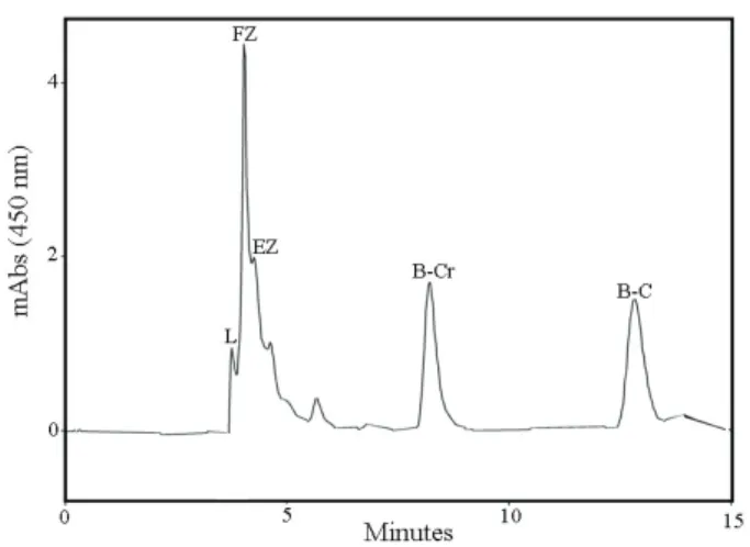
Carotenoid composition (lutein, zeaxanthin,
β-cryptoxanthin and trans-β-carotene) showed a
variation, which was influenced by the culture conditions of the tetrasporophytes. Figure 4 shows a For total soluble protein analyses, the algal
biomass (75 mg, n=3) was ground with liquid nitrogen, and extractions were carried out at 4 °C using 0.2 M phosphate buffer (pH 8.0) containing 5 mM EDTA and 1 mM DTT. Buffer was added in the ratio of 10 ml g-1
fresh biomass and the homogenates were centrifuged at 12000 rpm for 15 min. Total soluble protein contents were determined according to Bradford (1976), using a Bio-Rad protein assay kit and BSA as standard.
Data analysis
Data were analyzed by bifactorial Analysis of Variance (ANOVA) and the Student-Newman-Keuls´ test (Zar, 1999). All statistical analyses were performed using the Statistica software package (Release 6.0), considering p≤0.05. Homogeneity of the variance was tested using Levene’s test.
Results
Effects of the interaction of PFD and VSES concentration on the growth rates of tetrasporophytes of Gracilaria domingensis were not significant, while high PFD stimulated the growth (Figure 1, Table 1).
Figure 1. Growth rates (% d-1) of apical segments of tetrasporophytes of Gracilaria domingensis cultured in von Stosch medium (25% and 50% VSES) under 50 (white bars) and 100 (black bars) µmol photons m-2 s-1 for four weeks. Values are averages±SD (n= 6), with six segments of 2 cm
per replicate. Different letters indicate significant differences according to the Student-Newman-Keuls´ test (p≤0.05).
Capital letters indicate significant differences between VSES concentrations and lower case letters indicate differences between irradiance levels.
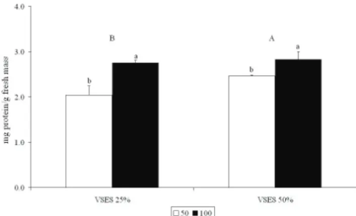
Figure 2. Total soluble protein content in apical segments of tetrasporophytes of Gracilaria domingensis cultured in von Stosch medium (25% and 50% VSES) under 50 (white bars) and 100 (black bars) µmol photons m-2 s-1 for four weeks. Values are averages±SD (n=3), with six segments of 2 cm
per replicate. Different letters indicate significant differences according to the Student-Newman-Keuls´ test (p≤0.05).
Capital letters indicate significant differences between VSES concentrations and lower case letters indicate differences between irradiance levels.
Table 2. Two-away ANOVA of total soluble protein content of tetrasporophytes of Gracilaria domingensis cultured in von
Stosch medium (25 and 50% VSES) under photon lux densities
(PFD) of 50 and 100 µmol photons m-2 s-1 for four weeks.
Variable df F p
VSES 1 9.543 0.014
PFD 1 44.622 0.0001
VSES x PFD 1 4.684 0.062
typical HPLC profile for the carotenoids.
The interaction between PFD and VSES concentration did not affect lutein and trans-β-carotene concentrations (Table 4). Concentrations of both compounds were only influenced by PFD, with the highest concentrations detected at high photon flux density (Figure 5A, E, Table 4). Free zeaxanthin and
β-cryptoxanthin concentrations were influenced by the
interaction between PFD and VSES concentration (Table 4), while treatment with low nutrient concentrations
(25% VSES) under high PFD stimulated the highest accumlation of free zeaxanthin (Figure 5B) and high nutrient concentrations (50% VSES) under high PFD
stimulated the highest β-cryptoxanthin concentration
(Figure 5D). For tetrasporophytes cultured under low PFD, the areas of the esterified zeaxanthin peaks were too small to allow quantification. At high PFD, the esterified zeaxanthin concentrations were the highest at low nutrient concentrations (25% VSES, Figure 5C).
Figure 3. Photosynthetic pigment concentrations in apical segments of tetrasporophytes of Gracilaria domingensis cultured in von Stosch medium (25% and 50% VSES) under 50 (white bars) and 100 (black bars) µmol photons m-2 s-1 for four weeks. Values are averages±SD (n=3), with six segments of 2 cm per replicate. Different letters indicate significant differences
according to the Student-Newman-Keuls´ test (p≤0.05). Capital letters indicate significant differences between VSES
concentrations and lower case letters indicate differences between irradiance levels.
Table 3. Two-away ANOVA of photosynthetic pigment concentrations of tetrasporophytes of Gracilaria domingensis cultured in
von Stosch medium (25 and 50% VSES) under photon lux densities (PFD) of 50 and 100 µmol photons m-2 s-1 for four weeks.
Variable df Phycoerythrin Phycocyanin Allophycocyanin Chlorophyll a
F p F p F p F p
VSES 1 0.905 0.369 1.699 0.228 2.993 0.121 0.201 0.665
PFD 1 18.196 0.002 10.214 0.012 8.038 0.021 4.921 0.0573
Figure 4. HPLC chromatogram for carotenoids detected in tetrasporophytes of Gracilaria domingensis cultured in 25% VSES under 100 µmol photons m-2 s-1. Detection wavelength 450 nm. L, lutein; FZ, free zeaxanthin; EZ, esteriied zeaxanthin; β-Cr, β -cryptoxanthin; β-C, trans-β-carotene.
Discussion
Irradiance was a limiting factor for growth of G. domingensis and the highest growth rate was observed at high PFD. G. domingensis is found in the intertidal region and it is exposed to high irradiances. Moreover, growth of G. domingensis was not inluenced by nutrient concentrations. Similarly, growth rates of G. foliifera var. angustissima (Harv.) Taylor (Lapointe, 1981) were
not inluenced signiicantly by an increase in nitrate
concentrations.
Light (PFD) inluenced phycobiliprotein
concentrations in Gracilaria domingensis. The highest concentrations of phycoerythrin, phycocyanin and allophycocyanin were observed in tetrasporophytes cultured at low PFD. Similar results have also been reported for other Gracilaria species (Zou & Gao, 2009) and could be related to a photoacclimation process at low irradiance. Concentrations of chlorophyll a in G. domingensis were
not inluenced by irradiance and nutrient levels and were
lower than the phycobiliprotein contents.
Figure 5. Carotenoid concentrations in apical segments of tetrasporophytes of Gracilaria domingensis cultured in von Stosch medium (25% and 50% VSES) under 50 (white bars) and 100 (black bars) µmol photons m-2 s-1 for four weeks. Values are averages±SD (n=6), with six segments of 2 cm per replicate. Different letters indicate significant differences according to
the Student-Newman-Keuls´ test (p≤0.05). Capital letters indicate significant differences between VSES concentrations, and
The relationship between pigment content and nutrient availability has been investigated in several studies (Jones et al., 1996; Godillo et al., 2006), indicating that Gracilaria species are able to assimilate nitrogen and, when present in excess, nitrogen is stored as proteins or pigments (Kosovel & Talarico, 1979). However, this is not the only factor determining the pigment concentrations. Light plays a key role and exerts an effect opposite to that of nitrogen (Talarico & Maranzana, 2000), as observed in our results. According to Lapointe (1981), the interaction between these two factors affects the pigment concentrations and increases or decreases in these concentrations are related to the photosynthetic capacity.
Accumulation of total soluble protein was
inluenced by the interaction between PFD and nutrient
availability. The highest protein concentration was observed at high nutrient concentration (50% VSES) under high PFD. As discussed previously, Gracilaria species have the capacity to store nitrogen during periods of high nitrogen availability. Under optimal conditions for photosynthesis, there is no need to store nitrogen as phycobiliproteins and G. domingensis tetrasporophytes then store the excess nitrogen preferentially in the form of proteins. Andria et al. (1999) observed that the total soluble protein content of Gracilaria sp. decreased when the species was cultured under conditions of limited availabilityof nitrogen.
High levels of irradiance stimulated carotenoid accumulation in G. domingensis tetrasporophytes. Carotenoid concentrations were lower than
phycobiliprotein concentrations, relecting the carotenoid
function of protecting the photosynthetic apparatus. Our
results show that the β-carotene pathway was more active, since the concentrations of zeaxanthin, β-cryptoxanthin and β-carotene were higher than that of lutein. According
to Demming-Adams (1990), zeaxanthin is a key pigment involved in the photoprotective response to the stress caused by high irradiance, because xanthophyll is
more eficient at dissipating the excess energy (Frank
et al., 2001). The high content of zeaxanthin during the acclimation to high irradiances suggests a response to the stress caused by excess light in G. domingensis. In a similar experiment, Carnicas et al. (1999) also observed an increase in zeaxanthin concentrations with increasing irradiance in G. tenuistipitata var. liui Zhang & B.M. Xia.
The decrease in concentration of free zeaxanthin in G. domingensis cultured under conditions of low nutrient availability (25% VSES) and low PFD and the
reduction in β-cryptoxanthin concentration under low
nutrient availability (25% VSES) and high PFD could be a photoacclimation response to low nutrient availability.
In conclusion, our results indicate that light is a limiting factor for G. domingensis growth, that variations in phycobiliprotein contents under different irradiance levels are related to photoacclimation process, and that higher carotenoid contents at high irradiances are due to photoprotection mechanism.
Acknowledgments
The authors thank the Conselho Nacional de
Desenvolvimento Cientíico e Tecnológico (CNPq, Brazil)
for grants (Proc. 485927/07-1) and fellowships (Proc. 381175/2007-3, 382175/2008-5, 140824/2009-0) to the
irst author. This study is part of the thesis presented by the irst author to the Graduate Programme in Plant
Biodiversity and Environment, Institute of Botany, São Paulo, Brazil.
References
Andersson M, Schubert H, Pedersén M, Snoeijs P 2006. Different
patterns of carotenoid composition and photosynthesis acclimation in two tropical red algae. Mar Biol 149: 653-665.
Andria JR, Vergara JJ, Perez-Llorens JL 1999. Biochemical responses and photosynthetic performance of Gracilaria
sp. (Rhodophyta) from Cádiz, Spain, cultured under different inorganic carbon and nitrogen levels. Eur J Phycol 34: 497-504.
Bradford MM 1976. A rapid and sensitive method for the quantitation of microgram quantities of protein utilizing the principle of protein-dye binding. Anal Biochem 72: 248-254.
Brinkhuis BH 1985. Growth patterns and rates. In Littler MM, Littler DS (eds.) Handbook of Phycological Methods. London: Cambridge University Press, p. 461-478. Carnicas E, Jimenez C, Niell FX 1999. Effects of changes of
irradiance on the pigment composition of Gracilaria tenuistipitata var. liui Zhang et Xia. J Photoch Photobiol 50: 149-158.
Demming-Adams B 1990. Carotenoids and photoprotection in plants: a role for the xanthophyll zeaxanthin. Biochim Table 4. Two-away ANOVA of carotenoid concentrations of tetrasporophytes of Gracilaria domingensis cultured in von Stosch
medium (25 and 50% VSES) under photon lux densities (PFD) of 50 and 100 µmol photons m-2 s-1 for four weeks.
Variable df Lutein Free zeaxanthin β-criptoxanthin β-carotene
F p F p F p F p
VSES 1 2.742 0.136 8.565 0.022 62.623 0.000047 0.343 0.574
PFD 1 327.370 0.000 135.584 0.000008 643.148 0.000 357.044 0.000
Biophys Acta 1020: 1-24.
Denault M, Stieve E, Valiela I 2000. Effects of nitrogen load and irradiance on photosynthetic pigment concentrations in
Cladophora vagabunda and Gracilaria tikvahiae in estuaries of Waquoit Bay. Biol Bull 199: 223225. Edwards P 1970. Illustrated guide to the seaweeds and seagrass
in the vicinity of Porto Aransas, Texas. Contrib Mar Sc Austin 15: 1-228.
Ferreira LB, Barui JB, Plastino EM 2006. Growth of red and
green strains of the tropical agarophyte Gracilaria cornea J. Agardh (Gracilariales, Rhodophyta) in laboratory. Rev Bras Bot 29: 187-192.
Frank H, Das SK, Bautista S, Bruce D, Vasil’ev S, Crimi M, Croce R, Bassi R 2001. Photochemical behaviour of xanthophylls in the recombinant photosystem II antena complex, CP26. Phytochemistry 40: 1220-1225. Godillo FJ, Aguilera J, Jiménez C 2006. The response of nutrient
assimilation and biochemical composition of Artic seaweeds to a nutrient input in summer. J Exp Bot 57: 2661-2671.
Gressler V, Yokoya NS, Fujii MT, Colepicolo P, Mancini Filho
J,Torres RP, Pinto E 2010. Lipid, fatty acid, protein, amino acid and ash contents in four Brazilian red algae species. Food Chem 120: 585-90.
Guimarães M, Plastino EM, Oliveira EC 1999. Life-history, reproduction, and growth of Gracilaria domingensis
(Gracilariales, Rhodophyta) from Brazil. Bot Mar 42: 481-486.
Guimarães M, Plastino EM, Destombe C 2003. Green mutant frequency in natural populations of Gracilaria domingensis (Gracilariales, Rhodophyta) from Brazil.
Eur J Phycol 38: 165-169.
Hulshof PJM, Kosmeijer-Schuil T, West CE, Hollman PCH
2007. Quick screening of maize kernels for provitamin A content. J Food Comp Anal 20: 655-661.
Jeffrey SW, Humphrey GF 1975. New spectrophotometric equations for determining chlorophylls a, b, c1 e c2 in higher plants, algae and natural phytoplankton. Biochem
Physiol Pl 167: 191-194.
Jones AB, Dennison WC, Stewart GR 1996. Macroalgal responses to nitrogen source and availability: amino acid
metabolic proiling as a bioindicator using Gracilaria edulis (Rhodophyta). J Phycol 52: 757-766.
Kakita H, Kamishima H 2006. Effects of environmental factors and metal ions on growth of the red alga Gracilaria chorda Holmes (Gracilariales, Rhodophyta). J Appl Phycol 18: 469-474.
Kosovel V, Talarico L 1979. Seasonal variation of photosynthetic
pigments in Gracilaria verrucosa (Huds.) Papenfuss (Florideophyceae, Gigartinales). B Soc Adriat Scien 63: 5-15.
Kuhnen S, Lemos PMM, Campestrini LH, Ogliari JB, Dias PF, Maraschin M 2009. Antiangiogenic properties of carotenoids: a potential role of maize as functional food.
J Funct Foods 1: 284-290.
Kursar TA, van der Meer J, Alberte RS 1983. Light-harvesting system of red alga Gracilaria tikvahiae. I. Biochemical analyses of pigment mutations. Plant Physiol 73: 353-360.
Lapointe BE 1981. The effects of light and nitrogen on growth, pigment content, and biochemical composition of
Gracilaria foliifera v. angustissima (Gigartinales, Rhodophyta). J Phycol 17: 90-95.
Plastino EM, Guimarães M, Matioli SR, Oliveira EC 1999. Codominant inheritance of polymorphic color variants of Gracilaria domingensis (Gracilariales, Rhodophyta).
Gen Mol Biol 22: 105-108.
Scott, CE, Eldridge, AL 2005. Comparison of carotenoid content in fresh, frozen and canned corn. J Food Comp Anal 18: 551-559.
Talarico L, Maranzana G 2000. Light and adaptive responses in red macroalgae: an overview. J Photoch Photobiol B 56: 1-11.
Ursi S, Plastino EM 2001. Growth of reddish and light green strains of Gracilaria sp. (Gracilariales, Rhodophyta) in two culture media: analysis of different reproductive phases. Rev Bras Bot 24: 587-594.
Wilson AJ, Critchley AT 1997. Studies on Gracilaria gracilis
(Stackhouse) Steentoft, Farnham and Irvine and
Gracilaria aculeata (Hering) Papenfuss from southern Africa. I. The influence of temperature, irradiance, salinity and nitrogen-nutrition on growth. S Afr J Bot
63: 465-473.
Zar JH 1999. Biostatistical analysis. New Jersey: Prentice Hall. Zou D, Gao K 2009. Effects of elevated CO2 on the red seaweed
Gracilaria lemaneiformis (Gigartinales, Rhodophyta) grown at different irradiance levels. Phycologia 48: 510-517.
*Correspondence
Nair S. Yokoya
Núcleo de Pesquisa em Ficologia, Instituto de Botânica Caixa Postal 3005, 01031-970 São Paulo-SP, Brazil [email protected]