CdtR Regulates TcdA and TcdB Production in
Clostridium difficile
Shelley A. Lyon, Melanie L. Hutton, Julian I. Rood, Jackie K. Cheung‡, Dena Lyras‡*
Infection and Immunity Program, Biomedicine Discovery Institute and Department of Microbiology, Monash University, Clayton, Victoria, Australia
‡JKC and DL are joint senior authors *[email protected]
Abstract
Clostridium difficileis a global health burden and the leading cause of antibiotic-associated diarrhoea worldwide, causing severe gastrointestinal disease and death. Three well charac-terised toxins are encoded by this bacterium in two genetic loci, specifically, TcdB (toxin B) and TcdA (toxin A) in the Pathogenicity Locus (PaLoc) and binary toxin (CDT) in the geno-mically distinct CDT locus (CdtLoc). Toxin production is controlled by regulators specific to each locus. The orphan response regulator, CdtR, encoded within the CdtLoc, up-regulates CDT production. Until now there has been no suggestion that CdtR influences TcdA and TcdB production since it is not carried by all PaLoc-containing strains and CdtLoc is not linked genetically to PaLoc. Here we show that, in addition to CDT, CdtR regulates TcdA and TcdB production but that this effect is strain dependent. Of clinical relevance, CdtR increased the production of TcdA, TcdB and CDT in two epidemic ribotype 027 human strains, modulating their virulence in a mouse infection model. Strains traditionally from ani-mal lineages, notably ribotype 078 strains, are increasingly being isolated from humans and their genetic and phenotypic analysis is critical for future studies on this important pathogen. Here we show that CdtR-mediated toxin regulation did not occur in other strain back-grounds, including a ribotype 078 animal strain. The finding that toxin gene regulation is strain dependent highlights the regulatory diversity betweenC.difficileisolates and the importance of studying virulence regulation in diverse lineages and clinically relevant strains. Our work provides the first evidence that TcdA, TcdB and CDT production is linked by a common regulatory mechanism and that CdtR may act as a global regulator of viru-lence in epidemic 027 strains.
Author Summary
Clostridium difficileis the leading cause of antibiotic-associated diarrhoea. The TcdB, TcdA and binary toxins produced byC.difficileare encoded in two genomically distinct loci: TcdB and TcdA in the Pathogenicity Locus (PaLoc) and binary toxin (CDT) in the CDT locus (CdtLoc). Toxin production is primarily controlled by regulators specific to each locus. Because the presence of these loci varies amongst different strains ofC.difficile,
a11111
OPEN ACCESS
Citation:Lyon SA, Hutton ML, Rood JI, Cheung JK, Lyras D (2016) CdtR Regulates TcdA and TcdB Production inClostridium difficile. PLoS Pathog 12(7): e1005758. doi:10.1371/journal.ppat.1005758
Editor:Steven R. Blanke, University of Illinois, UNITED STATES
Received:March 20, 2016
Accepted:June 18, 2016
Published:July 14, 2016
Copyright:© 2016 Lyon et al. This is an open access article distributed under the terms of the
Creative Commons Attribution License, which permits unrestricted use, distribution, and reproduction in any medium, provided the original author and source are credited.
Data Availability Statement:All relevant data are within the paper and its Supporting Information files.
Funding:This work was supported by grants from the Australian National Health and Medical Research Council (GNT1065985, awarded to JIR,
no rational link for their co-regulation has ever been proposed. Here we have shown that the regulator of CDT production, CdtR, also regulates production of TcdA and TcdB in a strain dependent manner. These results represent the first evidence that TcdA and TcdB production is linked to the production of CDT by a common regulatory mechanism. Col-lectively, our results establish CdtR as an important virulence regulator in two clinically important, epidemic strains ofC.difficile, and further highlights the need to investigate regulatory mechanisms of important virulence factors in diverse strain backgrounds.
Introduction
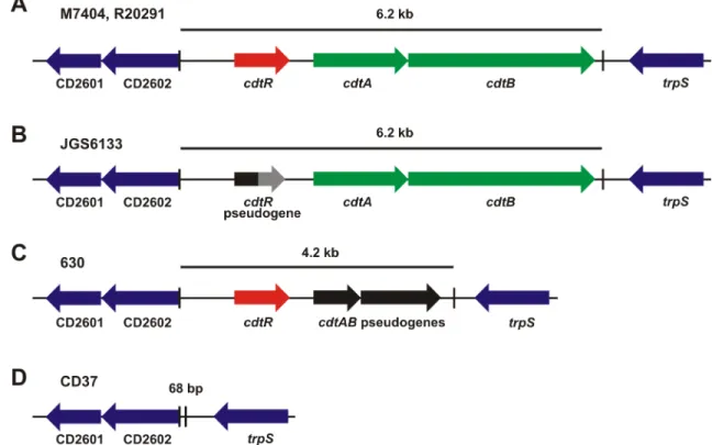
C.difficileantibiotic-associated diarrhoea is a toxin mediated disease [1,2]. During infection, TcdA, TcdB and CDT are secreted into the colonic epithelium by this bacterium, leading to diarrhoea that can progress to serious, life threatening inflammatory diseases, including pseu-domembranous colitis and toxic megacolon [3]. The production of these toxins varies between strains. TcdB is the most commonly encoded toxin and is most often co-located with the TcdA gene in the PaLoc region [4], both toxins act as monoglucosyltransferases that irreversibly modify Rho family members [3]. PaLoc variants that produce TcdB and not TcdA are, how-ever, becoming increasingly common, for example, they represented 23% of strains in one recent study of human strains in China [5]. CDT is encoded in a specific locus, CdtLoc (Fig 1) [6] the carriage of which has also increased significantly over the last decade; in 2004 6% of clinical isolates encoded CDT whereas 33.5% now encode this toxin [7,8]. CDT is an ADP-ribosyltransferase that is not essential for disease, but may be important for colonisation during an infection [9]. CdtLoc is not carried by all PaLoc-containing strains and it is not linked genet-ically to PaLoc.
Regulation of toxin production inC.difficileis somewhat strain dependent, suggesting that toxin regulatory mechanisms have evolved independently to modulate pathogenesis [10–13]. The TcdR alternative sigma factor and TcdC anti-sigma factor, which are encoded withtcdA
andtcdBin the PaLoc, act as the primary mechanism controlling the production of these toxins [14,15]. TcdA and TcdB regulation has also been linked to many important cellular processes in theC.difficilelife cycle, including sporulation, by Spo0A, the master sporulation regulator, motility,viathe flagella regulator SigD, and nutrient acquisition, by the regulators of carbon and amino acid metabolism, CcpA and CodY [10,11,16–20]. The ribotype 027 strains associ-ated with epidemics of severe CDI appear to be more virulent than strains previously isolassoci-ated, a phenotype that has been partly attributed to increased TcdA and TcdB production [21–23]. By comparison, little is known about the regulation of CDT production beyond the involve-ment of CdtR, and until now, no link had been identified between the control of CDT, TcdA and TcdB production. The difficulty in genetically manipulating strains from differentC. diffi-cileclonal lineages has also prevented a broader analysis of the role of this regulator across dif-ferent strain types.
In this study, we investigated the role of CdtR in different strains ofC.difficileincluding two epidemic ribotype 027 strains. As expected, CdtR was found to regulate the production of CDT. Surprisingly, however, CdtR also regulated the production of the PaLoc encoded toxins, TcdA and TcdB, in the two ribotype 027 strains. Further analysis showed that regulation occurred at the transcriptional level and probably resulted from indirect regulation of the posi-tive regulator of PaLoc gene expression, TcdR. Importantly, further analysis showed the impor-tance of CdtR forC.difficilepathogenesis, withcdtRmutants causing less severe disease than the wild type strain in a mouse infection model. To determine whether CdtR function is
CdtR-mediated Toxin Regulation inClostridium difficile
conserved across evolutionarily diverse isolates, ribotype 078 (JGS6133) and 012 (630) strains were investigated. CdtR regulated CDT production in the ribotype 078 strain; the ribotype 012 strain does not encode CDT. Notably, and in contrast to the ribotype 027 strains, CdtR did not regulated TcdA or TcdB production in either strain background, highlighting the regulatory variation of key virulence factors betweenC.difficilestrains. These results highlight the impor-tance of investigating regulatory mechanisms in clinically important strains ofC.difficileand suggest that CdtR-mediated toxin regulation is an important virulence mechanism in the epi-demic ribotype 027 strains.
Results
CdtR regulates production of CDT, TcdA and TcdB in two epidemic,
ribotype 027 strains of
C
.
difficile
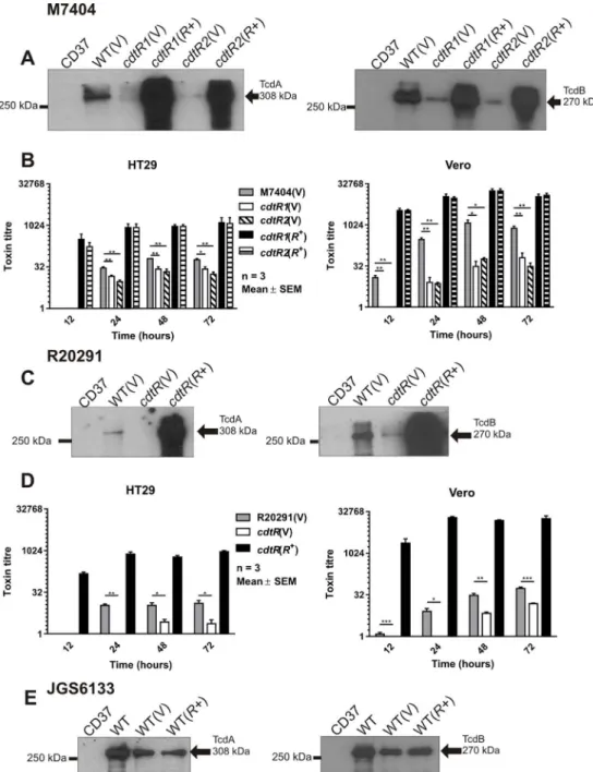
To investigate the role of CdtR in the regulation of CDT production in the epidemic ribotype 027 strains, we constructed two independentcdtRmutants in the Canadian isolate M7404 and acdtRmutant in the UK isolate R20291 and confirmed their genotype by Southern hybridisa-tion (S1 Fig). Western blot analysis showed that thecdtRmutants produced less CDTa and CDTb compared to the wild type and that complementation withcdtRintransresulted in over-expression of both toxin subunits (Fig 2A and 2B). Consistent with these results, ADP-ribosyltransferase assays demonstrated that thecdtRmutants had significantly reduced levels of CDT activity compared to the wild type, while the complementedcdtRmutants showed
Fig 1. Schematic representation of the CDT loci from representative strains. a,The full length CdtLoc from the ribotype 027 strains (M7404 and R20291).b,The CdtLoc from the ribotype 078 strain, JGS6133. ThecdtRpseudogene is shown in black and is grey after the premature stop codon.c,The CdtLoc from the ribotype 012 strain 630 carryingcdtAB
pseudogenes (shown in black).d,The CdtLoc in the CDT negative strain CD37 is replaced with a 68 bp sequence. The boundaries of the CdtLoc are indicated with vertical lines and the flanking genes are blue.
CDT activity greater than that of the wild type strain (Fig 2D and 2E). Overall, these data show, for the first time, that CdtR is important for regulating CDT production in epidemic ribotype 027C.difficilestrains.
Unexpectedly, Western blots performed using TcdA- and TcdB-specific antibodies showed that thecdtRmutants produced less TcdA and TcdB than the wild type, while the comple-mentedcdtRderivatives expressed high levels of both toxins (Fig 3A and 3C). Cytotoxicity assays were performed using HT29 and Vero cells to measure the activity of TcdA and TcdB, respectively, in the culture supernatants from the isogenic panel of M7404 and R20291 strains. The TcdA and TcdB activities of all of strains increased over time and, consistent with the Western blot results, showed lower activity in supernatants from thecdtRmutants compared to the wild type, confirming that thecdtRmutants were less cytotoxicin vitro(Fig 3B and 3D). TcdA and TcdB activity of the complementedcdtRmutants was consistently higher than the
cdtRmutants and the wild type across all time points (Fig 3B and 3D). These data show that CdtR regulates the production of TcdA and TcdB in both M7404 and R20291, which is the first demonstration of a common regulator modulating the expression of all three toxins inC.
difficile.
CdtR positively regulates expression of genes from the pathogenicity
locus (PaLoc) of
C
.
difficile
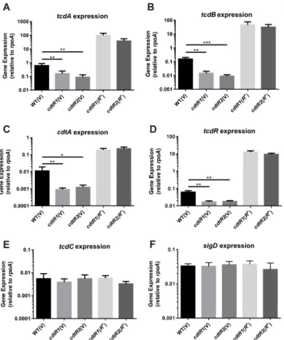
To investigate the molecular mechanism of regulation, we determined if CdtR controlled toxin production at the transcriptional level. Using the isogenic panel of M7404 strains, reverse-tran-scription droplet digital PCR (RT-ddPCR) analysis was employed to quantitatively compare
Fig 2. Analysis of CDT production. a–c,Western immunoblot using CDTa-specific and cross-reactive Ib-specific antibodies and precipitated supernatants from the strains indicated. CD37 (non-toxigenic), V = vector control,R+ =cdtRcomplemented. The arrows indicate the 48 kDa CDTa and 99 kDa CDTb proteins.d–f,
CDT activity assessed by ADP-ribosyltransferase assay. Samples were separated by SDS-PAGE and biotinylated (ADP-ribosylated) actin detected by HRP-streptavidin. Relative CDT activity was assessed by densitometry compared to the non-toxigenic control strain CD37. A = actin, A-A = ADP-ribosylated actin. Data represent the mean±SEM (n = 3).*, p0.05.
doi:10.1371/journal.ppat.1005758.g002
the level of expression of each of the toxin encoding genes (tcdA,tcdB,cdtA) and the PaLoc-encoded toxin regulators (tcdR,tcdC). The relative transcription of all three toxin genes and
tcdRwas significantly decreased in bothcdtRmutants compared to the wild type (Fig 4A–4D). Over-expression ofcdtRin the complemented strains resulted in a dramatic over-expression of
Fig 3. Analysis of TcdA and TcdB production. a, c, e,Western immunoblot using TcdA-specific and TcdB-specific antibodies with precipitated supernatant from the strains indicated. CD37 (non-toxigenic), V = vector control,R+ =cdtRcomplemented. Arrows indicate the 308 kDa TcdA and 270 kDa TcdB proteins.
Supernatants were collected at 12, 24, 48 and 72 hours post inoculation and assayed by doubling dilution cytotoxicity assays.b,The panel of M7404 strains were assayed using HT29 cells and Vero cells.d, R20291 panel of strains assayed using HT29 cells and Vero cells. Data represent the mean±SEM (n = 3–5).*, p0.05;**, p0.01;***, p0.001.
tcdA,tcdB,cdtAandtcdR(Fig 4A–4D). Although TcdC is predicted to be non-functional in the 027 strains [12], we analysed the expression oftcdCand found it to be similar in the iso-genic panel of strains (Fig 4E). Previous work in strain 630Δerm, a derivative of the non-027 historical strain 630 which belongs to ribotype 012, showed that the flagella synthesis regulator, SigD, is an important regulator of TcdA and TcdB productionviathe regulation oftcdR
expression [24]. Analysis of our isogenic panel of M7404 strains showed no change insigD
transcription, suggesting that CdtR does not influence the expression ofsigDin this strain background and that therefore the modulation oftcdAandtcdBexpression does not occurvia
SigD (Fig 4F). Collectively, these results indicate that regulation of all three toxins by CdtR in M7404 occurs at a transcriptional level and that the regulation oftcdAandtcdBoccursviathe upregulation oftcdRtranscription.
CdtR is important for modulating
C
.
difficile
virulence
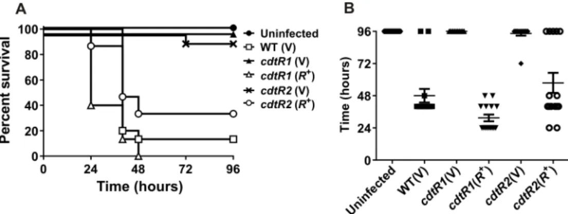
Our observation that CdtR increases toxin production in ribotype 027 strains has important implications for the virulence capacity of these clinically important strains. To determine if the modulatory effect of CdtR on toxin production influencesC.difficiledisease, we examined whethercdtRinactivation altered virulence in our mouse infection model [2]. It was previously
Fig 4. Transcriptional analysis of M7404cdtRmutant and complemented strains compared to wild-type.RNA was isolated from strains for analysis of (a)tcdA, (b)tcdB, (c)cdtA, (d)tcdR, (e)tcdCand (f)sigD
expression. Levels of gene expression were normalised torpoA. Data represent the mean normalised gene expression±SEM from five independent biological replicates.*, p0.05;**, p0.01;***, p0.001. doi:10.1371/journal.ppat.1005758.g004
shown that infection with acdtAmutant of M7404 resulted in disease that was indistinguish-able from the parent strain [2]. This mutant no longer produced CDT, but had an intactcdtR
gene and continued to produce TcdA and TcdB at wild type levels. A reduction in CDT levels is therefore not likely to have a major effect on disease in our animal model. All of the mice infected with the isogeniccdtR-series of M7404 derivatives were colonised withC.difficileat similar levels (S2 Fig). Wild type-infected mice rapidly lost weight and the majority were eutha-nized 40 to 48 hours post infection in accordance with animal ethics guidelines, with a mean time to death of 48 ± 5.1 hours and a survival rate of 13% (Fig 5). Mice infected with either of the two independentcdtRmutants had significantly higher survival rates (Mantel-Cox log rank test,P<0.0001) of 100% and 96% forcdtR1(V) andcdtR2(V), respectively, and showed no
overt signs of disease nor significant weight loss (Fig 5). By comparison, mice infected with either complemented mutant,cdtR1(R+) orcdtR2(R+), had a wild-type virulence phenotype, with marked weight loss, and a mean time to death of 31.5 ± 2.5 hours and 57.6 ± 7.5 hours, respectively, reflected in survival rates of 0% and 33% (Fig 5).
Damage to the colon and caecum ofC.difficile-infected mice results from the production of TcdA and TcdB [2]. We therefore performed histopathological analysis to assess the damage to colonic and caecal tissues collected from the groups of infected and uninfected mice under study here. All tissues were de-identified and independently scored using a previously defined set of scoring parameters that included overall tissue damage, polymorphonucleocyte (PMN) influx, crypt damage and oedema [2]. Tissues of uninfected mice only had minimal surface damage to the intestinal epithelia resulting from the disruption of microbiota by antibiotic pre-treatment and tissue processing (Fig 6A) with low colon and caecum damage scores of 4.7 and 3.8, respectively (Fig 6B and 6C). By comparison, wild type-infected mice had severely
inflamed tissues, with extensive damage to the epithelial surface, crypt branching and hyperpla-sia, goblet cell loss, significant PMN influx and mucosal and sub-mucosal oedema (Fig 6A). These histopathologies were reflected in the high damage scores of 12.9 and 13.6 for their colonic and caecal tissues, respectively (Fig 6B and 6C).
Mice infected with eithercdtRmutant had tissue architecture similar to that seen with the uninfected mice. Very little colonic and caecal damage was observed, with some surface epithe-lial damage to crypts occurring and no apparent crypt hyperplasia, and little PMN influx into the mucosa (Fig 6A), with correspondingly low colonic (6.0 and 5.3) and caecal (6.6 and 7.1) damage scores (Fig 6B and 6C). However, mice infected with eithercdtR-complemented
Fig 5. Virulence of M7404 wild-type,cdtRmutant and complemented strains in mice. a, Kaplan-Meier survival curve showing time from infection to euthanasia of mice infected with different strains ofC.difficilein hours. (n = 15).b, Time from inoculation of mice to death in hours. Data represents mean±S.E.M. (n = 15). Data represent the mean±SEM****, p0.0001.
Fig 6. Histopathology ofC.difficileinfected tissues.Representative images of sections of colon and caeca collected from uninfected mice or mice infected with different strains ofC.difficile, fixed and strained with PAS-Alcian blue. Red brackets ([) indicate crypt hyperplasia, arrow heads (▲) represent surface epithelial damage and asterisks (*) represent oedema and inflammation. Scale bars (200μm) are shown in yellow. Histopathology damage scores from uninfected or infected colons (b) and caeca (c). Data represent the mean±SEM****, p0.0001.
doi:10.1371/journal.ppat.1005758.g006
mutant had similar levels of tissue damage to mice infected with the wild-type strain. Severe crypt damage was observed in the majority of these tissues, particularly the caeca, with crypt hyperplasia, loss of goblet cells, PMN influx and severe oedema in the mucosa and sub-mucosa (Fig 6A) and high histopathological damage scores were determined for both the colon (11.8 and 9.6) and the caecum (14.5 and 11.4) (Fig 6B and 6C).
To confirm that the reduced virulence of thecdtRmutants can be attributed to reduced lev-els of TcdA and TcdB productionin vivo, the cytotoxicity of the intestinal contents collected from mice infected with the panel ofC.difficilestrains was assessed. No cytotoxicity was observed against HT29 or Vero cells by faecal samples collected from uninfected mice whereas mice infected with the wild type strain showed high levels of cytotoxicity against HT29 and Vero cells, indicating the production of TcdA and TcdBin vivo(S4 Fig). By comparison, sam-ples collected from mice infected with the twocdtRmutants (cdtR1(V) andcdtR2(V)) showed reduced cytotoxicity against HT29 or Vero cells, indicating decreased levels of TcdA and TcdB productionin vivocompared to the wild type strain (S4 Fig). Intestinal contents collected from mice infected with the two complementedcdtRmutants ((cdtR1(R+) andcdtR2(R+)) showed increased levels of cytotoxicity against HT29 and Vero cells, indicating restoredin vivoTcdA and TcdB production. Collectively, the survival data, histopathological andin vivocytotoxicity analysis support the hypothesis that CdtR modulates the virulence of theC.difficileribotype 027 strain M7404 due to the role that it plays in regulating TcdA and TcdB production.
CdtR differentially regulates toxin production in a ribotype 078 strain of
C
.
difficile
To investigate whether TcdA and TcdB regulation by CdtR occurs in other strains, we assessed this phenotype in a ribotype 078 animal isolate, JGS6133. Ribotype 078 strains are commonly isolated from animals, but have also been implicated in severe human infections [25,26]. Our sequencing analysis revealed that thecdtRgene in JGS6133 contains a naturally occurring stop codon mutation at codon 322, which has been described previously in other 078 strains [27], and results in a 142 amino acid truncation that is likely to result in a non-functional protein (Fig 1B). Western blots showed that JGS6133 complemented with the full lengthcdtRgenein trans, and hence producing functional CdtR, had significantly more CDTa and CDTb than the wild-type and vector control strains, which produced similar amounts of both proteins (Fig 2C). Increased CDT production by the JGS6133cdtR+derivative was reflected in increased toxin activity as assessed by ADP-ribosyltransferase assays (Fig 2F). By contrast, production of TcdA and TcdB was not altered in the JGS6133cdtR+strain compared to the wild type (Fig 3E). This result suggests that CdtR-mediated regulation of TcdA and TcdB production is not conserved within all strains ofC.difficile.
CdtR does not regulate TcdA and TcdB production in strain 630
To determine whether CdtR regulates TcdA and TcdB production in strains ofC.difficile
withcdtABpseudogenes we transferred thecdtRcomplementation vector, pJIR4218, into strain 630. Western blot analysis showed that derivatives of 630 over-expressingcdtRhad similar lev-els of TcdA and TcdB production as the isogenic vector control strains (S3A and S3B Fig). RT-ddPCR analysis confirmed thatcdtRwas over-expressed in these strains relative to the wild type and the vector control (S3C Fig). Taken together these results suggest that CdtR is not important for the regulation of TcdA and TcdB production in strain 630. Since CdtR is also not important for TcdA and TcdB regulation in a ribotype 078 strain, regulation of these toxins by CdtR may be specific to the ribotype 027 strains.
Discussion
The emergence of epidemic ribotype 027 strains over a decade ago prompted several investiga-tions of the genetic and phenotypic characteristics that may have led to the global dominance of these strains. These features may include higher sporulation rates, resistance to key antibiot-ics and unique aspects of toxin regulation [11,12,29–31]. The presence of a full length CdtLoc was also initially considered to be important in this regard because it encodes CDT [21,23], however, despite numerous studies, the importance of this toxin in virulence remains unde-fined [2,32,33]. The results of the work presented here suggest that CdtLoc, and specifically CdtR, may play an indirect but significant role in disease pathogenesis of the ribotype 027 strains by regulating TcdA and TcdB production.
Our work confirms that CdtR enhances CDT production. Strikingly, ribotype 078 strains produce CDT even though they contain a conserved mutation incdtRand they have CDT activity that is not significantly different from that of strains without this mutation [27,34]. Our data supports these observations since CDT production was detected from the ribotype 078 strain JGS6133 which contains this naturally occurringcdtRmutation. However, CDT pro-duction from JGS6133 was enhanced when functional CdtR was expressed in this strain. Even though CdtR is not essential for CDT production, our work suggests that the presence of this regulator increases the expression of this toxin.
Although CdtR was previously shown to be an important regulator of CDT production [6] there was no evidence to suggest that it played a role in TcdA or TcdB production, particularly since the pathogenicity loci encoding these toxins are not genomically linked. Our data clearly show that CdtR is an important regulator of TcdA and TcdB production, as well as CDT, in two ribotype 027 strains, and that this regulatory capacity plays a role in virulence since inacti-vation ofcdtRattenuated the virulence of strain M7404 in a mouse infection model. The results obtained with the mouse infection experiments are directly relevant to the disease-causing capacity of these strains. This is a significant finding as it is the first report of a regulatory link between the two pathogenicity loci, PaLoc and CdtLoc.
The ability to regulate toxin production through a variety of mechanisms may provide a selective advantage toC.difficilesince it may allow virulence factors to be produced in response to different and specific environmental cues. Ribotype 027 strains appear to have evolved to differ in their regulatory responses in comparison to otherC.difficilelineages.C.difficile regu-lates toxin production in response to many environmental stimuli, including metabolisable carbon sources and quorum signalling molecules, through several different regulatory proteins [20,35,36]. TcdR is the primary positive regulator of TcdA and TcdB production, while TcdC represses toxin production [12,15]. TcdR is highly conserved between strains and many regula-tors directly influence its expression [20,24,27,35]. Our data suggest that CdtR may regulate TcdA and TcdB production by controllingtcdRexpression. CdtR belongs to the LytTR family of DNA binding response regulators and may function by binding to thetcdRpromoter,
thereby regulating TcdR expression and, consequently,tcdAandtcdBexpression [6]. However, we could not identify canonical LytTR DNA binding sites upstream of thetcdR,tcdAortcdB
genes. Similarly, LytTR DNA binding sequences with the conserved sequence and spacing could not be identified within the promoters of other genes identified to regulate toxin produc-tion inC.difficilein other studies, includingsigD,codYorccpA. It may be that the CdtR bind-ing sites are too dissimilar to typical LytTR sites to be identified or that CdtR does not directly bind to these regions; instead, an unidentified, CdtR-controlled intermediate regulator may be modulating PaLoc toxin gene expression.
CdtR-mediated TcdA and TcdB regulation may have specifically evolved in ribotype 027C.
difficilestrains since the co-regulation of these toxins with CDT appears to be ribotype specific. CdtR does not play a similar role in other ribotypes tested here to that seen in ribotype 027 strains and this phenotype does not appear to be conserved between divergent strain back-grounds. The strains included in our assessment belong to three of the five defined evolution-ary clades ofC.difficile, specifically, clade 2 for ribotype 027 (M7404 and R20291), clade 5 for ribotype 078 (JGS6133) and clade 1 for ribotype 012 (630) [37]. Our observations in strain 630 are particularly relevant; many studies are performed using this isolate because it is relatively easy to genetically manipulate. It is clear from our research and other studies [10,11] that strain 630 characteristics may not always reflect those of other strains. Similar observations have been made for clade 5 ribotype 078 strains, which are genetically and phenotypically divergent from strains belonging to other clades [11,30,38,39].
Although the global regulators CodY and CcpA are conserved in strains ofC.difficile, it has been shown that these regulators control toxin production experimentally only in a strain 630 background and their role in other strains, including the 027 and 078 strains, is not known [19,20]. Similarly, several flagella structural and regulatory genes, including SigD, have only been linked to toxin production inC.difficilein strain 630 [24]. While the genetic organisation of the flagella genes within the F1 and F3 flagella regions are similar in strain 630 and the 027 strains, the sequence variation in these regions is thought to contribute to their different motil-ity phenotypes [30]. By comparison, the F3 region, which contains several genes involved in toxin regulation in strain 630, is absent in the 078 strains and is thought to explain the lack of motility in these strains [30]. It has been shown that several of the conserved flagella structural proteins encoded in the F1 and F3 flagella regions, including FliC, FliD and FlgE, are important for toxin production in strain 630 but do not contribute to toxin production in the 027 strain, R20291 [10]. Further research is required to determine if other conserved flagella genes, known to regulate toxin production in strain 630, play a similar role in 027 and 078 strain
backgrounds.
To date, only one study has investigated the regulation of toxin production in an 078 strain and showed that the master sporulation regulator, Spo0A, differentially regulates toxin produc-tion in an 078 strain, two epidemic 027 strains and a strain 630 derivative [11]. Dingle et al. [40] found that strains from clade 5, including the 078 strains, carry a PaLoc similar to that found in other ribotypes but that genes outside of this region are highly divergent. It was sug-gested that the 078 strains may have originated from a divergent, non-toxigenic strain that obtained the PaLoc in a separate event in comparison to other toxigenic lineages. We present data supporting the concept that strains from this background have evolved different toxin reg-ulatory mechanisms from the more commonly studied 027 strains and strain 630 derivatives.
In conclusion, we have provided the first evidence that TcdA and TcdB production is linked to the production of CDT by a common regulatory mechanism and that CdtR acts as a global regulator of toxin production and virulence in two ribotype 027 strains. The observed differ-ences in virulence between the ribotype 027 strains and other historical isolates have been attributed, in part, to elevated toxin production, mainly as a result of mutations in thetcdC
gene [21–23]. Another key genetic difference identified between these strains is the possession of a full length CdtLoc [30]. Our results suggest that possession of the CdtLoc in the 027 strains enhances virulence by the CdtR-mediated up-regulation of TcdA and TcdB production. There-fore, we postulate that the ability of the epidemic ribotype 027 strains to coordinate production of all knownC.difficiletoxins, CDT, TcdA and TcdB, by CdtR is a key factor in the increased virulence of these strains.
Methods
Bacterial strains and growth conditions
All bacterial strains are defined inTable 1. Culture media were from Oxoid or Becton Dickin-son (BD) and all antibiotics and supplements used are from Sigma-Aldrich, Amresco or Merck unless otherwise stated.E.coliandB.subtilisstrains were cultured at 37°C in 2xYT media [41] supplemented with either chloramphenicol (25 µg/ml forE.coli; 5μg/ml forB.subtilis) or
tet-racycline (10 µg/ml).C.difficilestrains were cultured in HIS broth[42] or on HIS agar supple-mented with 0.1% (w/v) L-cysteine and 0.375% (w/v) glucose or TY broth[1] with
D-cycloserine (250 µg/ml), thiamphenicol (10 µg/ml), lincomycin (50 µg/ml) or anhydrous tetra-cycline (50 ng/ml), as required.C.difficilecultures were grown in a Don Whitley A35 Anaero-bic Workstation in an atmosphere of 10% (v/v) H2, 10% (v/v) CO2and 80% (v/v) N2at 37°C.
Polymerase Chain Reaction (PCR)
All oligonucleotide primers are listed inTable 2. PCR cycling conditions (unless otherwise stated) were as follows: initial denaturation step at 94°C for 4 min, followed by 30 cycles of denaturation at 94°C for 30 sec, an annealing step at 50°C for 30 sec and an extension step at 72°C for 1 min per 1 kb. A final extension step was performed at 72°C for 10 minutes. PCRs were performed with Phusion DNA polymerase (New England Biolabs) and 2x Failsafe PCR buffer E (Epicentre Biotechnology). Splice-overlap extension (SOE)-PCR to re-target the Tar-getron was performed as described in the TargeTron Gene Knockout System users guide (Sigma-Aldrich) with modifications as previously described [43].
Isolation and manipulation of nucleic acids
Plasmid DNA was isolated fromE.coli,B.subtilisandC.difficileusing QIAprep spin miniprep columns (Qiagen) following the manufacturer’s instructions. Genomic DNA was isolated from
C.difficileas previously described [44]. Standard methods of DNA digestion, modification and ligation were used. DNA sequencing was carried out using BigDye Terminator v3.1 Ready Reaction Mix (Applied Biosystems) following the manufacturer’s instructions. Sequencing reactions were resolved on an Applied Biosystems 3730 DNA Analyzer. Sequences were ana-lysed using ContigExpress (Invitrogen).
Construction of recombinant plasmids
All plasmids are outlined inTable 1. Construction of thecdtRTargeTron plasmid was per-formed as previously described, with some modifications [43]. Briefly, the group II intron from pDLL45 was retargeted by SOE-PCR to insert between nucleotides 288 and 289 of thecdtR
gene using the primer pairs JRP5448 and JRP3867 and JRP5449 and JRP5450 (Table 2) to gen-erate a 350 bp product, which was digested withBsrGI andHindIII and cloned into the corre-sponding sites of pDLL45, resulting in pJIR4135. AStuI-HindIII fragment was then sub-cloned from pJIR4135 into the corresponding sites of pDLL55, resulting in pJIR4153.
ThecdtRcomplementation plasmid was constructed by PCR amplifying thecdtRgene and approximately 300 bp of its promoter region fromC.difficileM7404 using the primers JRP5632 and JIR5633 (Table 2). The resulting 1.1 kb fragment was purified using a PCR
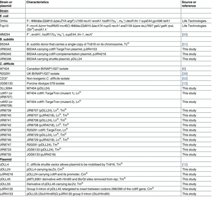
Table 1. Bacterial strains and plasmids.
Strain or plasmid
Characteristics Source or
reference
Strain
E.coli
DH5α F–Φ80dlacZΔM15Δ(lacZYA-argF)U169 recA1 endA1 hsdR17(rK–
, mK+)deoR thi-1 supE44 gyrA96 relA1 Life Technologies Top10 F–mcrAΔ(mrr-hsdRMS-mcrBC)Φ80lacZΔM15ΔlacX74 nupG recA1 araD139Δ(ara-leu)7697 galU galK rpsL
(StrR)endA1λ– Life Technologies
MM294 F–
,endA1,hsdR17(rK–
mK+),supE44,thi-1,recA+ [50]
B.subtilis
BS34A B.subtilisdonor that carries a single copy of Tn916on its chromosome, TcR [51]
JIR6342 BS34A carryingcdtRTargeTron plasmid, pJIR4153 This study
JIR6345 BS34A carryingcdtRcomplementation plasmid, pJIR4218 This study
JIR6346 BS34A carrying shuttle plasmid, pDLL24 This study
C.difficile
M7404 Canadian BI/NAP1/027 isolate [6]
R20291 UK BI/NAP1/027 isolate [39]
CD37 Non-toxigenicC.difficileisolate [52]
JGS6133 Porcine ribotype 078 isolate [12]
DLL3094 M7404 (pDLL24) This study
cdtR1(or JIR8707)
M7404cdtR::TargeTron (mutant 1), LnR This study
cdtR2(or JIR8708)
M7404cdtR::TargeTron (mutant 2), LnR This study
JIR8739 JIR8707 (pDLL24), LnR, TmR This study
JIR8740 JIR8707 (pJIR4218), LnR, TmR This study
JIR8741 JIR8708 (pDLL24), LnR, TmR This study
JIR8742 JIR8708 (pJIR4218), LnR, TmR This study
JIR8729 R20291cdtR::TargeTron, LnR This study
JIR8745 JIR8729 (pDLL24), LnR, TmR This study
JIR8746 JIR8729 (pJIR4218), LnR, TmR This study
JIR8747 R20291 (pDLL24), TmR This study
JIR8733 JGS6133 (pDLL24), TmR This study
JIR8735 JGS6133 (pJIR4218) This study
Plasmid
pDLL4 C.difficileshuttle vector allows plasmid to be mobilised by Tn916, TmR [12]
pDLL24 pDLL4 carryinglacZα, CmR This study
pJIR4218 pDLL24 carryingcdtRand its promoter, CmR This study
pDLL45 pMTL9361 derivative withHindIII andBsrGI sites removed fromrep; TmR This study
pDLL55 Derivative of pDLL45 carryinglacZα; TmR This study
purification kit (Qiagen) following the manufacturer’s instructions, digested withBamHI and
PstI and cloned into the corresponding sites of pDLL24, resulting in pJIR4218.
Transfer of plasmid DNA into
C
.
difficile
by conjugation
Plasmid DNA was introduced into theB.subtilisconjugative donor strain BS34A as previously described [45]. The resulting strain was used as the donor for the conjugative transfer of plas-mid DNA intoC.difficilestrains as before [11].
Isolation of
cdtR
mutants
C.difficile cdtRmutants were isolated using the method previously described [11] and con-firmed by PCR and Southern hybridisation analysis. Complementation of the mutation was achieved using thecdtRcomplementation plasmid, pJIR4218. The cloning vector, pDLL24, was transferred into thecdtRmutant and the wild-type strain to construct vector (v) controls.
Detection of TcdA, TcdB, CDTa and CDTb by western blotting
Toxins were partially purified and concentrated eight-fold from 72 hourC.difficileTY culture supernatants by methanol-chloroform precipitation [11]. Protein concentrations were deter-mined using the BCA protein assay kit (Pierce) as per the manufacturer’s instructions. Concen-trated supernatant proteins (10 µg) were separated by 10% (v/v) sodium dodecyl sulfate-polyacrylamide gel electrophoresis (SDS-PAGE) [46] and transferred onto a nitrocellulose membrane (Whatman). Membranes were analysed as previously described [43]. TcdA and
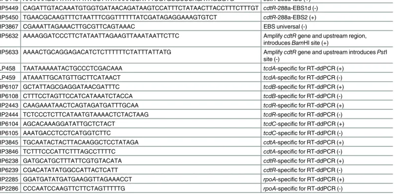
Table 2. Oligonucleotide primers.
Primer Sequence (5’- 3’) Use
JRP5448 AAAAAAGCTTATAATTATCCTTATAAAACCATTTCGTGCGCCCAGATAGGGTG cdtR-288a-IBS (+) JRP5449 CAGATTGTACAAATGTGGTGATAACAGATAAGTCCATTTCTATAACTTACCTTTCTTTGT cdtR-288a-EBS1d (-) JRP5450 TGAACGCAAGTTTCTAATTTCGGTTTTTTATCGATAGAGGAAAGTGTCT cdtR-288a-EBS2 (+)
JRP3867 CGAAATTAGAAACTTGCGTTCAGTAAAC EBS universal (-)
JRP5632 AAAAGGATCCCTTCTATAATTAGAAGTTAAATAATTCTTC AmplifycdtRgene and upstream region, introducesBamHI site (+)
JRP5633 AAAACTGCAGGAGACATCTCTTTTTTCTATTTATTATG AmplifycdtRgene and upstream introducesPstI site (-)
DLP458 TAATAAAAATACTGCCCTCGACAAA tcdA-specific for RT-ddPCR (+)
DLP459 ATAAATTGCATGTTGCTTCATAACT tcdA-specific for RT-ddPCR (-)
JRP6107 GCTATTAGCGAGGATAACGATTTC tcdB-specific for RT-ddPCR (+)
JRP6108 CTTTCCTAGTTCCATCATAAATCTACCA tcdB-specific for RT-ddPCR (-)
JRP2443 CAAGAAATAACTCAGTAGATGATTTGCAA tcdR-specific for RT-ddPCR (+)
JRP2444 TCTCCCTCTTCATAATGTAAAACTCTACTAAG tcdR-specific for RT-ddPCR (-)
JRP6104 AGCACAAAGGATATTGCTCTACT tcdC-specific for RT-ddPCR (+)
JRP6105 AAATGACCTCCTCATGGTCTTC tcdC-specific for RT-ddPCR (-)
JRP3845 TGCAATACTACTTACAAGGCTCCTATAGA cdtA-specific for RT-ddPCR (+)
JRP3846 TCTTTCCCATTCTTTAGCCTTTTC cdtA-specific for RT-ddPCR (-)
JRP6238 GATGCATGCTTTATTCGTGTACATA cdtR-specific for RT-ddPCR (+)
JRP6239 CGACATATATGGCCATTACTCATT cdtR-specific for RT-ddPCR (-)
JRP2285 GGATGATATGATGAAGGTTAGAAACCT rpoA-specific for RT-ddPCR (+)
JRP2286 CCCAATCCAAGTTCTTCTAGTTTTTG rpoA-specific for RT-ddPCR (-)
(+) = forward primer, (-) = reverse primer
doi:10.1371/journal.ppat.1005758.t002
TcdB were detected using TcdA-specific monoclonal and TcdB-specific polyclonal antibodies (tgcBIOMICS), respectively. CDTa and CDTb were detected, respectively, using a CDTa-spe-cific antibody andC.perfringensIb-specific antibody that is cross reactive with CDTb [47]. CDTa, CDTb and TcdB-bound antibodies were detected using horseradish peroxidase conju-gated anti-rabbit goat antibodies (Millipore) and TcdA-bound antibodies were detected using anti-mouse goat antibodies (Millipore). The Western Lightning Chemiluminescence reagent kit (Perkin-Elmer) was used to detect the bands, which were visualised by exposure to X-ray film or on a BioRad ChemiDoc XRS+system.
ADP-ribosyltransferase assays
Toxins were partially purified from culture supernatants by precipitation with 70% ammonium sulphate as described previously [6]. ADP ribosyltransferase assays were performed as previ-ously described [48]. Briefly, precipitated supernatant protein (50 µg) was incubated for 60 minutes at 37°C with 10 µg of actin in assay buffer (20 mM Tris-HCl pH 7.5, 1 µM dithiothrei-tol (DTT), 40 µM ATP, 40 µM CaCl2, 5 µM MgCl2) and 10 µM of biotinylated NAD+ (Trevi-gen). The reaction was heat inactivated at 95°C for 5 minutes in 4x SDS sample buffer (240 mM Tris-Cl (pH 6.8), 40% glycerol (v/v), 8% SDS (w/v), 5% (v/v) 2-mercaptoethanol, 0.05% (v/v) bromophenol blue and separated by 10% SDS-PAGE. Proteins were transferred onto a nitrocellulose membrane and biotinylated proteins were detected with horseradish peroxidase-conjugated streptavidin (GE Healthcare Life Sciences) and the Western Lightning Chemilumi-nescence reagent kit (Perkin-Elmer), following the manufacturer’s instructions. Relative band intensities were determined by densitometry using Image Lab Software (Bio-Rad). Data were analysed using GraphPad Prism 6 and statistical significance assessed using an unpaired t-test with a 95% confidence interval.
Vero and HT29 cell cytotoxicity assays
C.difficilestrains were grown overnight in 20 ml of HIS broth with thiamphenicol and linco-mycin, as required. The cultures then were used to inoculate 50 ml of TY broth with selection, such that each culture had a starting OD600of approximately 0.05. Aliquots (5 ml) were taken at 12, 24, 48 and 72 hours, pelleted by centrifugation (10,000g, 10 min, room temperature) and the supernatants filter sterilised through 0.45 µM and 0.2 µM filters (Sartorius). Supernatants were stored on ice until use. Vero cell and HT29 cell cytotoxicity assays were performed using the filteredC.difficilesupernatants as previously described [43]. The levels of TcdA and TcdB produced by theC.difficilestrainsin vivowas assessed by determining the cytotoxicity of the intestinal contents collected 24 hours post infection against HT29 and Vero cells. Intestinal samples were resuspended in 100 mg/ml in PBS, diluted one in eight, filter sterilised and applied to Vero and HT29 cells, as described previously [2]. The endpoint (toxin titre) was scored as the last dilution with 100% cytopathic effect (CPE). Data were analysed using Graph-Pad Prism 6 and statistical significance assessed using an unpaired t-test with a 95% confidence interval.
RT-ddPCR analysis of
C
.
difficile
gene expression
Digital PCR System (BioRad) using QX200 ddPCR EvaGreen Supermix and 0.1–5 µg of total cDNA and specific primers (Table 2) at a concentration of 200 nM. Transcription levels of each gene was normalised to transcription levels of the housekeeping generpoA. Data were analysed using GraphPad Prism 6 and statistical significance assessed using a Mann Whitney U test.
C
.
difficile
virulence trials
Groups of five male six to eight week old C57BL/6 mice were used inC.difficilevirulence trials as previously described [2], except mice were switched back to plain drinking water on the day of infection. Mice were administered 106C.difficilespores by oral gavage and were humanely euthanised at the onset of severe disease or at the end of the experiment, as previously defined [2]. Animal handling and experimentation were performed in accordance with institutional guidelines (Monash University animal ethics committee numbers MARP/2014/135 and SOBSB/M/2010/25). Faecal samples were taken daily to monitorC.difficileshedding using HIS agar supplemented with 0.1% (w/v) cysteine, 0.1% (w/v) taurocholate, 0.375% (w/v) glucose, 250 µg/ml D-cycloserine, 8 µg/ml cefoxitin, 10 µg/ml erythromycin, 12 µg/ml norfloxacin, 32 µg/ml moxalactam. Data were analysed using GraphPad Prism 6 and statistical significance assessed using a log-rank (Mantel-Cox) test.
Histology
The entire colon and caecum were collected from each mouse and Swiss-rolled [49] prior to fixation to allow for cross-sectional examination of the entire length of the colon. Tissues were stained with PAS-Alcian blue and histopathological assessment of damage and scoring of tis-sues was performed blind by independent observers using a previously defined set of parame-ters [2].
Supporting Information
S1 Fig. Confirmation of M7404 and R20291cdtRmutants by Southern hybridisation anal-ysis.Schematic diagram of thecdtRgenomic location and surrounding genes in (a) wild-type M7404 or R20291 and (b) TargeTron-derivedcdtRinsertion mutants. Southern hybridisation using acdtRspecific probe (red) showed a size increase from a 3.2 kbAvaII fragment in the wild type (black arrow) to a 5.0 kbAvaII fragment in the independentcdtRmutants (white arrow) in both (c) M7404 and (e) R20291 strain backgrounds. Hybridization of anermBprobe (purple) to a 5.0 kbAvaII fragment (black arrow) in the (d) M7404cdtRmutants and (f) R20291cdtRmutant confirmed the TargeTron insertion.
(TIF)
S2 Fig. Colonisation of mice infected withC.difficilewild type,cdtRmutant and comple-mented strains.Colonisation efficiencies are shown as total colony forming unit (CFU) ofC.
difficileisolated per gram of faeces collected from mice at 24 and 48 hours. Mice surviving beyond 48 hours had similar levels of colonisation. Data represent the mean ± SEM (n = 6–15). (TIF)
S3 Fig. Analysis of TcdA and TcdB production in 630 stain derivatives over-expressing CdtR.Western immunoblots were performed using precipitated supernatant proteins from the CD37 non-toxigenic strain, two 630 vector control strains and two 630 strains carrying the
cdtR+complementation vector, pJIR4218, and detected using antibodies specific for (a) TcdA and (b) TcdB.c, Expression ofcdtRin 630, 630 carrying the vector control and 630 carrying
thecdtR+complementation vector normalised torpoAexpression. (TIF)
S4 Fig.In vivocytotoxicity ofC.difficilewild type,cdtRmutant and complemented strains. Faecal samples collected from uninfected andC.difficileinfected mice 24 hours post infection were assayed for cytotoxicity by doubling dilution cytotoxicity assays using (a) HT29 cells and (b) Vero cells. Data represent the mean ± SEM (n = 5).
(TIF)
Acknowledgments
We thank M. Popoff for providing CDTa-specific and Ib-specific antibodies, P. Howarth for technical assistance, S. Larcombe for the construction of pDLL24, S. Mileto for helpful discus-sions and B. Cunningham for construction of DLL3094 and technical assistance.
Author Contributions
Conceived and designed the experiments: SAL MLH JIR JKC DL. Performed the experiments: SAL MLH JKC. Analyzed the data: SAL MLH JIR JKC DL. Contributed reagents/materials/ analysis tools: SAL MLH JIR JKC DL. Wrote the paper: SAL MLH JIR JKC DL.
References
1. Lyras D, O'Connor JR, Howarth PM, Sambol SP, Carter GP, et al. (2009) Toxin B is essential for viru-lence ofClostridium difficile. Nature 458: 1176–1179. doi:10.1038/nature07822PMID:19252482 2. Carter GP, Chakravorty A, Pham Nguyen TA, Mileto S, Schreiber F, et al. (2015) Defining the Roles of
TcdA and TcdB in Localized Gastrointestinal Disease, Systemic Organ Damage, and the Host Response duringClostridium difficileInfections. MBio 6: e00551. doi:10.1128/mBio.00551-15PMID:
26037121
3. Awad MM, Johanesen PA, Carter GP, Rose E, Lyras D (2014)Clostridium difficilevirulence factors: Insights into an anaerobic spore-forming pathogen. Gut Microbes 5: 579–593. doi:10.4161/19490976. 2014.969632PMID:25483328
4. Rupnik M, Janezic S (2016) An Update onClostridium difficileToxinotyping. J Clin Microbiol 54: 13–
18. doi:10.1128/JCM.02083-15PMID:26511734
5. Yan Q, Zhang J, Chen C, Zhou H, Du P, et al. (2013) Multilocus sequence typing (MLST) analysis of 104Clostridium difficilestrains isolated from China. Epidemiol Infect 141: 195–199. doi:10.1017/ S0950268812000453PMID:22475233
6. Carter GP, Lyras D, Allen DL, Mackin KE, Howarth PM, et al. (2007) Binary toxin production in Clostrid-ium difficileis regulated by CdtR, a LytTR family response regulator. J Bacteriol 189: 7290–7301. PMID:17693517
7. Goncalves C, Decre D, Barbut F, Burghoffer B, Petit JC (2004) Prevalence and characterization of a binary toxin (actin-specific ADP-ribosyltransferase) fromClostridium difficile. J Clin Microbiol 42: 1933–1939. PMID:15131151
8. Snydman DR, McDermott LA, Jacobus NV, Thorpe C, Stone S, et al. (2015) U.S.-Based National Senti-nel Surveillance Study for the Epidemiology ofClostridium difficile-Associated Diarrheal Isolates and Their Susceptibility to Fidaxomicin. Antimicrob Agents Chemother 59: 6437–6443. doi:10.1128/AAC. 00845-15PMID:26239985
9. Schwan C, Stecher B, Tzivelekidis T, Van Ham M, Rohde M, et al. (2009)Clostridium difficiletoxin CDT induces formation of microtubule-based protrusions and increases adherence of bacteria. PLoS Pathog 5.
10. Baban ST, Kuehne SA, Barketi-Klai A, Cartman ST, Kelly ML, et al. (2013) The role of flagella in Clos-tridium difficilepathogenesis: comparison between a non-epidemic and an epidemic strain. PLoS One 8: e73026. doi:10.1371/journal.pone.0073026PMID:24086268
12. Carter GP, Douce GR, Govind R, Howarth PM, Mackin KE, et al. (2011) The anti-sigma factor TcdC modulates hypervirulence in an epidemic BI/NAP1/027 clinical isolate ofClostridium difficile. PLoS Pathog 7: e1002317. doi:10.1371/journal.ppat.1002317PMID:22022270
13. Cartman ST, Kelly ML, Heeg D, Heap JT, Minton NP (2012) Precise manipulation of theClostridium dif-ficilechromosome reveals a lack of association between thetcdCgenotype and toxin production. Appl Environ Microbiol 78: 4683–4690. doi:10.1128/AEM.00249-12PMID:22522680
14. Matamouros S, England P, Dupuy B (2007)Clostridium difficiletoxin expression is inhibited by the novel regulator TcdC. Mol Microbiol 64: 1274–1288. PMID:17542920
15. Mani N, Lyras D, Barroso L, Howarth P, Wilkins T, et al. (2002) Environmental response and autoregu-lation ofClostridium difficileTxeR, a sigma factor for toxin gene expression. J Bacteriol 184: 5971–
5978. PMID:12374831
16. Saujet L, Monot M, Dupuy B, Soutourina O, Martin-Verstraete I (2011) The key sigma factor of transition phase, SigH, controls sporulation, metabolism, and virulence factor expression inClostridium difficile. J Bacteriol 193: 3186–3196. doi:10.1128/JB.00272-11PMID:21572003
17. McKee RW, Mangalea MR, Purcell EB, Borchardt EK, Tamayo R (2013) The second messenger cyclic Di-GMP regulatesClostridium difficiletoxin production by controlling expression ofsigD. J Bacteriol 195: 5174–5185. doi:10.1128/JB.00501-13PMID:24039264
18. Martin MJ, Clare S, Goulding D, Faulds-Pain A, Barquist L, et al. (2013) Theagrlocus regulates viru-lence and colonization genes inClostridium difficile027. J Bacteriol 195: 3672–3681. doi:10.1128/JB. 00473-13PMID:23772065
19. Antunes A, Martin-Verstraete I, Dupuy B (2011) CcpA-mediated repression ofClostridium difficiletoxin gene expression. Mol Microbiol 79: 882–899. doi:10.1111/j.1365-2958.2010.07495.xPMID:
21299645
20. Dineen SS, McBride SM, Sonenshein AL (2010) Integration of metabolism and virulence byClostridium difficileCodY. J Bacteriol 192: 5350–5362. doi:10.1128/JB.00341-10PMID:20709897
21. Loo VG, Poirier L, Miller MA, Oughton M, Libman MD, et al. (2005) A predominantly clonal multi-institu-tional outbreak ofClostridium difficile—Associated diarrhea with high morbidity and mortality. N Engl J Med 353: 2442–2449. PMID:16322602
22. Warny M, Pepin J, Fang A, Killgore G, Thompson A, et al. (2005) Toxin production by an emerging strain ofClostridium difficileassociated with outbreaks of severe disease in North America and Europe. Lancet 366: 1079–1084. PMID:16182895
23. McDonald LC, Killgore GE, Thompson A, Owens RC Jr, Kazakova SV, et al. (2005) An epidemic, toxin gene-variant strain ofClostridium difficile. N Engl J Med 353: 2433–2441. PMID:16322603
24. El Meouche I, Peltier J, Monot M, Soutourina O, Pestel-Caron M, et al. (2013) Characterization of the SigD regulon ofC.difficileand its positive control of toxin production through the regulation oftcdR. PLoS One 8: e83748. doi:10.1371/journal.pone.0083748PMID:24358307
25. Keel K, Brazier JS, Post KW, Weese S, Songer JG (2007) Prevalence of PCR ribotypes among Clos-tridium difficileisolates from pigs, calves, and other species. Journal of Clinical Microbiology 45: 1963–
1964. PMID:17428945
26. Goorhuis A, Bakker D, Corver J, Debast SB, Harmanus C, et al. (2008) Emergence ofClostridium diffi-cileinfection due to a new hypervirulent strain, polymerase chain reaction ribotype 078. Clinical Infec-tious Diseases 47: 1162–1170. doi:10.1086/592257PMID:18808358
27. Bouvet PJ, Popoff MR (2008) Genetic relatedness ofClostridium difficileisolates from various origins determined by triple-locus sequence analysis based on toxin regulatory genestcdC,tcdR,and cdtR. J Clin Microbiol 46: 3703–3713. doi:10.1128/JCM.00866-08PMID:18832125
28. Sebaihia M, Wren B. W., Mullany P., Fairweather N. F., Minton N., Stabler NRT R., Roberts A. P., Cer-deno-Tarraga A. M., Wang H., Holden AW M. T., Churcher C., Quail M. A., Baker S., Bason N., Brooks TC K., Cronin A., Davis P., Dowd L., Fraser A., Feltwell ZH T., Holroyd S., Jagels K., Moule S., Mungall K., Price, E. C., et al. (2006) The multidrug-resistant human pathogenClostridium difficilehas a highly mobile, mosaic genome. Nat Gen 38: 779–786.
29. Merrigan M, Venugopal A, Mallozzi M, Roxas B, Viswanathan VK, et al. (2010) Human hypervirulent
Clostridium difficilestrains exhibit increased sporulation as well as robust toxin production. J Bacteriol 192: 4904–4911. doi:10.1128/JB.00445-10PMID:20675495
30. Stabler RA, He M, Dawson L, Martin M, Valiente E, et al. (2009) Comparative genome and phenotypic analysis ofClostridium difficile027 strains provides insight into the evolution of a hypervirulent bacte-rium. Genome Biol 10.
31. Vohra P, Poxton IR (2011) Comparison of toxin and spore production in clinically relevant strains of
Clostridium difficile. Microbiology 157: 1343–1353. doi:10.1099/mic.0.046243-0PMID:21330434
32. Geric B, Carman RJ, Rupnik M, Genheimer CW, Sambol SP, et al. (2006) Binary toxin-producing, large clostridial toxin-negativeClostridium difficilestrains are enterotoxic but do not cause disease in ham-sters. J Infect Dis 193: 1143–1150. PMID:16544255
33. Schwan C, Kruppke AS, Nolke T, Schumacher L, Koch-Nolte F, et al. (2014)Clostridium difficiletoxin CDT hijacks microtubule organization and reroutes vesicle traffic to increase pathogen adherence. Proc Natl Acad Sci U S A 111: 2313–2318. doi:10.1073/pnas.1311589111PMID:24469807 34. Metcalf DS, Weese JS (2011) Binary toxin locus analysis inClostridium difficile. J Med Microbiol 60:
1137–1145. doi:10.1099/jmm.0.028498-0PMID:21459907
35. Antunes A, Camiade E, Monot M, Courtois E, Barbut F, et al. (2012) Global transcriptional control by glucose and carbon regulator CcpA inClostridium difficile. Nucleic Acids Res 40: 10701–10718. doi:
10.1093/nar/gks864PMID:22989714
36. Darkoh C, DuPont HL, Norris SJ, Kaplan HB (2015) Toxin synthesis byClostridium difficileis regulated through quorum signaling. MBio 6: e02569. doi:10.1128/mBio.02569-14PMID:25714717
37. Knight DR, Elliott B, Chang BJ, Perkins TT, Riley TV (2015) Diversity and Evolution in the Genome of
Clostridium difficile. Clin Microbiol Rev 28: 721–741. doi:10.1128/CMR.00127-14PMID:26085550 38. Elliott B, Dingle KE, Didelot X, Crook DW, Riley TV (2014) The complexity and diversity of the
Pathoge-nicity Locus inClostridium difficileclade 5. Genome Biol Evol 6: 3159–3170. doi:10.1093/gbe/evu248
PMID:25381663
39. Stabler RA, Gerding DN, Songer JG, Drudy D, Brazier JS, et al. (2006) Comparative phylogenomics of
Clostridium difficilereveals clade specificity and microevolution of hypervirulent strains. J Bacteriol 188: 7297–7305. PMID:17015669
40. Dingle KE, Griffiths D, Didelot X, Evans J, Vaughan A, et al. (2011) ClinicalClostridium difficile: clonal-ity and pathogenicclonal-ity locus diversclonal-ity. PLoS One 6: e19993. doi:10.1371/journal.pone.0019993PMID:
21625511
41. Sambrook J, Russell David W (1989) Molecular cloning: a laboratory manual. Vol. 3: Cold spring har-bor lahar-boratory press.
42. Smith CJ, Markowitz SM, Macrina FL (1981) Transferable tetracycline resistance inClostridium diffi-cile. Antimicrobial Agents and Chemotherapy 19: 997–1003. PMID:7271279
43. Carter GP, Larcombe S, Li L, Jayawardena D, Awad MM, et al. (2014) Expression of the large clostridial toxins is controlled by conserved regulatory mechanisms. International Journal of Medical
Microbiology.
44. O'Connor JR, Lyras D, Farrow KA, Adams V, Powell DR, et al. (2006) Construction and analysis of chromosomalClostridium difficilemutants. Molecular Microbiology 61: 1335–1351. PMID:16925561 45. Anagnostopoulos C, Spizizen J (1961) Requirements for Transformation inBacillus subtilis. J Bacteriol
81: 741–746. PMID:16561900
46. Laemmli UK (1970) Cleavage of structural proteins during the assembly of the head of bacteriophage T4. Nature 227: 680–685. PMID:5432063
47. Perelle S, Gibert M, Bourlioux P, Corthier G, Popoff MR (1997) Production of a complete binary toxin (actin-specific ADP- ribosyltransferase) byClostridium difficileCD196. Infect Immun 65: 1402–1407. PMID:9119480
48. Yonogi S, Matsuda S, Kawai T, Yoda T, Harada T, et al. (2014) BEC, a novel enterotoxin ofClostridium perfringensfound in human clinical isolates from acute gastroenteritis outbreaks. Infect Immun 82: 2390–2399. doi:10.1128/IAI.01759-14PMID:24664508
49. Moolenbeek C, Ruitenberg EJ (1981) The "Swiss roll": a simple technique for histological studies of the rodent intestine. Lab Anim 15: 57–59. PMID:7022018
50. Backman K, Ptashne M, Gilbert W (1976) Construction of plasmids carrying thecI gene of bacterio-phage lambda. Proc Natl Acad Sci U S A 73: 4174–4178. PMID:1069307
51. Roberts AP, Hennequin C, Elmore M, Collignon A, Karjalainen T, et al. (2003) Development of an inte-grative vector for the expression of antisense RNA inClostridium difficile. J Microbiol Meth 55: 617–
624.