Mycobacterium massiliense
in Mice
Shaobin Shang1, Sara Gibbs1, Marcela Henao-Tamayo1, Crystal A. Shanley1, Gerald McDonnell2, Rafael Silva Duarte3, Diane J. Ordway1*, Mary Jackson1*
1Mycobacteria Research Laboratories, Department of Microbiology, Immunology and Pathology, Colorado State University, Fort Collins, Colorado, United States of America,2STERIS Limited, Basingstoke, England,3Instituto de Microbiologia, Universidade Federal do Rio de Janeiro, Rio de Janeiro, Brazil
Abstract
Background:Chronic pulmonary disease and skin/soft tissue infections due to non-tuberculous mycobacteria (NTM) of the
Mycobacterium chelonae-abscessus-massiliensegroup is an emerging health problem worldwide. Moreover, the cure rate for the infections this group causes is low despite aggressive treatment. Post-surgical outbreaks that reached epidemic proportions in Brazil recently were caused byM. massilienseisolates resistant to high-level disinfection with glutaraldehyde (GTA). Understanding the differences in the virulence and host immune responses induced by NTM differing in their sensitivity to disinfectants, and therefore their relative threat of causing outbreaks in hospitals, is an important issue.
Methodology/Principal Finding: We compared the replication and survival inside macrophages of a GTA-susceptible reference Mycobacterium massiliense clinical isolate CIP 108297 and an epidemic strain from Brazil, CRM-0019, and characterized the immune responses of IFNcknockout mice exposed to a high dose aerosol with these two isolates.
CRM-0019 replicated more efficiently than CIP 108297 inside mouse bone marrow macrophages. Moreover, the animals infected with CRM-0019 showed a progressive lung infection characterized by a delayed influx of CD4+ and CD8+ T cells, culminating in extensive lung consolidation and demonstrated increased numbers of pulmonary CD4+Foxp3+regulatory T cells compared to those infected with the reference strain. Immunosuppressive activity of regulatory T cells may contribute to the progression and worsening of NTM disease by preventing the induction of specific protective immune responses.
Conclusions/Significance:These results provide the first direct evidence of the increased virulence in macrophages and mice and pathogenicity in vivoof the Brazilian epidemic isolate and the first observation that NTM infections can be associated with variable levels of regulatory T cells which may impact on their virulence and ability to persist in the host.
Citation:Shang S, Gibbs S, Henao-Tamayo M, Shanley CA, McDonnell G, et al. (2011) Increased Virulence of an Epidemic Strain ofMycobacterium massiliensein Mice. PLoS ONE 6(9): e24726. doi:10.1371/journal.pone.0024726
Editor:J. Ross Fitzgerald, University of Edinburgh, United Kingdom
ReceivedFebruary 23, 2011;AcceptedAugust 19, 2011;PublishedSeptember 12, 2011
Copyright:ß2011 Shang et al. This is an open-access article distributed under the terms of the Creative Commons Attribution License, which permits
unrestricted use, distribution, and reproduction in any medium, provided the original author and source are credited.
Funding:Supported in part by the National Institutes of Health/National Institute of Allergy and Infectious Diseases grant AI089718, the College of Veterinary Medicine and Biomedical Sciences Research Council CRC 0014 (Colorado State University) and the STERIS Foundation. The STERIS Foundation provided funds for equipment and materials purchased for the study. In addition, Dr. McDonnell, an employee of STERIS, assisted in the technical aspects of disinfectant-susceptibility testing and aided in the presentation of these results in the manuscript. The STERIS Foundation and the Research Council CRC 0014 had no role in the decision to publish.
Competing Interests:I have read the journal’s policy and have the following conflict. Dr. Gerald McDonnell is an employee of STERIS. This affiliation does not alter our adherence to all the PLoS ONE policies on sharing data and materials.
* E-mail: [email protected] (MJ); D.Ordway-Rodriguez@ colostate.edu (DJO)
Introduction
Non-tuberculous mycobacteria (NTM) are ubiquitous in the environment and are responsible for colonization, infection and pseudo-outbreaks in health care settings throughout the world. Most NTM nosocomial outbreaks caused by rapidly growing mycobac-teria (RGM) have involved the speciesM. abscessus,M. chelonae,M. fortuitum and two recently recognizedMycobacteriumspecies closely related to M. abscessus: M. massiliense and M. bolletii [1,2]. These pathogens can cause a wide range of clinical diseases including skin and soft tissue infections, pulmonary, central nervous system and disseminated infections [3,4,5,6,7,8,9,10,11,12,13,14]. While dis-seminated infections caused by RGM essentially occur in immunodeficient patients and pulmonary infections are widely thought to be opportunistic and to occur in patients with struc-tural lung disease (e.g., chronic obstructive pulmonary disease,
bronchiectasis, cystic fibrosis), numerous cases of skin and soft tissue infections have been reported in otherwise immunocompetent patients, caused by contaminated materials and invasive procedures involving catheters, non-sterile surgical procedures, injections and implantations of foreign bodies. Recent published reports [15,16] and outbreaks in Brazil [13,17] suggest that the prevalence of infections caused by these microorganisms is increasing and that infections due to RGM are particularly problematic due to their ubiquitous presence in hospitals’ water sources and the difficulty of treating some of the infections they cause.
of M. abscessus by antigen-presenting cells such as alveolar macrophages and dendritic cells [20,21]. Autocrine and paracrine activation of dendritic cells by IL-12 p40 and migration of the infected dendritic cells to the regional mediastinal lymph nodes and subsequent mycobacterial antigen presentation to naive T cells results in differentiation and further activation of effector T cells. The accumulation in the lung of protective T cells capable of expressing IFNcand TNFa are crucial for phagocyte activation and intracellular killing [19,22]. The development of a pulmonary granuloma is orchestrated by chemokines and cytokines produced by local cells causing a continuous recruitment of lymphocytes, macrophages, dendritic cells and granulocytes to the lung. Control of the infection relies on this granulomatous response, which is an organized cellular network acting to restrain NTM growth and limit dissemination. In the context ofM. tuberculosispersistence and chronic disease, it has been shown that CD4+Foxp3+regulatory T cells are important for regulation of immunopathology [23,24,25]. The induction of CD4+Foxp3+regulatory T cells has never been reported in the case of Mycobacterium chelonae-abscessus-massiliense group persistence and we propose it is one of the strategies adopted by these microorganisms to enhance their persistence in the human host. The immunosuppressive activity of regulatory T cells may contribute to the progression and worsening of clinical disease caused by NTMs by preventing the induction of specific protective immune responses required to kill the bacterium.
The most important outbreaks – reaching epidemic proportions - of nosocomial M. massiliense infections ever reported occurred between 2004 and 2007 in Brazil. Interestingly, these outbreaks seem to have been caused by one single strain of M. massiliense, named BRA100 [12,13,17], which displays high level resistance to glutaraldehyde (GTA), the chemical used for disinfection of surgical instruments in all of the hospital sites that had confirmed cases. While the resistance of this particular strain to high-level disinfection has been advanced as the most likely reason for its selection and dissemination across the country [12,13,17], the unusual size of the outbreaks also suggests that BRA100 may represent a particularly invasive and pathogenic strain of M. massiliense. Such variability in pathogenicity amongM. tuberculosis clinical isolates has been well documented [23,26,27,28]. The potential increase in pathogenicity of the epidemic BRA100 isolates is further supported by our recent findings that one of the mechanisms through which RGM species develop high-level resistance to GTA involves a remodeling of the composition and/ or organization of their cell surface susceptible of affecting their intracellular survival and interactions with the host [29].
We undertook this study with the goal of comparing the M. massilienseBRA100 epidemic isolate from Brazil to the glutaralde-hyde-susceptible reference strain, M. massiliense CIP 108297, in their abilities to replicate inside macrophages and to infect, replicate, persist, induce organ pathology and immune responses in mice.
Results
Morphological traits, disinfectant resistance and in vitro growth characteristics of theM. massilienseisolates used in this study
M. massilienseCIP 108297, the type strain used in this study, was isolated from the sputum and the bronchoalveolar fluid of a patient with hemoptoic pneumonia in France in 2004 [2]. CRM-0019 is a BRA100 M. massiliense isolate from Brazil. It was responsible for a nosocomial infection following hepatic tumor resection by videolaparoscopy, the supposed index case of the epidemic in the state of Rio de Janeiro [13]. The female patient
was 45 years old and presented the initial symptoms 90 days after the videosurgery. She was submitted to clarithromycin-based antimicrobial therapy and two additional laparotomies for granuloma resections, resulting in complete recovery.M. massiliense CIP 108297 and CRM-0019 display different pulse field gel electrophoresis (PFGE) patterns confirming their lack of genetic relationship [13]. The species assignment of both isolates was confirmed by partial sequencing of their hsp65 and rpoB genes (view Information S1).
The plating of CIP 108297 yielded uniform colonies interme-diate between rough and smooth, as described earlier [2]. Plating of CRM-0019 onto 7H11-OADC agar, in contrast, yielded a mixed population of nonphotochromogenic rough and smooth colonies. Suggestive of genetic instability, the culturing and re-plating of individual rough and smooth CFUs led to colonies with mixed phenotypes. Comparison of the growth rate of the two strains in 7H9-OADC broth at 30uC revealed only a marginal delay in growth associated to the BRA100 isolate during exponential phase (view Information S1). Their growth rate on 7H11-OADC agar plates was comparable. Suspension suscepti-bility tests confirmed the high level resistance of the BRA100 isolate, CRM-0019, to 2.2% GTA (view Information S1). Smooth colonies of CRM-0019 displayed the same level of resistance as the rough ones.
Growth ofM. massiliensestrains in mouse bone marrow derived macrophages (BMDM)
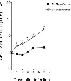
We investigated the capacity of M. massiliense reference strain CIP 108297 and theM. massilienseoutbreak strain CRM-0019 to grow in BMDM to evaluate their virulence. The growth of each isolate was determined by the method of plating to measure colony-forming units (CFU; Figure 1). TheM. massilienseoutbreak strain CRM-0019 compared to the reference strain CIP 108297 demonstrated a significantly increased ability to replicate in BMDM after 24 hours of infection and steadily increased peaking at 7 days of infection. The reference strain CIP 108297 demonstrated nominal bacterial replication for 48 hours of infection and thereafter began to steadily increase in bacterial numbers in BMDM. Lastly, we evaluated the BMDM cellular viability the percentage of propidium iodide positive dead cells induced by theM. massiliensereference strain CIP 108297 and the M. massilienseoutbreak strain CRM-0019 was below 6.0% for the BMDM (data not shown).
Bacterial loads in GKO mice
TNFablockers are at increased risk for soft tissue infections and pulmonary NTM infections, including the RGM [21]. In addition, it has been shown that individuals infected with the human immunodeficiency virus show reduced T cell IFNcproduction and
TH1 immunosuppression by CD4+Foxp3+ regulatory T cell [31,32]. Lastly, the choice of using GKO mice instead of C57BL/ 6 ones owes to the reported difficulty of infecting through the aerosol route wild-type C57BL/6 mice that eliminate the bacteria very rapidly, whereas GKO mice aerosolized with about 1,000 CFU develop a successful infection [33].
Following a high dose aerosol infection withM. massiliensereference strain CIP 108297 and Brazilian outbreak strain CRM-0019 in GKO mice (,1,000 bacilli per animal), the bacterial loads in the lungs and spleens were quantified on days 1, 30 and 60 after the challenge. Aerosol infection with the outbreak strainM. massilienseCRM-0019 resulted in a sustained infection in the lungs and spleens with more than 103 organisms recovered from both organs at 60 days (Figures 2A and 2B). In comparison, GKO mice infected with the reference strain ofM. massilienseshowed complete clearance of this strain in the lungs and spleens by day 60 (Figures 2A and 2B).
Lung histopathology in GKO mice after aerosol challenge withMycobacterium massiliense
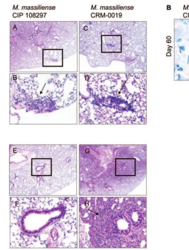
Lung histopathology following high dose aerosol challenge with the reference and outbreakM. massiliensestrains was characterized in GKO mice (Figure 3). At day 30 following the challenge, when there was bacterial persistence in the lungs for both strains, lung tissues demonstrated a local inflammatory response denoted by peribronchiolar inflammatory infiltrates (Figure 3, panel A, A-D, arrow). However, by day 60, while the reference M. massiliense strain showed complete bacterial clearance and healthy lung tissue (Figure 3, panel A, E–F), the lungs of the CRM-0019-infected GKO mice showed large areas of consolidation (Figure 3, panel A, G–H). In addition, CRM-0019-infected mice showed peribron-chiolar inflammatory infiltrates with large aggregates of foamy cells and the presence of macrophages with multiple intracellular acid fast staining bacilli (Figure 3B).
Macrophage and dendritic cell cytokine expression duringMycobacterium massiliense infection
Given the clear differences seen in cell influx indicated by the histological analysis, we conducted a comparative flow cytometric
Figure 1. Increased intracellular replication of the Brazilian outbreak strain M. massiliense CRM-0019 in mouse bone marrow macrophages. Intracellular growth of M. massiliense
reference strain CIP 108297 andM. massilienseoutbreak strain CRM-0019 in BMDM. BMDM were infected withM. massiliensestrains at a MOI of 5, and the numbers of intracellular bacteria were determined using the bacterial colony count method (CFU) immediately after 2 hours of infection or at 1, 2, 3, 4, 5, 6 and 7 days after infection. Values shown are the mean6SD from two independent experiments. Growth of theM. massilienseoutbreak strain CRM-0019 was significantly higher than the other isolate (*,P,0.05).
doi:10.1371/journal.pone.0024726.g001
Figure 2. Increased bacteria in the lungs and spleens of the Brazilian epidemic strainM. massilienseCRM-0019 infected IFNc-KO
mice.Bacterial counts in the lungs (A) and spleens (B), on days 1, 30 and 60 from IFNc-KO mice infected with a high dose aerosol of the epidemic strainM. massilienseCRM-0019 (open circle) andM. massilienseCIP 108297 (solid square) were compared. Results are expressed as the average (n = 5) of the bacterial load in each group expressed as Log10CFU,6standard error mean (SEM). Studentt-test, *p,0.050.
analysis of alveolar macrophages (CD11b+) and dendritic cell (CD11c+) populations from lungs and spleens of M. massiliense CRM-0019-infected mice compared to CIP 108297-infected mice. Figure 4 A shows representative dot plots of lung cells from the isotype control, reference CIP 108297 and the outbreak M. massiliense CRM-0019 strain after 30 days of infection in mice primarily gated on CD11b+macrophages cells expression of IL-12. As shown in Figure 4 B and C, macrophages and dendritic cells from the CRM-0019-infected mice showed reduced numbers of lung and spleen IL-12 and TNFa producing macrophage and dendritic cells compared to the reference strain CIP 108297.
CD4+ and CD8+T cell cytokine expression during
Mycobacterium massiliense infection
We next sought to investigate the differences in the CD4+and CD8+T cells in the lungs and spleens of mice infected with the epidemic strain M. massilienseCRM-0019 compared to reference strain CIP 108297 after 30 and 60 days of infection. We evaluated CD4+and CD8+T cells ability to produce the protective cytokine TNFa required for efficient granuloma formation [34]. We also evaluated CD4+and CD8+T cells ability to induce immunosup-pressive cytokines such as IL-4 which is considered to increase host susceptibility to intracellular pathogens [20].
Figure 5 A shows representative dot plots of lung cells from the isotype control, reference CIP 108297 and the outbreak M.
massiliense CRM-0019 strain after 30 days of infection in mice primarily gated on CD4+ T cells expression of TNFa. Mice infected with CRM-0019 showed significant reduced numbers of lung and spleen TNFa–expressing CD4+ T cells by day 30 of infection compared to the reference strain (Figure 5 B-C). However, animals infected with the epidemic strain during chronic infection showed similar level of TNFa–expressing CD4+ and CD8+ T cells in the spleen as the reference strain (Figure 5 C). The two types of infected mice did not differ in lung and spleen IL-4–expressing CD4+ and CD8+ T cells (data not shown).
CD4+Foxp3 regulatory T cell expression during
Mycobacterium massilienseinfection
To try to understand these differences seen between the epidemic strain CRM-0019 and the referenceM. massiliensestrain CIP 108297, we performed studies in which we re-examined the T cell response. Figure 6 A shows representative dot plots of lung cells from the isotype control, reference CIP 108297 and the outbreakM. massilienseCRM-0019 strain after 30 days of infection in mice primarily gated on CD4+T cells expression of Foxp3+. The results in Figure 6 B clearly show an increased number of CD4+Foxp3+IL-10+ T cells in the lungs and spleens of mice infected with the epidemic strain compared to the reference strain at both time points. In fact, compared to CIP 108297-infected
Figure 3. Lung histology fromM. massiliense infected IFNc-KO mice.Panel A, A-H shows representative lung histopathology from the
M. massilienseCIP 108297- and epidemic CRM-0019-infected animals on day 30 and 60 of the infection. Tissue sections are stained with hematoxylin and eosin. The lower photograph depicts the magnified region denoted by the square (upper photograph). CRM-0019-infected mice (panel A: D and H) showed an increased rate of granuloma (arrows) progression and involvement of larger areas of the lung compared to the control mice (panel A: B and E). Panel B shows representative lung acid fast staining bacilli at day 60 after M. massilienseCIP 108297 and CRM-0019 infection. Total magnification, panel A [A, C, E, G] = 10x; panel A [B, D, F, H] = 20x; panel B = 100x.
mice, those infected with CRM-0019 demonstrated increased numbers of both CD4+Foxp3+T cells and CD4+Foxp3+IL10
+T cells in the lungs and spleens during the course of infection.
Discussion
Owing to its relatively recent recognition as a novel Mycobac-terium species [2], not much is known about the virulence and pathogenicity ofM. massiliense. The limited information presently available suggests that the clinical manifestations of the diseases caused byM. massiliensein humans are similar to those caused by M. abscessus [14]. The results of this study clearly show thatM. massiliense clinical isolates with different genetic backgrounds can significantly differ in their ability to replicate and persist inside macrophages and induce distinct immune responses contributing to the different virulence phenotypes in mice. The Brazilian epidemic BRA100 strain M. massiliense CRM-0019 clearly replicates more efficiently than the reference strain CIP 108297 in mouse bone marrow macrophages resulting in rapid cell lysis. It is also more virulent for GKO mice than is strain CIP 108297, persisting in the lungs and spleens and inducing more severe lung and spleen pathology. Mice infected with strain CIP 108297
eventually clear the infection showing an absence of residual bacilli and organ pathology 60 days post-infection. Several reasons may account for the enhanced persistence of CRM-0019 in vivo including the increased ability of this isolate to replicate and survive inside macrophages and, potentially, our novel observation here that this particularM. massilienseisolate expands a regulatory T cell response that attempts to dampen the progression of organ pathology associated with bacterial persistence but also suppresses the protective TNFa and IL-12 [TH1] response required for mycobacterial containment and eventual elimination.
In this regard, we found evidence that immunodeficient GKO mice administered the epidemic BRA100 strain M. massiliense CRM-0019 through the aerosol route expand a weak TH1 response, compared to the reference strain CIP 108297. Recent studies have shown that immunocompetent C57BL/6 and BALB/ c mice infected with a large intravenous dose of aM. massiliense BRA100 isolate closely related to CRM-0019 developed a robust TH1 response consisting of macrophage, dendritic cell and natural killer cell activation, induced by IL-12 and IL-17 [35]. However, there are important differences between their study and ours which raise new interesting questions. First, they intravenously infected their immunocompetent mice with a very large inoculum
Figure 4. Decreased macrophage and dendritic cell expression of IL-12 and TNFain the lungs and spleens of IFNc-KO mice infected
with the epidemic strainM. massilienseCRM-0019.Mice infected withM. massiliense CIP 108297 (closed square) and the epidemic strain
M. massilienseCRM-0019 (open circle) were assayed by flow cytometry on days 30 and 60. Panel A shows representative dot plots of lung cells from the isotype control, reference CIP 108297 and the outbreakM. massilienseCRM-0019 strain after 30 days of infection in mice primarily gated on CD11b+macrophages cells expression of IL-12. Panels B and C show the total number of macrophage (CD11b+) and dendritic (CD11c+) cells expressing IL-12 or TNFain the lungs (A) and spleens (B) of both types of infected mice. Results are expressed as the mean number of CD11b+IL-12+, CD11b+TNF
of M. massiliense CRM-0019 (16106 organisms per mice, with concentration level reaching 107CFU in the liver, spleen and lungs [35]. Thus, although their murine model of intravenous infection revealed important immunological findings, it may not reflect the typical bacterial numbers nor immune responses elicited by immunodeficient humans during a nosocomial, wound or aerosol infection [7,21]. Prior studies have shown that increased doses of NTM can induce increasingly robust immune responses [20]. Second, an intravenous infection which kinetically spreads to all organs rapidly causing rapid bacterial dissemination does not reflect bacilli deposited in soft tissue during a surgical procedure nor a pulmonary infection [36]. Our model was developed to use a dose of bacteria and an immunodeficient animal model which best reflects those individuals which generally become infected with
such opportunistic pathogens as those from the M. abscessus-M. massiliensegroup.
The apparent lack of TH1 immunity during aerosol infection with the outbreak strain M. massiliense CRM-0019 was due to reduced numbers of CD11b+
cells producing TNF-a and IL-12 cells at the site of infection. In addition, CD11c+ dendritic cells were also expressing less TNF-aand IL-12. Importantly, in our comparative evaluation of T cell responses inM. massiliense CRM-0019 and CIP 108297-infected mice, we observed reduced numbers of CD4+ T cells secreting TNFa in the lungs and spleens of CRM-0019-infected mice at 30 days, while at the same time increased numbers of CD4+Foxp3+IL-10+
CD4 cells was observed. This is the first observation that non-tuberculous rapidly-growing mycobacteria are capable of inducing the
Figure 5. Decreased early CD4+and CD8+T cell expression of TNFain the lungs and spleens of IFNc-KO mice infected with the
epidemic strainM. massilienseCRM-0019.Mice infected with an aerosol dose ofM. massilienseCIP 108297 (closed square) and CRM-0019 (open circle) were assayed by flow cytometry on days 30 and 60. Panel A shows representative dot plots of lung cells from the isotype control, reference CIP 108297 and the outbreakM. massilienseCRM-0019 strain after 30 days of infection in mice primarily gated on CD4+T cells expression of TNFa. Panels B and C show decreased CD4+and CD8+T cell expression of TNFain the lungs (A) and spleens (B) of mice infected with the epidemic strain CRM-0019 compared to CIP 108297 (6SEM, n = 5). Results are expressed as the mean number of CD4+
TNFa+, CD8+TNFa+, (6SEM, n = 5) T cells in the lungs and spleens. Studentt-test, *p,0.050.
development of immunosuppressive regulatory T cells. Earlier reports have shown that clinical isolates ofM. tuberculosis vary in their ability to induce regulatory T cells with possible consequenc-es on virulence and host protective immunity [23,37]. Moreover, there is evidence that patients with active tuberculosis have raised levels of circulating regulatory T cells [38,39]. Thus, regulatory T cell induction may be inherent in mycobacterial host immunity and not just responding to the acute lung damage caused by highly
virulentM. tuberculosis. Our study suggests that IL-10, a product of regulatory T cells as well as macrophages, may play a role in maintaining the stability of persistent NTM disease as has also been suggested in the case ofBordetella pertussisinfection [40].
A likely correlate of our important findings is that the ability of someM. massilienseisolates and, perhaps, closely related Mycobac-terium species, to persist in vivothrough increased resistance to the bactericidal mechanisms of macrophages and the modulation of T
Figure 6. Increased CD4+Foxp3+IL-10+regulatory T cell expression in the lungs and spleens of IFNc-KO mice infected withM.
massilienseCRM-0019.Lung cells obtained from mice infected withM. massilienseCIP 108297 (closed square) and CRM-0019 (open circle) were assayed by multi-parametric flow cytometry on days 30 and 60. Panel A shows representative dot plots of lung cells from the isotype control, reference CIP 108297 and the outbreakM. massilienseCRM-0019 strain after 30 days of infection in mice primarily gated on CD4+T cells expression of Foxp3+. Panel B shows that mice infected withM. massilienseCRM-0019 had a significant increase in CD4+Foxp3+and CD4+Foxp3+IL-10+regulatory T cells in the lungs and spleens at 30 and 60 days of infection compared toM. massilienseCIP 108297-infected mice. Results are expressed as the mean number of CD4+Foxp3+and CD4+
regulatory cells may account, at least in part, for the extreme difficulty of treating the infections they cause [7] and the lack of correlation between their susceptibility to drugs under axenic conditions andin vivo.
At present the only available vaccine against tuberculosis, M. bovisBacillus Calmette-Gue´rin (BCG), has proven unreliable in being able to protect against pulmonary tuberculosis in adults [41]. It has been proposed by some epidemiological studies that BCG protects the host in countries where low levels of environmental NTM exposure occurs compared to lack of BCG protection in developing countries where high levels of environmental NTM exposure occurs [42]. We propose that the widely varying degrees of BCG efficacy present in different studies may be due to environmental NTM exposure and pre-sensitization of individuals to regulatory T cell expansion resulting in cross reactivity during M. tuberculosis[24] disease.
Further research is required to link the virulence and molecular mechanisms underlying the increased pathogenicity of BRA100M. massilienseepidemic isolates, whether related to changes in surface composition under the selective pressure of GTA disinfection or otherwise, which are presently being investigated in our laboratories.
Materials and Methods
Bacterial Cultures
CIP 108297 (theM. massiliensetype strain) [2] and the BRA100 isolate, CRM-0019 [13], were routinely grown at 30uC in liquid Middlebrook 7H9-OADC medium (Difco) supplemented with 0.05% Tween 80 or on solid Middlebrook 7H11-OADC medium.
GTA-susceptibility testing
The susceptibility ofM. massilienseto glutaraldehyde in suspension tests was determined as described by Griffithset al. [43].
Mice
Specific-pathogen-free female GKO mice, from 6 to 8 weeks old, were purchased from the Jackson Laboratories, Bar Harbor, Maine. Mice were maintained in the Biosafety Level III animal laboratory at Colorado State University, and were given sterile water, mouse chow, bedding, and enrichment for the duration of the experiments. The specific pathogen-free nature of the mouse colonies was demonstrated by testing sentinel animals. All experimental protocols were approved by the Animal Care and Usage Committee of Colorado State University. The CSU animal assurance welfare number is A3572-01.
Experimental infection of BMDM and bacterial enumeration
Macrophages were infected with mycobacteria, and 2 hours later, the monolayers were washed to remove extracellular bacilli. The numbers of intracellular mycobacteria were measured by plating. Briefly, monolayers were washed at each time point to remove extracellular bacilli and 1 ml double-distilled H2O
containing 0.05% Tween 80 was added to monolayers and incubated for 10 min to lyse macrophages. After passing through a 26-gauge needle five times, the lysates were serially diluted and plated onto Middlebrook 7H11 agar plates. Monolayers that weren’t lysed were replenished with fresh medium.
Cellular viability assays
BMDM cell death was assayed by trypan blue exclusion and determined by flow cytometry using BDTM Viability Counting
Beads, as described by the manufacturer (BD PharMingen, San Jose, CA USA 95131) cell viability-staining methods [32].
Experimental infections in mice
Mice were challenged with reference strain Mycobacterium massiliense CIP 108297 and a clinical epidemic BRA100 strain, Mycobacterium massiliense CRM-0019, using a Glas-Col (Terre Haute, Inc.) aerosol generator calibrated to deliver either a HDA of ,1,000 bacilli per animal. At days 1, 30 and 60 following infection, bacterial loads in the lungs and spleen, lung histology, and mononuclear and lymphocytic cellular expressions were determined. Bacterial counts were determined by plating serial dilutions of homogenates of lungs on nutrient 7H11 agar and counting colony-forming units after 5-10 days incubation at 30uC. A total of five animals were infected for each time point.
Histological analysis
The accessory lobe of the lung from each mouse was fixed with 10% formalin in phosphate buffered saline (PBS). Tissue sections were stained using haematoxylin and eosin and acid-fast stains as previously reported [20,23].
Lung cell digestion
Briefly, single cell suspensions were prepared as described previously [20]. Lungs and spleens were aseptically removed, teased apart and treated with a solution of deoxyribonuclease IV (DNAse) (Sigma Chemical, 30mg/ml) and collagenase XI (Sigma Chemical, 0.7 mg/ml) for 45 min at 37uC. To obtain a single-cell suspension, the organs were gently passed through cell strainers (Becton Dickinson, Lincoln Park, NJ). The remaining erythrocytes were lysed with Gey’s solution (0.15 M NH4Cl, 10 mM KHCO3)
and the cells were washed with Dulbecco’s modified Eagle’s minimal essential medium. Cell suspensions from each individual mouse were incubated with monoclonal antibodies labeled with fluorescein isothiocyanate (FITC), phycoerythrin (PE), peridinin chlorophyll-a protein (PerCP), or allophycocyanin (APC) at 4uC for 30 minutes in the dark as described previously [20]. Total cell numbers were determined by flow cytometry using BDTMLiquid Counting Beads, as described by the manufacturer (BD PharMin-gen, San Jose, CA USA 95131). All analyses were performed with an acquisition of at least 100,000 total events.
Intracytoplasmic cytokine staining
Cells were first stained for cell surface markers as indicated above and thereafter the same cell suspensions were prepared for intracellular staining as described before [20,23]. For flow cytometry analysis, single-cell suspensions of lung from each mice were re-suspended in PBS (Sigma-Aldrich) containing 0.1% of Sodium Azide (PBS+Na/Az). Cells were incubated in the dark for 25 min at 37uC with pre-determined optimal titrations of specific antibody (directly conjugated to fluorescein isothiocyanate (FITC), phycoerythrin (PE), peridin-cholorophyll-protein (PerCP), allo-phycocyanin (APC), Pacific Blue, Alexa 700); or after biotin antibody incubations washed and incubated for 25 minutes more with streptavidin Qdot800 (Invitrogen), followed by two washes in PBS containing 4% sodium azide. Measurement of intracellular cytokines was conducted by pre-incubating lung cells with monensin (3mM) (Golgi Stop, BD PharMingen), anti-CD3 and anti-CD28 (both at 0.2mg/106 cells) for 4 h at 37uC, 5% CO2.
TB 15), or its respective isotype controls (BD Pharmingen) for a further 30 min. All the samples were run on a Becton Dickinson LSR-II and data were analyzed using FACSDiva v5.0.1 software. Cells were gated on lymphocytes based on characteristic forward and side scatter profiles. Individual cell populations were identified according to their presence of specific fluorescent-labeled anti-bodies. All the analyses were performed with a minimum acquisition of 100,000 events. Our laboratory routinely uses the Becton-Dickinson Company LSR II flow cytometer fluorescence minus one (FMO) technology comparing the isotype control verses the fluorescence of the subpopulations evaluated as an internal quality control in addition to the standard isotype verses isotype control. We have verified that no significant differences exist in values between our isotype verse isotype control compared to our isotype verses the fluorescence of the subpopulations evaluated in our study.
Statistical analysis
Data are presented using the mean values from 5 mice per group and from values from replicate samples and duplicate or triplicate assays. The Student t-test test was used to assess statistical significance between groups of mice.
Supporting Information
Information S1 (A) Partial sequencing of thehsp65and
rpoB genes of M. massiliense CIP 108297 and CRM-0019.The corresponding sequences ofM. abscessusATCC 19977 are included in the alignments as a reference. PCR amplification were performed as described by Ringuetet al.(1999) and Kirshner et al. (1993) (see references below). (B) Growth rates of
M. massiliense CIP 108297 and CRM-0019 in 7H9-OADC-Tween 80 broth at 306C. M. massilienseCIP 108297 (diamonds); M. massiliense CRM-0019 (rectangles). (C) GTA susceptibility of M. massilienseCIP 108297 and CRM-0019.Results are expressed as CFU counts upon exposure of the test organisms to 2.2% GTA (under the formulated form of CidexH, Johnson & Johnson) for 0 to 30 min. M. massilienseCIP 108297 (diamonds);M. massilienseCRM-0019 (rectangles). (PDF)
Author Contributions
Conceived and designed the experiments: RSD GM DJO MJ. Performed the experiments: SS SG MHT CAS. Analyzed the data: SS SG MHT CAS. Contributed reagents/materials/analysis tools: RSD GM DJO MJ. Wrote the paper: RSD GM DJO MJ. Revised the manuscript critically for important intellectual content: DJO MJ.
References
1. Adekambi T, Berger P, Raoult D, Drancourt M (2006)rpoBgene sequence-based characterization of emerging non-tuberculous mycobacteria with descriptions ofMycobacterium bolletii,Mycobacterium phocaicumandMycobacterium aubagnense. Int J Syst Evol Microbiol 56: 133–143.
2. Adekambi T, Reynaud-Gaubert M, Greub G, Gevaudan M-J, La Scola B et al (2004) Amoebal coculture of ‘‘Mycobacterium massiliense’’ from the sputum of a patient with hemoptoic pneumonia. J Clin Microbiol 42: 5493–5501. 3. Wallace Jr. RJ, Brown BA, Griffith DE (1998) Nosocomial
outbreaks/pseudo-outbreaks caused by nontuberculous mycobacteria. Annu Rev Microbiol 52: 453–490.
4. Phillips MS, Fordham von Reyn C (2001) Nosocomial infections due to nontuberculous mycobacteria. Clinical Infectious Diseases 33: 1363–1374. 5. Holland SM (2001) Nontuberculous mycobacteria. Am J Med Sci 321: 49–55. 6. Brown-Elliott BA, Wallace Jr. RJ (2002) Clinical and taxonomic status of pathogenic nonpigmented or late-pigmenting rapidly growing mycobacteria. Clin Microbiol Rev 15: 716–746.
7. De Groote M, Pace NR, Fulton K, Falkinham III JO (2006) Relationships between mycobacterium isolates from patients with pulmonary mycobacterial infection and potting soils. Appl Environ Microbiol 72: 7602–7606. 8. Petrini B (2006) Mycobacterium abscessus: an emerging rapid-growing potential
pathogen. APMIS 114: 319–328.
9. Kim H-Y, Kook Y, Yun Y-J, Park CG, Lee NY, et al. (2008) Proportions of Mycobacterium massiliense and Mycobacterium bolletii strains among Korean Mycobacterium chelonae-Mycobacterium abscessusgroup isolates. J Clin Microbiol 46: 3384–3390.
10. Kim H-Y, Yun Y-J, Park CG, Lee DH, Cho YK, et al. (2007) Outbreak of Mycobacterium massilienseinfection associated with intramuscular injections. J Clin Microbiol 45: 3127–3130.
11. Talati NJ, Rouphael N, Kuppalli K, Franco-Paredes C (2008) Spectrum of CNS disease caused by rapidly growing mycobacteria. Lancet Infect Dis 8: 390–398. 12. Cardoso AM, de Sousa EM, Viana-Niero C, de Bortoli FB, Pereira das Neves ZC, et al. (2008) Emergence of nosocomial Mycobacterium massiliense infection in Goias, Brazil. Microbes and Infection 10: 1552–1557.
13. Duarte RS, Lourenco MCS, de Souza Fonseca LS, Leao SC, de Lourdes T, et al. (2009) An epidemic of postsurgical infections caused byMycobacterium massiliense. J Clin Microbiol 47: 2149–2155.
14. Zelazny AM, Root JM, Shea YR, Colombo RE, Shamputa IC et al (2009) Cohort study of molecular identification and typing ofMycobacterium abscessus, Mycobacterium massiliense, and Mycobacterium bolletii. J Clin Microbiol 47: 1985–1995.
15. Prevots DR, Shaw PA, Strickland D, Jackson LA, Raebel MA, et al. (2010) Nontuberculous mycobacterial lung disease prevalence at four integrated health care delivery systems. Am J Respir Crit Care Med 182: 970–976.
16. Moore JE, Kruijshaar ME, Ormerod LP, Drobniewski F, Abubakar I (2010) Increasing reports of non-tuberculous mycobacteria in England, Wales and Northern Ireland, 1995-2006. BMC Public Health 10: 612.
17. Leao SC, Viana-Niero C, Matsumoto CK, Lima KVB, Lopes ML, et al. (2010) Epidemic of surgical-site infections by a single clone of rapidly growing mycobacteria in Brazil. Future Microbiol 5: 971–980.
18. Seiler P, Aichele P, Raupach B, Odermatt B, Steinhoff U, et al. (2000) Rapid neutrophil response controls fast-replicating intracellular bacteria but not slow-replicatingMycobacterium tuberculosis. J Infect Dis 181: 671–680.
19. Rottman M, Catherinot E, Hochedez P, Emile J-F, Casanova J-L , et al. (2007) Importance of T cells, gamma interferon, and tumor necrosis factor in immune control of the rapid grower Mycobacterium abscessusin C57BL/6 mice. Infect Immun 75: 5898–5907.
20. Ordway D, Henao-Tamayo M, Shanley C, Smith EE, Palanisamy G, et al. (2008) Influence of Mycobacterium bovisBCG vaccination on cellular immune response of guinea pigs challenged withMycobacterium tuberculosis. Cellul Vaccine Immunol 15: 1248–1258.
21. Chan ED, Bai X, Kartalija M, Orme IM, Ordway DL (2010) Host immune response to rapidly-growing mycobacteria, an emerging cause of chronic lung disease. Am J Respir Cell Mol Biol 43: 387–393.
22. Byrd TF, Lyons CR (1999) Preliminary characterization of a Mycobacterium abscessusmutant in human and murine models of infection. Infect Immun 67: 4700–4707.
23. Ordway D, Henao-Tamayo M, Harton M, Palanisamy G, Troudt J, et al. (2007) The hypervirulentMycobacterium tuberculosisstrain HN878 induces a potent TH1 response followed by rapid down-regulation. J Immunol 179: 522–531. 24. Shafiani S T, Kariyone A, Takatsu K, Urdahl KB (2010) Pathogen-specific
regulatory T cells delay the arrival of effector T cells in the lung during early tuberculosis. J Exp Med Jun 7: 1409–1420.
25. Cooper AM (2009) T cells in mycobacterial infection and disease. Curr Opin Immunol 21: 378–384.
26. Ordway DJ, Sonnenberg MG, Donahue SA, Belisle JT, Orme IM (1995) Drug-resistant strains ofMycobacterium tuberculosisexhibit a range of virulence for mice. Infect Immun 63: 741–743.
27. Basu S, Orenstein E, Galvani AA (2008) The theoretical influence of immunity between strain groups on the progression of drug resistant tuberculosis epidemics. J Infect Dis 198: 1502–1513.
28. Sinsimer D, Huet G, Manca C, Tsenova L, Koo MS, et al. (2008) The phenolic glycolipid of Mycobacterium tuberculosis differentially modulates the early host cytokine response but does not in itself confer hypervirulence. Infect Immun 76: 3027–3036.
29. Svetlı´kova´ Z, Sˇkovierova´ H, Niederweis M, Gaillard J-L, McDonnell G, et al. (2009) The role of porins in the susceptibility of Mycobacterium smegmatisand Mycobacterium chelonae to aldehyde-based disinfectants and drugs. Antimicrob Agents Chemother 53: 4015–4018.
30. Matsumoto CK, Bombarda S, Duarte RS, Leao SC (2011) Diversity of pulsed-field gel electrophoresis patterns ofMycobacterium abscessustype 2 clinical isolates. J Clin Microbiol 49: 62–68.
31. Cao W, Hultin LE, Hultin PM, Detels R (2009) Regulatory T cell expansion and immune activation during untreated HIV type 1 infection are associated with disease progression. AIDS Res Hum Retroviruses 25: 183–191.
32. Kinter A, Horak R, Sion M, Riggin L, McNally J, et al. (2007) CD25+
33. Ordway D, Henao-Tamayo M, Smith E, Shanley C, Harton M, et al. (2008) Animal model of Mycobacterium abscessus lung infection. J Leukoc Biol 83: 1502–1511.
34. Lin PL, Myers A, Smith L, Bigbee C, Bigbee M, et al. (2010) Tumor necrosis factor neutralization results in disseminated disease in acute and latent Mycobacterium tuberculosis infection with normal granuloma structure in a cynomolgus macaque model. Arthritis Rheum 62: 340–350.
35. Sousa M, Bonfim M, Bortoli F, Amaral EP, Batista AC, et al. (2010) Acute immune response toMycobacterium massiliensein C57BL/6 and BALB/c mice. Infect Immun 78: 1571–1581.
36. Cardona PJ, Cooper A, Luquin M, Ariza A, Filipo F, et al. (1999) The intravenous model of murine tuberculosis is less pathogenic than the aerogenic model owing to a more rapid induction of systemic immunity. Scand J Immunol 49: 362–366.
37. Sharma PK, Saha PK, Singh A, Sharma SK, B. Ghosh, et al. (2009) Foxp3+
regulatory T cells suppress effector T-cell function at pathologic site in miliary tuberculosis. 179: 1061–1070.
38. Chiacchio T, Casetti R, Butera O, Vanini V, Carrara S, et al. (2009) Characterization of regulatory T cells identified as CD4(+)CD25(high)CD39(+) in patients with active tuberculosis. Clin Exp Immunol 156: 463–470. 39. Garg A, Barnes PF, Roy S, Quiroga MF, Wu S, et al. (2008) Mannose-capped
lipoarabinomannan- and prostaglandin E2-dependent expansion of regulatory T cells in humanMycobacterium tuberculosisinfection. Eur J Immunol 38: 459–469. 40. Higgins SC, Lavelle EC, McCann C, Keogh B, McNeela E, et al. (2003) Toll-like receptor 4-mediated innate IL-10 activates antigen-specific regulatory T cells and confers resistance to Bordetella pertussis by inhibiting inflammatory pathology. J Immunol 171: 3119–3127.
41. Behr MA, Small PM (1997) Has BCG attenuated to impotence? Nature 389: 133–134.
42. Fine P (1995) Variation in protection by BCG: implications of and for heterologous immunity. Lancet 346: 1339–1345.