Universidade Federal de São Paulo – Escola Paulista de Medicina Mailing address: Ana Fátima Salles – Rua Cantagalo, 229 - 03319-000 – São Paulo, SP, Brazil
Ana Fátima Salles, Japy Angelini O liveira Fº
São Paulo, SP - Brazil
Adaptation to Exercise Following Cardiac Transplantation
Cardiac transplantation has been the treatment of choice for patients with terminal cardiac insufficiency,
incre-asing survival time by more than 80% in the first year 1 and
by more than 50% over ten years 2. Following cardiac
trans-plantation, the quality of life improves considerably, and many transplanted patients return to work becoming
reinte-grated into the community 3. During regular activity,
trans-planted subjects have shown physical conditioning similar
to that of healthy individuals 4-7. Starling’s axiom, “today’s
physiology will be tomorrow’s medicine” 8, emphasizes the
need for knowledge about post-transplantation cardiovas-cular adaptations, to serve as the basis for clinical treatment and rehabilitation. The present article has the aim of discus-sing the state-of -the-art of this subject.
Aerobic capacity – Aerobic capacity is the total
amo-unt of O2 capable of being metabolized by an organism.
Aerobic potency is the amount of 02 consumed per unit of
time (VO2). Maximal O2 consumption (VO2 max.) or maximal
aerobic potency is the maximum VO2 obtained in an
endu-rance (of progressive loads) test, in which VO2 reaches a
maximal value without additional increase due to an additio-nal work load. In tests in which the patient does not reach maximum oxygen consumption, as frequently occurs in
car-diopathy patients and transplanted subjects, peak VO2 is
defined as the highest value of VO2obtained. Following
cardiac transplantation, patients progress with a reduction
of peak VO29-15 of 30-50% 16-22. In our study 6,7, deficits were
32.4% and 25.7% at peak exercise and at the anaerobic
threshold, respectively. Marzo et al 18 found a 35% reduction
in the absolute values of the anaerobic threshold. Degré et
al 23 reported an early and intense accumulation of lactate
during exercise, attributed to increased production in active tissues and reduced clearance secondary to decreased
blo-od flow in the liver and other inactive tissues. In our study 7,
VO2 in light to moderate submaximal exercise (40 watt load)
below the anaerobic threshold was 12.34 and 12.38ml/kg/ min, in transplanted and healthy subjects, respectively, wi-thout significant differences between these groups. Meyer
et al 24, working with a load of 50 watts, reported a VO
2 of
0.96 ±0.1 and 0.95±0.08 L/min-1 in transplanted and control
subjects, respectively.
The reduction of peak VO2 is due to multiple factors,
both central and peripheral. Chronotropic incompetence and
alteration in diastolic function are central factors 21,22,25,26. At
the peripheral level, reduction of peripheral oxygen extraction
occurs 17,21,22,27-29. An exaggerated neuroendocrine
respon-se 19 and reduced capacity of pulmonary diffusion 20,30 also
seem to be involved in decreased tolerance to exercise.
Heart Rate – Heart rate values at rest have been obser-ved to be higher in transplanted compared with healthy in-dividuals due to the absence of parasympathetic innerva-tion and corresponding to the intrinsic frequency of the
sinus node 17,23,31-35. The resting heart rate of transplanted
subjects is 14 to 15 bpm above that of their controls paired
by sex and age 36, and 0 to 26 bpm above controls paired by
sex, age, weight and height 6,7. At the beginning of exercise,
the heart rate rises slowly with a pattern of a depressed cur-ve relaticur-ve to that in healthy individuals; frequency accele-ration is restricted to about one third of that of healthy
indi-viduals 37.It has been reported that this pattern persists for
10 years following transplantation 38. Due to
posttransplan-tation denervation, heart rate is controlled by the humoral route dependent of the levels of catecholamines released
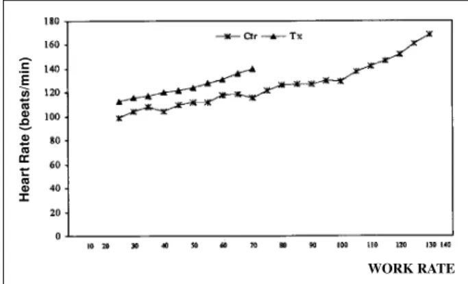
from the suprarenal gland 9,10. Nevertheless, submaximal
heart rate remains significantly higher in transplanted pati-ents relative to controls up to levels of 50% of maximal
exercise (fig.1) 39. Such higher levels during submaximal
exercise could reflect increased plasma levels of
catechola-mines, increased density of beta-receptors 40, as well as an
in-trinsic positive chronotropic effect at the pacemaker induced
by venous return 41-43. At peak exercise, the heart rate in
transplanted individuals is 20 to 25% lower than that in heal-thy controls. This chronotropic deficiency is attributed to
the absence of sympathetic innervation of the sinus node 35.
Persistence of chronotropic incompetence has been
obser-ved 2 to 6 years following cardiac transplantation 22. Later
transplanted patients have better chronotropic responses
than recent ones 44. The improved chronotropic response
during exercise of some patients six months following car-diac transplantation suggests sympathetic efferent
reinnervation 44. Evidence of late reinnervation in some
transplanted subjects has been demonstrated
immunoche-mically 45. Also, the reappearance of the circadian rhythm of
to reinforce the hypothesis of partial reinnervation in some
transplanted patients 46-48. In the early phase of cardiac
pos-transplantation, cardiac frequency keeps increasing during the first two minutes of the phase of recovery from exercise, despite the immediate decrease of circulating catecholami-nes 23,34 . This delayed deceleration is possibly due to an
in-creased sensitivity of the denervated heart to
catecholami-nes 49 (fig.2). Individuals examined from one to ten years
after transplantation had an immediately decreased heart
rate in the first minute of recovery 23. Kavanagh and Yacoub
reported a reduction in resting heart rate and increased peak cardiac frequency after two years of physical training. Ho-wever, resting heart rate remained higher and peak heart rate lower when compared to with that in controls. The mecha-nism responsible for the adaptation of heart rate following
conditioning has not been clarified 49.
Ventricular function – Reduction in systolic volume during rest and exercise has been reported in transplanted
compared with healthy individuals. Kao et al 22 submitted
transplanted patients to an invasive exercise test in associ-ated with oxygen consumption and radioisotope effort ven-triculography, obtaining direct measurements of the ejection
fraction and ventricular volume. They reported lower systolic volume in transplanted individuals during rest in the orthostatic position, submaximal and maximal exercise, and related this finding to reduced final diastolic volume
secondary to the alteration of diastolic function 21. They
demonstrated that the relationship between pulmonary capillary pressure and the index of final diastolic volume (PCP/IVDf) had higher values in transplanted subjects du-ring rest and dudu-ring exercise, indicating decreased
ventri-cular complacency 21. Martin et al 26 found similar results in
supine, and orthostatic resting, and at 20% maxímal oxygen consumption in transplanted subjectes. The pathogenesis of diastolic dysfunction has not been clarified. Preservation techniques, length of time of graft ischemia, occurrence of rejection, systemic arterial hypertension, arterial coronary disease and the use of cyclosporine have been suggested
as etiological factors 21,26.
During exercise, increased systolic volume, seconda-ry to the Frank-Starling mechanism and elevations in heart rate and contractility, secondary to the release of catechola-mines by the adrenal glands, occur sequentially in trans-planted patients; the Frank-Starling mechanism is apparent in the initial phase. In the healthy heart these events occur
simultaneously 50. Despite the chronotropic incompetence
at peak exercise, transplanted subjects make less use of the Frank-Starling mechanism to increase systolic volume than
do healthy individuals, due to diastolic dysfunction 21.
Follow-up evaluation of these patients did not show
impro-vement of diastolic function 22.
The evaluation of systolic function via ejection fraction has shown conflicting results. The ejection fraction of transplanted patients has values similar to those in healthy individuals during rest and exercise in the
orthosta-tic position 8.5±3.9 months following transplantation 21.
Follow-up evaluation of these patients (2-6 years following transplantation) showed ejection fraction values sig-nificantly higher during rest, and similar to normal values
during exercise 22 . Pflufelder et al 51 analyzed ejection
fraction 11 months following transplantation and found values similar to those in healthy patients, both at rest and
during supine exercise. Tischler et al 52 in a serial evaluation
of ventricular function, found normal ejection fraction values after one month and after one and four years following transplantation.
During the first year following transplantation, the cardiac index reaches significantly lower values in trans-planted patients at peak exercise, mainly because of a chro-notropic deficit; in submaximal exercise, this occurs at the
cost of systolic volume 17,21. Two to six years following
transplantation, cardiac index values remain significantly
below normal in such patients 22.
Peripheral oxygen extraction – Peripheral factors play a relevant role in functional limitation after cardiac
trans-plantation. Bussières et al 53 demonstrated an inverse
corre-lation (p<0.001) between posttransplantation oxygen
arte-riovenous difference (D(a-v) O2) and functional aerobic
Fig. 1 – Variation in heart rate during an exercise test of a 44- year-old male patient five months following cardiac transplantation due to dilated myocardiopathy. Ctr: healthy control; Tx; transplanted patient; bpm: beats per min 6.
WORK RATE
Heart Rate (beats/min)
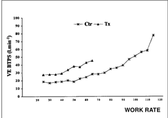
Fig. 2 – Pulmonary ventilation during an exercise test in a 38-year-old male patient, 47months following cardiac transplantation due to dilated myocardiopathy. Ctr: healthy control; Tx; transplanted patient; VE BTPS- pulmonary ventilation at body temperature and pressure and under saturation with steam 6.
TIME
Heart Rate (beats/min)
Exercise
deficit (FAI: max.pred.VO2 - peak VO2/max.pred.VO2 x 100),
(r= - 0.66). Savin et al 54 reported a D(a-v)O
2 significantly
lower than that of healthy controls at peak exercise. At sub-maximal exercise levels, a tendency towards higher values of
D(a-v)O2 was found and attributed to a compensatory
mecha-nism in view of the reduction of cardiac output. Kao et al 21
reported D(a-v)O2 values significantly reduced at peak
exercise but similar to control values at rest or submaximal
exercise. Mettauer et al 55 described similar results. The
na-ture of these abnormalities has not been clarified. Irre-versible alterations due to congestive cardiac insufficiency, physical deconditioning and prolonged corticoid treatment
may possibly be interfering with these results 56-60.
Vascular resistance – Transplanted patients pro-gress with a 45 to 92% incidence of arterial hypertension
(In-ternational Registry of Cardiac Transplantation) 61.
Borto-lotto et al 62 observed arterial hypertension in 58.5% of
patients 30 days following surgery, increasing to 93% after one year. Despite high blood pressure levels during rest, the patients’ mean arterial pressure reached values
significan-tly lower than those of healthy controls at peak exercise 21.
In our study, we found significantly higher levels of diasto-lic arterial pressure at rest and at peak exercise in transplan-ted subjects relative to that in controls,and no differences
of systolic arterial pressure between groups 6,7. The etiology
of this complication seems to be multifactorial, having as a common final route the elevation of systemic vascular
resis-tance 63-67. The reason for the attenuated pressure response
at peak exercise has not been clarified 18.
In view of the high incidence of posttransplantation arterial hypertension, comparative studies of cardiac trans-plants should include subgroups of apparently healthy and hypertensive patients. The majority of studies relating to physiological adaptation of transplanted patients to exerci-se rely on controls of apparently healthy individuals.
In transplanted subjects, levels of systemic vascular resistance relative to apparently healthy controls are persis-tently elevated both at rest and at exercise; however, resting values undergo marked reduction during exercise, a
beha-vior similar to that of healthy individuals 21,26. Bocchi et al 68
noted a fall in systemic vascular resistance during exercise in the supine position. Raised systemic vascular resistance could be attributed to the persistence of a pre-transplanta-tion abnormality, secondary to congestive cardiac insuffi-ciency (physical deconditioning, deficient mechanisms of
peripheral vasodilatation due to Na+ and H
20 retention) and
special posttransplantation conditions (physical decondi-tioning, use of cyclosporine and neuroendocrine
abnorma-lities) 21.
Following transplantation, mean pulmonary arterial pressure is significantly higher at rest and during exercise. Similarly to that in healthy controls, values rise during the
ef-fort test 21,26. Pulmonary vascular resistance is significantly
elevated in transplanted patients at rest and during exercise and decreases during effort in the same manner as in healthy individuals. These findings have been associated with the
irreversibility of vascular pulmonary alterations due to
chronically elevated pressure in the pulmonary artery 21.26.
Pulmonary ventilation - Several studies have pointed to the excessive ventilation work of the transplanted patient, characterized by higher values of the ventilatory equivalents
for 02 and CO2 at submaximal exercise 18,19,23. Pulmonary
ventilation at peak exercise is significantly reduced in
trans-planted subjects relative to that in healthy controls 6,7,11,18,19,26
(fig.3). However, when analyzing values of pulmonary ven-tilation, ventilatory equivalent for oxygen, ventilatory equivalent for carbon dioxide at the anaerobic threshold and at the 40W potency, no significant differences between
healthy and transplanted patients are apparent 7. The
me-chanism responsible for the excessive ventilation response in transplanted individuals has not been clarified. Pope et al and Savin et al attributed this response to the attenuated cardiac output curve during exercise secondary to cardiac denervation with an altered ventilation/perfusion ratio and
an increased physiological dead space 9,54. Marzo et al 18
de-monstrated that in transplanted patients, the ventilatory equivalent for carbon dioxide at rest was not significantly different in relation to that in controls. In this situation car-diac output was similar to the normal. Another explanation for the excessive ventilatory response could be muscular respiratory dysfunction consequent to hypoperfusion and
muscular fatigue 67,70. Kavanagh et al 71 reported significant
improvement in the respiratory response during exercise following physical training.
Neuroendocrine response – An exaggerated neuro-endocrine response in transplanted vs control subjects who performed the same relative levels of exercise has been
reported. Braith et al 19 found neuroendocrine hyperactivity
at rest characterized by significantly higher values of plas-ma renin and atrial natriuretic peptide. At 70 and 100% peak
VO2, plasma renin activity and estimates of atrial natriuretic
peptide, vasopressin and norepinephrine were significantly
elevated in the transplanted group 19. The neuroendocrine
profile of the transplanted patients could be attributed to the
Fig. 3 – Variation in heart rate during an exercise test of a 30-year-old female patient 16 months following cardiac transplantation due to dilated myocardiopathy. The exercise peak is indicated by *; bpm: beats per min.
use of cyclosporine 72,73 and hypertensive medication 74,
ske-letal muscle deconditioning 28,75 and cardiac denervation 19.
Cardiac denervation causes loss of the afferent stimulation of atrial stretch receptors due to disconnection of the heart from the brain, with consequent reflex inhibition of
neuro-hormones 19. Chronic neuroendocrine hyperactivity has
been associated with the incidence and seriousness of
arte-rial hypertension following cardiac transplantation 19.
Exercise test – Relative to cinecoronariography, the conventional exercise test shows poor postcardiac trans-plantation performance in the detection of coronary artery disease. Sensitivity and positive prediction values were
respectively 21% and 21% (Smart et al 76) and 0% and 0%
(Ehrman et al 77). The detection and evaluation of the
se-riousness of coronary artery disease by myocardial scinti-lography has been considered class IIB following cardiac
transplantation due to reduced sensitivity and specificity 78.
Ehrman et al 77 attribute the low sensitivity of the exercise
test for the detection of myocardial ischemia to the low car-diac frequency reached at peak exercise and to the high pre-valence of complete right branch blockade. Rodney and
Johnson 79 reported the diffuse nature of coronary artery
disease at grafting as the most probable cause of the low sensitivity of studies of myocardial perfusion.
The exercise test is used in the prescription of exercise in supervised rehabilitation programs. Cardiac frequency and arterial pressure responses during effort of transplan-ted persons are often modest, and other parameters like per-ceived effort are better for the estimation of the degree of the exercise. Exercise test protocols must have increments of lower intensity to give the denervated heart time to respond
to circulatory catecholamines 80. Steady state protocols are
more appropriate because they permit better hormonal and
metabolic adaptation. The VO2 peak is not altered with this
type of protocol 81. Modified forms of the Bruce or
Naugh-ton 49 protocols attaining peak effort in 8-10 min by 1 to 2
MET increments have been used. In the cycloergometer, increments of 50 or 100 Kpm/min at each minute have been
used 49. We have used increasing load protocols with
incre-ments of 5 watts/minute after an initial stage of 3 min at 25
W, maintaining an average of 50 rotations per min 6,7. The
electrocardiogram is continuously monitored, and the arte-rial pressure is measured every two min, at the peak of the
exercise and during recovery. Measurements of VE, VO2,
VCO2, RER, PEO2 and PECO2 are made in expired air at each
respiration. Borg’s scale of perceived effort is used at every
stage 49. Special attention should be paid to symptoms of
dyspnea, dizziness, weakness and electrocardiographic signs, in view of the incapacity of transplanted patients to manifest angina pectoris.
Cardiac rehabilitation – Long periods of periopera-tion inactivity, lack of motivaperiopera-tion, anxiety, depression, inse-curity, corticoid-induced skeletal muscle atrophy, recur-rence of rejection, and reduction in cardiorespiratory per-formance of the transplanted patient justify the prescription of physical exercise. A number of physical conditioning
programs have been described 72,82-88. In 1983, Squires et al 82
started a two-month supervised program six weeks follo-wing cardiac transplantation in two patients. The training was performed on a treadmill and bicycle three times per week for 30min using Borg’s scale of perceived effort
between 12 and 13. Kavanagh et al 71 effected a program of
walking and light running five times per week with sessions
of 45 min at 60-70% maximal VO2 and 14 on Borg’s scale. The
training lasted 16±7 months. Ferraz and Arakaki 87
establi-shed a supervised rehabilitation program with calisthenics on a stationary bicycle, short walks or runs and recreational games like adapted volleyball three times a week with sessions lasting 45 min at the 80% of anaerobic threshold and Borg’s scale between 13 and 15. The program lasted on
average 14 months. Romano et al 86 trained transplanted
subjects for 6 to 10 months and compared them with a group
of untrained subjects. In the trained group, VO2 was raised
by 85% (vs. 45% in the untrained group). In general, transplanted subjects should exercise three to five times a
week at between 50 and 75% VO2 and Borg’s scale between
13 and 15. Innumerable benefits of posttransplantation physical conditioning have been reported: reduced heart rate and arterial pressure during rest, decreased heart rate, arterial pressure, ventilartory equivalent for oxygen, ventilatory equivalent for carbon dioxide in submaximal exercise; increased heart rate, systolic arterial pressure,
VO2, pulmonary ventilation, and reduction in arterial
diasto-lic pressure at peak exercise; increased aerobic threshold, retardation of lactate elevation during exercise, reduction in
effort perception by the Borg scale 72,82-87.
References
1. Hunt SA, Bristow MR, Kubo SH, O’Connel JB, Young JB. Task Force 8: Training in heart failure and transplantation. J Am Coll Cardiol 1995; 25: 29-31. 2. Kaye MP. The Registry of the International Society for Heart and Lung
Transplantation: Tenth official report-1993. J Heart Lung Transplant 1993; 12: 541-8.
3. Paris W, Woodbury A, Thompson S, et al. Returning to work after heart transplan-tation. J Heart Lung Transplant1993; 12: 46-54.
4. Salles AF, Carvalho AC, Almeida DR, et al. Avaliação funcional cardio
respirató-ria durante o exercício em portadores de transplante cardíaco. Arq Bras Cardiol 1992; 59(supl. II): 156.
5. Salles AF, Carvalho AC, Almeida DR, et al. Cinética do consumo de oxigênio no exercício em indivíduos submetidos a transplante ortotópico de coração. Arq Bras Cardiol 1993; 61(supl. II): 99.
7. Salles AF, Oliveira Filho JA, Barros Neto TL, et al. Respostas cardiorrespiratórias durante exercício em portadores de transplante cardíaco. Análise ergoespiromé-trica comparativa com indivíduos normais. Arq Bras Cardiol 1998; 70: 15-18. 8. Starling EH. —Apud: Valenti PF, Mazzei ES, Masnatta G. Medicina Interna.
Vol.I. 7 ed. Barcelona: Marin, 1970: XVII.
9. Pope SE, Stinson EB, Daughters GT, Schroeder JS, Ingels NB, Alderman E. Exer-cise response of the denervated heart in long-term cardiac transplant recipients. Am J Cardiol 1980; 46: 213-8.
10. Savin WM, Haskell WL, Schroeder JS, Stinson EB. Cadiorespiratory responses of cardiac transplant patients to graded, symptom-limited exercise. Ciculation 1980; 62: 55-60.
11. Cerretelli P, Grassi B, Colombini A, Caru B, Marconi C. Gas exchange and metabo-lic transients in heart transplant recipients. Resp Physiology 1988; 74: 355-71. 12. Banner NR, Lloyd MH, Hamilton RD, Innes JA, Guz A, Yacoub MH. Cardiopul-monary response to dynamic exercise after heart and combined heart-lung trans-plantation. Br Heart J 1989; 61: 215-23.
13. Stevenson LW, Sietsema K, Tillisch JH, et al. Exercise capacity for survivors of cardiac transplantation or sustained medical therapy for stable failure. Circula-tion 1990; 81: 78-85.
14. Mandak JS, Donchez LJ, Mull RL, Mancini DM. Serial assesment of exercise capa-city post cardiac transplantation. Circulation 1993; 88(suppl.): I-591. 15. Quigg R, Salyer J, Mohanty PK, Simpson P. Impaired exercise capacity late after
cardiac transplantation: Influence of chronotropic incompetence, hypertension, and calcium channel blockers. Am Heart J 1998; 136: 465-73.
16. Hidalgo R, Alegriá E, Castelló R, et al. Stress testing in patients one year after or-thotopic cardiac transplantation. Angiology 1989; 40: 650-5.
17. Jensen RL, Yanowitz FG, Crapo RO. Exercise hemodynamics and oxygen delive-ry measurements using rebreathing techniques in heart transplant patients. Am J Cardiol 1991; 68: 129-33.
18. Marzo KP, Wilson JR, Mancini DM. Effects of cardiac transplantation on venti-latory response to execise. Am J Cardiol 1992; 69: 547-53.
19. Braith RW, Wood CE, Limacher MC, et al. Abnormal neuroendocrine responses during exercise in heart transplant recipients. Circulation 1992; 86: 1453-63. 20. Braith RW, Limacher MC, Staples ED, Pollock ML. Blood gas dynamics on the
onset of exercise in heart transplant recipients. Chest 1993; 103: 1692-8. 21. Kao AC, Trigt PV, Shaeffer-McCall GS, et al. Central and peripheral limitations to
upright exercise in untrained cardiac transplant recipients. Circulation 1994; 89: 2605-15.
22. Kao AC, Trigt PV, Shaeffer-McCall GS, et al. Allograft diastolic dysfunction and chronotropic incompetence limit cardiac output response to exercise two to six years after heart transplantation. J Heart Lung Transplant 1995; 14: 11-22. 23. Degré SGL, Niset GL, De Smet JM, Ibrahim T, Stoupel E. Cadiorespiratory
res-ponse to early exercise testing after orthotopic cardiac transplantation. Am J Car-diol 1987; 60: 926-28.
24. Meyer M, Cerretelli P, Cabrol C, Piiper J. O2 transport during exercise after cardiac
transplantation. In: Erdmann W & Bruley DP, ed,- Oxygen transport to tissue XIV. New York, Plenum Press, 1992; P: 491-6.
25. Paulus WJ, Brauzwaer JGF, Felice H, Kishan N, Welleus F. Deficient acceleration on the left ventricular relaxation during exercise after transplantation. Circulati-on 1992; 86: 1175-85.
26. Martin TW, Gaucher J, Pupa LE, Seaworth JF. Response to upright exercise after cardiac transplantation. Clin Cardiol 1994; 17: 292-300.
27. Horber FF, Hoppeler HS, Cheidegger JR, Gruning BE, Howald H, Frey FJ. Impact of physical training on the ultrastructure of midthigh muscle in normal subjects and in patients treated with glucocorticoids. J Clin Invest 1987; 79: 1181-90. 28. Massie B, Conway M, Rajagopalan B, et al. Skeletal muscle metabolism during
exercise under ischemic conditions in congestive heart failure. Evidence for ab-normalities unrelated to blood flow. Ciculation 1988; 78: 320-6.
29. Sinoway L, Minotti J, Davis D, et al. Delayed reversal of impaired vasodilation in congestive heart failure after heart transplantation. Am J Cardiol 1988; 61: 1076-9. 30. Ravenscraft AS, Gross CR , Kubo SH, et al. Pulmonary function after successful
heart transplantation. Chest 1993; 103:54-8.
31. Beck W, Bernard CN, Schrive V. Heart rate after cardiac transplantation. Circula-tion 1968; 40: 437-45.
32. Campeau L, Pospisil L, Grondin P, Dyrda I, Lepage G. Cardiac catheterization findings at rest and after exercise in patients following cardiac transplantation. Am J Cardiol 1970; 25: 523-8.
33. Jose A, Collision D. The normal range and determinants of the intrinsic heart rate in man. Cadiovasc Res 1970; 4: 160-7.
34. Yusuf S, Theodoropoulos S, Dhalla N, Mathias C, Yacoub M. Effect of betablo-ckade on dynamic exercise in human heart transplant recipients. Heart Transplant 1985; 4: 314-21.
35. Marneffe M, Jacobs P, Haardt R, Englert M. Variations of normal sinus node func-tion in relafunc-tion to age: role of autonomic influence. Eur Heart J 1986; 7: 662-72. 36. Gaer J. Physiological consequences of complete cardiac denervation. British
Jour-nal of Hospital Medicine 1992; 48: 220-5.
37. Rudas L, Pflugfelder P, Menkis AH, Novick RJ, Mckenzie FN, Kostuck WJ. Evo-lution of heart rate responsiveness after orthotopic heart transplatation. Am J Cardiol 1991; 68: 232-6.
38. Degré S, Niset G, Coustry C. Apud: Degré SGLG. Are cardiac transplant recipi-ents still suffering cardiac failure? Acta Cardiol 1993; XLVIII: 1-9. 39. Degré SGLG. Are cardiac transplant recipients still suffering cardiac failure?
Acta Cardiol 1993; 48: 1-9.
40. Yusuf S, Theodoropoulos S, Mathias CJ, et al. Increased sensitivity of the dener-vated transplanted human heart to isoprenaline both before and after betaadre-nergic blockade. Circulation 1987; 75: 696-704.
41. Blinks JR. Positive chronotropic effect of increasing right atrial pressure in the isolated mammalian heart. Am J Physiol 1956; 186: 299-303.
42. Shaver JA, Leon DF, Gray S, Leonard JJ, Bahnson HT. Hemodynamic observations after cardiac transplantation. N Engl J Med 1969; 281: 822-7.
43. Bexton RS, Milne JR, Cory-Pearce R, English TAH, Camm AJ. Effect of betablo-ckade on exercise response after cardiac transplantation. Br Heart J 1983; 49: 584-8.
44. Scott CD, Dark JH, McComb JM. Evolution of the chronotropic response to exer-cise after cardiac transplantation. Am J Cardiol 1995; 76:1292-6.
45. Wharton J, Polak JM, Gordon L, et al. Immunohistochemical demonstration of human cardiac innervation before and after transplantation. Circ Res 1990; 66: 900-12.
46. Mohanty P, Thames M, Capehart J, Kawaguchi A, Ballon B, Lower R. Afferent reinnervation of the autotransplanted heart in dogs. J Am Coll Cardiol 1986: 7: 414.
47. Burke MN, Mc Ginn AL, Homans DC, Christensen BV, Kubo SH, Wilson RF. Evidence for functional sympathetic reinnervation of left ventricle and coronary arteries after orthotopic cardiac transplantation in humans. Circulation 1995; 91: 72.
48. Stark RP, Mc Ginn AL. Chest pain in cardiac- transplant recipients: evidence of sensory reinnervation after cardiac transplantation. N Engl J Med 1991; 324: 1791.
49. Kavanagh T, Yacoub MH. Exercise training in patients after heart transplantati-on. Annals Academy of Medicine 1992; 21: 372-8.
50. Griepp RB, Stinson EB, Dong E, Clark DA, Shumway NE. Hemodynamic perfor-mance of the transplanted human heart. Surgery 1971; 70: 88-96.
51. Pflugfelder PW, Purves PD, Mckenzie FN, Kostuk WJ. Cardiac dynamics during supine exercise in cyclosporine-treated orthotopic heart transplant recipients: assesment by radionuclide angiography. J Am Coll Cardiol 1987; 10: 336-41. 52. Tischler MD, Lee RT, Plappert T, Mudge GH, Sutton MJ, Parker JD. Serial
asses-ment of left ventricular function and mass after orthotopic heart transplantation: A 4 year longitudinal study . J Am Coll Cardiol 1992; 19: 60-6.
53. Bussières LM, Pflugfelder PW, Menkis AH, et al. Basis for aerobic impairment in patients after heart transplantation. J Heart Lung Transplant 1995; 14: 1073-80. 54. Savin WM, Schroeder JS, Haskell WL. Reponse of cardiac transplant recipients
to static and dynamic exercise: A review. Heart Transplant 1983; 1: 72. 55. Mettauer B, Lampert E, Petitjean P, et al. Persistent exercise intolerance
follo-wing cardiac tranplantation despite normal oxygen transport. Int J Sports Med 1996; 17: 277-86.
56. Klausen K, Andersen LB, Pelle I. Adaptive changes in work capacity training and detraining. Acta Physiol Scand 1981; 113: 9-16.
57. Chi MMY, Hintz CS, Coyle EF, et al. Effects of detraining on enzymes of energy metabolism in individual human muscle fibers. Am J Physiol 1983; 244: C276-C87.
58. Horber FF, Scheidegger JR, Gruning BE, Frey FJ. Thigh muscle mass and function in patients treated with glucocorticoids. Eur J Clin Invest 1985; 15:302-07. 59. Ruff RL. Endocrine myopathies (hyper-and hipofunction of adrenal, thyroid,
pi-tuitary, and parathyroid glands and iatrogenic steroid myopathy). In: Engel AG, Banker BQ, eds. Myology. New York: Mc Graw-Hill, 1986: 1871-9. 60. Sullivan MJ, Green HJ, Cobb FR. Skeletal muscle biochemistry and histology in
ambulatory patients with long-term heart failure. Circulation 1990; 81: 518-27. 61. Registry of the International Society of Heart Tranplantation. Transplantation
around the world. J Heart Transplant 1986; 5: 1-88.
62. Bortolotto LA, Silva HB, Bocchi EA, Bellotti G, Stolf N, Jatene AD. Evolução a longo prazo e complicações da hipertensão arterial após transplante cardíaco. Arq Bras Cardiol 1997; 69: 317-21.
63. Schachter M. Cyclosporine A and hypertension. J Hypertension 1986; 6: 511-6. 64. Luke RG. Mechanism of cyclosporine-induced hypertension. Am J Hypertens
1989; 4: 468-71.
65. Starling RC, Cody RJ. Cardiac transplant hypertension. Am J Cardiol 1990; 65: 106-11.
66. Scott JP, Higenbottam TW, Hutter JA, Large S, Wallwork J. Effects of the immuno-suppressant cyclosporine on the circulation of the heart transplant recipients. Am J Cardiol 1991; 67: 628-32.
68. Bocchi E, Vilas-Boas F, Bacal F, et al. Avaliação hemodinâmica durante exercício isotônico em pacientes submetidos a transplante cardíaco ortotópico. Arq Bras Cardiol 1994: 63: 7-12.
69. Killian K, Jones N. Respiratory muscle and dyspnea. Clin Chest Med 1988; 9: 237-48.
70. Mancini DM, Ferraro N, Nazzaro D, Chance B, Wilson J. Demonstration of respi-ratory muscle deoxygenation during exercise in patients with heart failure using near- infrared spectroscopy. J Am Coll Cardiol 1991b; 18: 492-8.
71. Kavanagh T, Yacoub MH, Mertens DJ, Kennedy J, Cambell RB, Sawyer P. Car-diorespiratory responses to exercise training after orthotopic cardiac transplan-tation. Circulation 1988; 77: 162-71.
72. Schuler S, Thomas D, Hetzer R. Cyclosporine A - related nephrotoxicity after car-diac transplantation: The role of plasma renin activity. Transplant Proc 1987; 19: 3998-4001.
73. Scherrer U, Vissing S, Morgan B, et al. Cyclosporine- induced sympathetic acti-vation and hypertension after heart transplantation. N Engl J Med 1990; 323: 693-9.
74. Drieu L, Rainfray M, Cabrol C, Ardaillou R. Vasopressin, aldosterone, and renin res-ponses to volume depletion in heart transplant recipients. Clin Sci 1986; 70: 233-41. 75. Drexler H, Reide U, Munzel T, Konig H, Funke E, Just H. Alterations of skeletal
muscle in chronic heart failure. Circulation 1992; 85: 1751-9.
76. Smart FW, Ballantyne CM, Cocanougher B, et al. Insensitivity of noninvasive tests heart coronary artery vasculopathy after heart transplantation. Am J Cardiol 1991; 67: 243-7.
77. Ehrman JK, Keteyian SJ, Levine AB, Rhoads KL, Elder LR, Levine TB, Stein PD. Exercise stress tests after cardiac transplantation. Am J Cardiol 1993; 71: 1372-3.
78. ACC/AHA Task Force Report- Guidelines for clinical use of cardiac radionucli-de imaging. J Am Coll Cardiol 1995; 25: 521-47.
79. Rodney RA, Johnson LL. Myocardial perfusion scintigraphy to assess heart transplant vasculopathy. J Heart Lung Transplant 1992; 11: 574-8. 80. AHA Medical/Scientific Statement. Exercise Standars. Ciculation 1995; 91:
580-615.
81. Gullestad L, Myers J, Noddeland H, et al. Influence of the exercise protocol on hemodynamic, gas exchange and neurohormonal responses to exercise in heart transplant recipients. J. Heart Lung Transplant 1996; 15: 304-13.
82. Squires RW, Arthur PR, Gau GT, Muri A, Lambert WB. Exercise after cardiac trans-plantation: A report of two cases. J Cardiopulmonary Rehab 1983; 3: 570-4. 83. Degre S, Miset G, Desmet JM, et al. Effects du trainement physique sur le coeur
hu-man denervé après transplantation cardiaque orthotopique. Ann Cardiol Angeiol 1986; 35: 147-9.
84. Niset G, Counstry-Degre C, Degre S. Psychosocial and physical rehabilitation after heart transplantation. 1 year follow-up. Cardiology 1988; 75: 311-7. 85. Keteyian S, Ehrman J, Fedel F, Rhoads K. Rehabilitation following cardiac heart
transplantation. Med Sci Sports Exercise 1989; 21: 555.
86. Romano A, Stolf N, Bocchi E, Bellotti G. Contribuição do treinamento físico após transplante cardíaco. In: Yazbek Jr P, Battistella LR. Condicionamento Físico do Atleta ao Transplantado. São Paulo: Sarvier, 1994.
87. Ferraz AS, Arakaki H. Atividade física e qualidade de vida após transplante car-díaco. Rev Soc Cardiol ESP 1995; 6: 670-8.