Campus de São José do Rio Preto
PROGRAMA DE PÓS-GRADUAÇÃO EM GENÉTICA
Bruna Victorasso Jardim-Perassi
Avaliação da angiogênese em resposta ao tratamento com
melatonina no câncer de mama: Estudo
in vitro
e
in vivo
Tese apresentada para
obtenção do Título de Doutor em Genética
Avaliação da angiogênese em resposta ao tratamento com
melatonina no câncer de mama: Estudo
in vitro
e
in vivo
Orientadora: Profª. Drª. Debora Aparecida Pires de Campos Zuccari
São José do Rio Preto – SP
2014
Tese apresentada para
melatonina no câncer de mama: Estudo
in vitro
e
in vivo
Tese apresentada como parte dos requisitos para obtenção do título de Doutor em Genética, junto ao Programa de Pós-Graduação em Genética do Instituto de Biociências, Letras e Ciências Exatas da Universidade Estadual Paulista “Júlio de Mesquita Filho”, Campus de São José do Rio Preto.
BANCA EXAMINADORA
Profa. Dra. Debora Ap. Pires de Campos Zuccari Professor Doutor
Universidade Estadual Paulista (UNESP) – São José do Rio Preto - SP Orientador
Profa. Dra. Ana Elizabete Silva Professor Doutor
Universidade Estadual Paulista (UNESP) – São José do Rio Preto - SP Profa. Dra.Dorotéia Rossi Silva Souza
Professor Doutor
Faculdade de Medicina de São José do Rio Preto (FAMERP) – SP Prof. Dr. Samuel Cos Corral
Professor Doutor
Facultad de Medicina – Santander – Espanha Prof. Dr. Bruno Cogliati
Professor Doutor
Universidade de São Paulo (USP) – SP
Dedico esse trabalho a minha mãe Marlene, um grande exemplo
de força e coragem na minha vida.
Dedico também ao meu marido Fábio, que dividiu com muito
Muitas pessoas fizeram parte desse período e fico feliz de poder agradecê-las nesse momento...
Primeiramente gostaria de agradecer a minha orientadora Profa. Dra. Debora Zuccari pela oportunidade, confiança e todo apoio durante a realização desse trabalho. Agradeço imensamente pela amizade e incentivo que me fizeram vencer cada conquista e superar as dificuldades no caminho.
Aos amigos do laboratório: Marina, Gabriela, Thaiz, Lívia, Gustavo, Larissa, Naiane, Juliana, Giovanna, Rubens, Nathália, Tialfi, Jucimara e Suzana pela amizade e conhecimento compartilhados. Cada um de vocês contribuiu de alguma forma neste trabalho e só tenho a agradecê-los por isso, pois sozinha esta etapa da minha vida não teria sido tão produtiva.
Ao Dr. Ali Arbab, pela colaboração nesse projeto e por todo “hard work” que aprendi durante a realização de parte desse projeto em seu laboratório “Cellular and Molecular Imaging Laboratory” em Detroit nos Estados Unidos. Agradeço também a todos que conheci nesse período, especialmente ao Varma, que além de me auxiliar todos os dias no laboratório, tornou-se um grande amigo. Agradeço mais uma vez a Lívia e a Thaiz que compartilharam comigo essa experiência maravilhosa, tanto profissional como pessoal.
À banca examinadora da minha qualificação Profa. Dra. Ana Elizabete Silva e Prof. Dr. Newton Antonio Bordin Júnior pelas considerações que contribuíram com esse trabalho.
À banca examinadora da minha tese, Profa. Dra. Ana Elizabete Silva, Prof. Dr. Bruno Cogliati, Profa. Dra.Dorotéia Rossi Silva Souza e Prof. Dr. Samuel Cos Corral pelo aceite em participar da análise e enriquecimento deste trabalho.
A todos os professores e funcionários da Pós-Graduação em Genética do IBILCE que contribuíram com meu aprendizado. Agradeço especialmente a Profa. Mary Massumi Itoyama por ter me aceitado no estágio de docência, que complementou minha formação.
A FAMERP pela infra-estrutura e profissionais que possibilitaram a realização desse projeto.
A FAPESP pela concessão da bolsa de estudos no país e no exterior e auxílio financeiro, indispensáveis para a realização desse projeto.
A minha mãe Marlene, por toda luta e incentivo para que eu chegasse até aqui. Obrigada por acreditar que eu seria capaz!
Ao meu marido Fábio, que sempre esteve ao meu lado em todos os momentos, desde a faculdade, até hoje! Meu eterno companheiro, obrigada por todo apoio, amor e paciência.
Ao meu irmão Lucas, meu grande amigo!
À minha família, minhas tias, primas e primos que estiveram sempre presentes.
Aos meus sogros Marco e Valéria que me acolheram como uma filha e que estão sempre torcendo por mim.
Aos meus amigos, Marcela, Leandro, Larissa, Gustavo S., Carol e Gustavo M. que estão sempre me incentivando e proporcionando momentos de descontração.
Ao meu pai, que mesmo em memória sempre esteve ao meu lado dando força e me abençoando.
“A mente
que se abre para uma nova ideia jamais
voltará ao seu tamanho original"
Sumário
RESUMO ... 17
ABSTRACT ... 20
I. INTRODUÇÃO ... 23
1. Câncer de mama ... 23
2. Angiogênese ... 25
3. Melatonina ... 32
3.1. Síntese e degradação ... 32
3.2. Receptores da melatonina ... 35
3.3. Melatonina e câncer ... 36
II. OBJETIVOS ... 40
III. CAPÍTULOS ... 42
ARTIGO I - Effect of Melatonin on Tumor Growth and Angiogenesis in Xenograft Model of Breast Cancer...………..44
ARTIGO II - Melatonin regulates angiogenic factors under hypoxia in breast cancer cell lines……… ... 56
IV. DISCUSSÃO ... 93
V. CONCLUSÕES ... 102
VI. REFERÊNCIAS ... 104
Lista de abreviaturas e símbolos
[6(OH)M] Fórmula química do 6-hidroximelatonina
µg micrograma
µl micro-litro
µM micromol
13-HODE do inglês 13-Hydroxyoctadecadienoic Acid 5-HTP Fórmula química do 5-hidroxitriptofano AA-NAT Arilalquilamina N-acetiltransferase ACTB do inglês Beta Actin
Akt do inglês Protein Kinase B
ANOVA do inglês Analysis of Variance
bFGF do inglês Basic Fibroblast Growth Factor
cAMP 3`, 5-cyclic monophosphate cDNA DNA complementar
cm Centímetros
cm2 Centímetros quadrados
CO2 Fórmula química do gás carbônico
CoCl2 Fórmula química do Cloreto de Cobalto
DAB do inglês 3,3'-diaminobenzidine tetrahydrochloride DMBA 7,12 dimethylbenz(alpha)_anthracene
DMEM do inglês Dulbecco's Modified Eagle Medium
DMSO do inglês Dimethyl sulfoxide
EGF do inglês Epidermal Growth Factor
EGFR do inglês Epidermal Growth Factor Receptor
FAMERP Faculdade de Medicina de São José do Rio Preto
FAPESP Fundação de Amparo à Pesquisa do Estado de São Paulo
g Grama
GAPDH do inglês Glyceraldehyde 3-phosphate dehydrogenase
HER-2/neu Receptor do Fator de Crescimento Epidermal 2 HIF do inglês Hypoxia Inducible Factor
HIOMT Hidroxi-indol-O-metiltransferase HRP do inglês horseradish peroxidase
HUVEC do inglês Human Umbilical Vein Endothelial Cell
HYNIC do inglês Hydrazine Nicotinamide
IACUC do ingles Institutional Animal Care and User Committee IGF-I do inglês Insulin-like Growth Factor I
INCA Instituto Nacional do Câncer IP Intraperitoneal
Kg Quilogramas
Ki-67 do inglês antigen identified by monoclonal antibody Ki-67
LIMC Laboratório de Investigação Molecular no Câncer MOD do inglês Mean Optical Density
MAPK do inglês Mitogen-Activated Protein Kinase
mCi millicurie
mg Miligrama
mL Mililitro
mM Milimolar
mm2 Milímetros quadrados
mm3 Milímetros cúbicos
MMPs Matriz Metaloproteinases mRNA RNA mensageiro
MTT do inglês 3-(4,5-Dimethylthiazol-2-yl)-2,5-diphenyltetrazolium bromide
MVD do inglês micro-vessel density
NAS N-acetilserotonina
oC Graus Celsius
OMS Organização Mundial de Saúde PBS do inglês Phosphate Buffer Solution
PDGF-Ra do inglês platelet-derived growth factor receptor, alpha polypeptide
PGF do ingles PlacentalGrowthFactor
pH Potencial Hidrogeniônico
PHDs do inglês Hydroxylated by Prolyl-4-hydroxlyases PI3K do inglês phosphatidylinositol 3-kinase
PKC do inglês Protein Kinase C
pVHL do inglês Protein Von Hippel-Lindau
RANTES Quimiocina que regula expressãoe secreção por Linfócitos T RE Receptor de estrógeno
RENCA do inglês Mice Bearing Renal Adenocarcinoma
RNA Ácido Ribonucléico
ROI do inglês Regions of Interest
RP Receptor de Progesterona
RZR/ROR Receptor Z para Retinóide / Receptor Órfão para Retinóide S.E.M. Desvio Padrão
SDF-1α do inglês Stromal-Cell-Derived Factor-1α
SPECT do ingles Single-Photon Emission Computed Tomography
Tc-99m do inglês Technetium-99m
Tie-1 Receptor de Tirosina-quinase 1 Tie-2 Receptor de Tirosina-quinase 2
TNM Sistema de estadiamento clínico, T= tumor, N=linfonodo (do inglês node), M= metástase
TPH1 Enzima Triptofano Hidroxilase 1 u.a Unidades arbitrárias
VEGF do inglês Vascular Endothelial Growth Factor
RESUMO
O câncer de mama apresenta altas taxas de incidência e mortalidade, sendo a neoplasia mais comum entre as mulheres. O rápido crescimento do tumor resulta em hipóxia no microambiente tumoral, levando a uma cascata de eventos que induzem a angiogênese e conseqüente progressão do câncer. Assim, a identificação de agentes terapêuticos que possam inibir a angiogênese é essencial para o controle da progressão tumoral. Nesse sentido, tem sido demonstrado que a administração exógena da melatonina, um hormônio naturalmente secretado pela glândula pineal, apresenta diversos efeitos oncostáticos em diferentes tipos de câncer. Assim, o objetivo desse estudo foi avaliar a efetividade do tratamento com melatonina sobre a angiogênese do câncer de mama, em estudos in vitro e in vivo.
VEGFR2, VEGFR3, matriz metaloproteinase 9 (MMP-9) e Angiogenina em células MCF-7 (p < 0.05). Para linhagem MDA-MB-231, foi observada redução significante para as proteínas VEGFR2, receptor do fator de crescimento epidermal (EGFR) e Angiogenina (p < 0.05). No estudo in vivo os camundongos tratados com melatonina apresentaram menor crescimento tumoral em relação aos animais controle (144,90 ± 38,38 mm3 vs 282,00 ± 88,53 mm3; p < 0,05). Além disso, um animal apresentou regressão do tumor durante o tratamento com melatonina (Dia 7 = 27,38 mm3; Dia 14 = 8,79 mm3; Dia 21 = 4,8 mm3). Pela técnica de SPECT foi possível detectar menor radioatividade de Tc-99-HYNIC-VEGF-c e consequente expressão reduzida de VEGFR2/3 nos tumores tratados com melatonina (150,46 ± 17,06 % vs 183,55 ± 20,92 %) mas o nível de significância estatística não foi alcançado (p > 0,05). A diminuição de VEGFR2 nos tumores tratados com melatonina foi confirmada por imuno-histoquímica (p < 0,05), assim como a redução da microdensidade vascular (vWF) e da proliferação celular (Ki-67) (p < 0,05). Não houve alteração na expressão das demais proteínas avaliadas, no entanto umaumento significante de EGFR e do fator de crescimento semelhante a insulina tipo 1 (IGF-I) foi observado nos tumores tratados com melatonina (p < 0.05). Dessa forma, conclui-se que a melatonina apresenta importante ação anti-angiogênica in vitro e in vivo, evidenciando sua potencial ação terapêutica no câncer de mama.
ABSTRACT
Breast cancer has high rates of incidence and mortality, and it is the most common cancer among women. The rapid tumor growth results in hypoxia on tumor microenvironment, leading to a cascade of events that induce angiogenesis and subsequent cancer progression. Thus, the identification of therapeutic agents that can inhibit angiogenesis is essential for the control of tumor progression. Exogenous administration of melatonin, a hormone secreted by the pineal gland, has been shown several oncostatics effects on different types of cancers. The aim of this study was to evaluate the effectiveness of melatonin treatment on angiogenesis in breast cancer, in the in vitro and in vivo studies. In the in vitro study, breast cancer cell lines (MCF-7 and MDA-MB-231) were treated with melatonin under cobalt chloride (CoCl2)-induced hypoxic conditions. Cell
viability was measured by MTT assay, the expression of hypoxia-inducible factor 1-alpha (HIF-1α) and vascular endothelial growth factor (VEGF-A) was assessed by real-time PCR and immunocytochemistry. Additionally, other proteins involved in angiogenesis were evaluated by the Protein Array. In the in vivo study, the MDA-MB-231 cells were implanted in athymic nude mice, which were treated with melatonin (40 mg/kg) for 21 days. The tumor was measured weekly and evaluation of angiogenesis was performed by single-photon emission computed tomography (SPECT) with Tc-99m-HYNIC-VEGF-c, which is specific for VEGF receptors (VEGFR2/VEGF3). Moreover, VEGFR2, VEGFR3, von Willebrand factor (vWF) and cell proliferation marker (Ki-67) were evaluated in tumor tissue by immunohistochemistry, and other angiogenic proteins by Protein Array. Results from the in vitro study showed that 1 mM of melatonin under hypoxic conditions (200 µM CoCl2) led to decreased cell viability and levels of HIF-1α
compared to control animals (144.90 ± 38.38 mm3 vs 282.00 ± 88.53 mm3, p < 0.05). Furthermore, one animal showed tumor regression during melatonin treatment (Day 7 = 27.38 mm3, 8.79 mm3 Day = 14, mm3 Day 21 = 4.8). SPECT
detect less radioactivity of Tc-99m-HYNIC-VEGF-c and consequent reduced expression of VEGFR2/3 in tumors treated with melatonin (150,46 ± 17,06 % vs
183,55 ± 20,92 %) but statistical significance was not achieved (p > 0.05). The reduction of VEGFR2 in tumors treated with melatonin was confirmed by immunohistochemistry (p <0.05), as well as the reduction of micro-vessel density (vWF) and cell proliferation (ki-67) (p < 0.05). There was no change in the expression of the other proteins evaluated, however a significant increase in EGFR and Insulin-like growth factor (IGF-I) was observed in tumors treated with melatonin (p< 0.05). Taken together, our results showed that melatonin has a important anti-angiogenic effect, suggesting its potential therapeutic action in breast cancer.
I. INTRODUÇÃO
1. Câncer de mama
O câncer de mama apresenta alta incidência, sendo o tipo mais freqüente entre as mulheres. No Brasil, o Instituto Nacional do Câncer (INCA) estimou a ocorrência de 52.680 novos casos para o ano de 2012, e 57.120 para o ano de 2014 (INCA, 2014). A Organização Mundial da Saúde (OMS) estima que, por ano, ocorram mais de 1.050.000 novos casos de câncer de mama em todo o mundo (INCA, 2011).
Alguns fatores de risco incluem menarca precoce, menopausa tardia, nuliparidade, idade avançada, aumento do peso corporal, entre outros (FRANK; CRITCHFIELD, 2002; WU et al., 2007; LERTKHACHONSUK et al., 2013). Países do norte da Europa, Austrália, Nova Zelândia e América do Norte apresentam as taxas de incidência mais elevadas, seguido de taxas intermediárias na América do Sul e menores índices no continente asiático (JEMAL et al., 2011). Historicamente, a menor incidência atribuída à população asiática relaciona-se a idade mais avançada da menarca, baixo peso corporal e estilo de vida da população, que inclui atividade física regular e alimentação rica em vegetais (WU
et al., 2006; SIEGEL et al., 2014).
nos últimos 30 anos, essa doença é ainda responsável por quase meio milhão de mortes por ano (SNOUSSI et al., 2010).
O estabelecimento do prognóstico e do planejamento terapêutico do câncer de mama baseia-se no tipo, grau histológico do tumor e classificação pelo sistema de estadiamento TNM, que abrange a avaliação do tamanho tumoral, presença de metástase em linfonodos regionais e de metástase à distância (PEDERSEN et al., 2004). Além dos fatores clínico-patológicos, o conhecimento das características moleculares dos tumores tem contribuído com a avaliação mais precisa do prognóstico, e com o desenvolvimento e incorporação de novos agentes e estratégias terapêuticas (GONZALEZ-ANGULO et al., 2007; GRALOW et al., 2008; HICKS; KULKARNI, 2008; DUFFY et al., 2011). Atualmente, o status do receptor de estrógeno (RE), receptor de progesterona (RP) e do receptor do fator de crescimento epidermal 2 (HER-2/neu) são exemplos de características moleculares dos tumores mamários que foram incorporados à rotina clínica e permitem que se estabeleça um tratamento individualizado (HSIAO et al., 2010).
Nesse contexto, atualmente muitas pesquisas exploram os mecanismos moleculares que contribuem com a progressão do tumor, a fim de identificar novos alvos de terapias. Dentre esses mecanismos, o processo de formação de novos vasos sanguíneos no tecido tumoral é essencial para seu crescimento e disseminação metastática e, portanto, cresce o interesse pelo desenvolvimento de agentes terapêuticos que atuem nesse processo (HANAHAN; WEINBERG, 2011; GACCHE; MESHRAM, 2013).
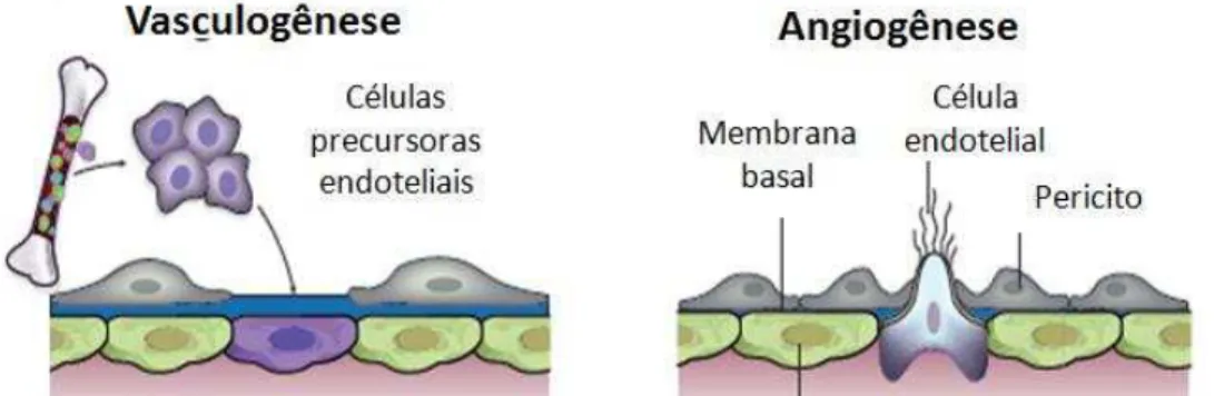
2. Angiogênese
Figura 1. Representação dos diferentes modelos de formação de novos vasos sanguíneos. Adaptado de (CARMELIET; JAIN, 2000).
A formação de novos vasos sanguíneos no tecido tumoral visa o fornecimento de nutrientes e oxigênio permitindo a proliferação das células e consequente crescimento e progressão do tumor (TAKAHASHI; SHIBUYA, 2005; GAVALAS et al., 2013). Além disso, permitem a retirada do gás carbônico (CO2) e dos resíduos metabólicos, e representam uma importante via de disseminação metastática (ZHANG et al., 2010; LIU; OUYANG, 2013).
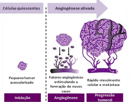
Figura 2: Representação da ativação da angiogênese a partir de células quiescentes. Adaptado de http://www.clinicaloptions.com/
Durante seu crescimento, o tumor pode alcançar aproximadamente 1-2 mm3 antes que suas demandas metabólicas sejam restritas devido ao limite de difusão de oxigênio e nutrientes no local (CARMELIET; JAIN, 2011). A baixa oxigenação é caracterizada como hipóxia e pode ocorrer devido à proliferação descontrolada das células e rápido crescimento do tumor, além da perfusão inadequada em parte do tecido resultante da estrutura caótica dos novos vasos sanguíneos formados (CARMELIET; JAIN, 2000; HARRIS, 2002; MILANI; HARRIS, 2008; VORDERMARK, 2010).
como a indução de fatores envolvidos no processo de angiogênese (KALLERGI et al., 2011).
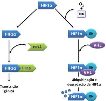
O fator de transcrição induzido por hipóxia (HIF-1) é essencial na manutenção da homeostase do oxigênio, e responsável por essas respostas adaptativas. O HIF-1 é um fator heterodímero composto de duas subunidades 1α e 1β. Em normóxia o 1β é constitutivamente expresso e o HIF-1α sofre degradação dependente de oxigênio (SEMENZA, 2002). O HIF-HIF-1α é hidroxilado e então reconhecido pela proteína supressora de tumor Von-Hippel-Lindau (pVHL) e ubiquitinilado para sofrer degradação proteossomal (DANG; SEMENZA, 1999). Em condições de hipóxia, essa degradação não acontece e então o HIF-1α migra para o núcleo, associando-se a HIF-1β, atuando como fator de transcrição de diversos genes (Figura 3) (SEMENZA, 2002; MIMEAULT; BATRA, 2013; TUNG et al., 2013).
HIF1β
HIF1α
HIF1α
HIF1β
HIF1α
Transcrição gênica
O2
+
PHD
OH
HIF1α
VHL
OH
HIF1α VHL
OH
HIF1α
Ubiquitinação e degradação de HIF1α
O HIF-1α pode regular mais de 100 genes envolvidos nos processos da eritropoiese, metabolismo do ferro e da glicose, proliferação celular, apoptose e angiogênese (KE; COSTA, 2006). Em condições de hipóxia, o principal alvo do HIF-1α é o fator pró-angiogênico denominado fator de crescimento endotelial vascular (VEGF) (KE; COSTA, 2006).
O VEGF é um potente mitógeno que atua em diferentes etapas do processo angiogênico, promovendo o aumento da permeabilidade vascular, estimulação da migração, proliferação e invasão de células endoteliais (HOEBEN et al., 2004; DELLI CARPINI et al., 2010; GREENBERG; RUGO, 2010; SHIBUYA, 2011). Esse fator foi primeiramente descrito em células endoteliais e, portanto, denominado “fator de crescimento endotelial vascular”, no entanto, o VEGF pode exercer ação mitogênica em outros tipos celulares (FERRARA; DAVIS-SMYTH, 1997). O VEGF é composto por uma família de cinco isoformas denominadas VEGF-A, VEGF-B, VEGF-C, VEGF-D e fator de crescimento placentário (PGF), os quais ligam-se a receptores específicos do tipo tirosina quinase, promovendo uma cascata de eventos intracelulares (BRITO et al., 2011; FINLEY et al., 2013).
tumorais, e podem estimular o crescimento celular de maneira autócrina (WEIS; CHERESH, 2011; ARBAB, 2012).
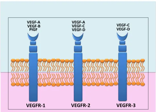
O VEGF–A se liga a dois receptores específicos, o VEGFR1 e o VEGFR2 enquanto o VEGF–B e PGF são reconhecidos apenas pelo receptor VEGFR1. O VEGF-C e VEGF-D se ligam ao VEGFR2 e também são reconhecidos pelo VEGFR3 (Figura 4) (RAHIMI, 2012). A ligação entre o VEGF-A e VEGFR2 é considerada o mais importante passo do processo de angiogênese (WOOLARD et al., 2004; MATSUURA et al., 2009; COULON et al., 2011), enquanto a ligação de VEGF-C com VEGFR3 está envolvida no processo de linfangiogênese (BRITO et al., 2011).
VEGFR-1 VEGFR-2 VEGFR-3
VEGF-A VEGF-A
VEGF-B VEGF-C VEGF-C
VEGF-D VEGF-D
PIGF
Figura 4. Esquema da ligação entre as isoformas de VEGF e seus receptores.
Metaloproteinases de matriz (MMPs), fator de crescimento derivado de plaquetas (PDGF), fator de crescimento de fibroblastos básico (bFGF), fator de crescimento epidermal (EGF), fator de crescimento dependente de insulina (IGF-I), citocinas pró-inflamatórias, entre outros (ARBAB, 2012).
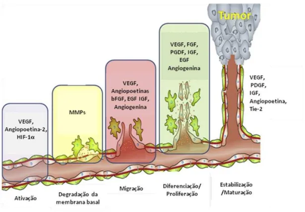
Inicialmente, ocorre a ativação de células endoteliais quiescentes por fatores liberados pelas células tumorais em resposta a condições adversas como privação de nutrientes e oxigênio. Alguns fatores como VEGF e Angiopoitina-2 atuam na desestabilização inicial dos vasos pré-existentes e aumento da permeabilidade vascular. Em seguida ocorre a degradação da membrana basal e da matriz extracelular, permitindo a migração de células endoteliais pelo espaço intersticial e eventual liberação de fatores pró-angiogênicos ligados a matriz. Subsequentemente ocorre a migração e proliferação de células endoteliais, bem como a formação do tubo, recrutamento e diferenciação de células de suporte perivascular (pericitos). Nessa etapa inúmeros fatores estão envolvidos como VEGF, Angiogenina, PDGF, bFGF, EGF, entre outros. Ao final, ocorre a maturação e estabilização dos novos vasos formados, com a participação de fatores como a angiopoitina-1 e seu receptor Tie-2 (Figura 5) (CLAPP et al., 2009; COULON et al., 2011; WEIS; CHERESH, 2011).
Figura 5. Representação das etapas do processo de angiogênese com a participação de inúmeros fatores pró-angiogênicos (GACCHE; MESHRAM, 2013).
Nesse contexto, dada a variedade de sinais envolvidos no processo angiogênico, diversos fatores podem ser considerados alvos terapêuticos, auxiliando no bloqueio da progressão do câncer (ARBAB, 2012; GACCHE; MESHRAM, 2013).
3. Melatonina
3.1. Síntese e degradação
diversas substâncias, como o cortisol e adrenalina, atuando sobre os ciclos de atividade-repouso e vigília-sono (SOUSA-NETO, 2005; MAGANHIN, 2008). Além disso, a melatonina atua sobre o sistema reprodutor, cardiovascular, sistema imunológico, crescimento e envelhecimento (MAGANHIN, 2008; PANDI-PERUMAL et al., 2013).
NH2 N
H
C C C OH H O H H NH2 N H
C C C OH H O H H NH2 N H C C H H H N H
C C N CH H H H N H HO HO HO
CH2O
H
C O H H
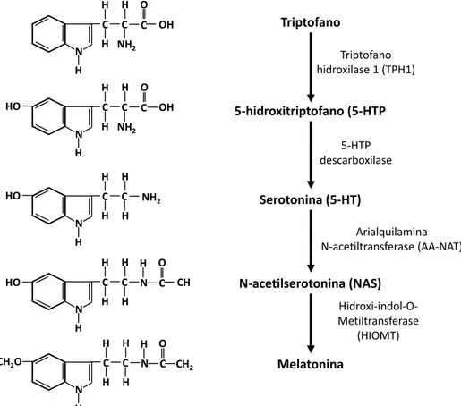
C C N CH2 H H H C O H H Triptofano 5-hidroxitriptofano (5-HTP Serotonina (5-HT) N-acetilserotonina (NAS) Melatonina Triptofano hidroxilase 1 (TPH1)
5-HTP descarboxilase Arialquilamina N-acetiltransferase (AA-NAT) Hidroxi-indol-O-Metiltransferase (HIOMT)
Figura 6. Esquema representativo da via de síntese da melatonina, desde o aminoácido triptofano até o último passo para a formação de melatonina pela enzima HIOMT.
Sua degradação ocorre principalmente no fígado, envolvendo a hidroxilação em 6-hidroximelatonina [6(OH)M], seguida dos processos de sulfatação ou glicuronidação, e posterior excreção na urina (REITER, 1991; FACCIOLA et al., 2001; SEMAK et al., 2008).
3.2. Receptores da melatonina
A melatonina pode atuar por meio de receptores de membrana acoplados a proteína G, denominados MT1 e MT2. Esses receptores são encontrados em diversos tecidos, incluindo sistema nervoso central, trato gastrointestinal, pulmão, pele, glândula adrenal, coração, vasos sanguíneos, células do sistema imune, entre outros (DUBOCOVICH; MARKOWSKA, 2005). Um terceiro receptor com menor afinidade pela melatonina é denominado MT3 e sua ativação ainda não possui papel fisiológico definido, no entanto apresenta 95 % de homologia com a enzima quinona redutase II, envolvida na detoxificação de radicais livres (FOSTER et al., 2000).
relacionadas à alteração de genes envolvidos na proliferação celular e apoptose (SANCHEZ-BARCELO et al., 2003).
3.3. Melatonina e câncer
Além de estar envolvida em muitas funções fisiológicas, a melatonina tem um importante papel em processos patológicos, incluindo o câncer. Os primeiros indícios de que a melatonina poderia ser útil na terapêutica oncológica surgiram em um estudo realizado por Cohen et al. (COHEN et al., 1978). Os autores propuseram que a diminuição da função da glândula pineal poderia aumentar o risco de desenvolvimento do câncer de mama, sugerindo que a ausência da síntese de melatonina poderia induzir a exposição prolongada ao estrógeno resultando no desenvolvimento do tumor mamário.
Em 1981, Tamarkin et al., (1981) demonstraram que o tratamento com melatonina inibiu o desenvolvimento de tumores mamários induzidos pelo carcinógeno 7,12-dimethylbenz(alpha)-anthracene (DMBA) em ratas Sprague-Dawley, e, ao contrário, a remoção da glândula pineal estimulou o desenvolvimento desses tumores. No mesmo ano, Bartsch et al., (1981) demonstraram que pacientes com câncer de mama apresentam diminuição nos níveis plasmáticos de melatonina.
sendo assim, associa-se o risco elevado da ocorrência desta doença em trabalhadores noturnos, e, de provável idêntica razão, baixo risco em mulheres cegas (STEVENS et al., 2013).
Na década de 90 alguns estudos clínicos foram realizados, demonstrando que a utilização da melatonina, em conjunto com terapias convencionais, apresenta efeitos benefícios em diferentes tipos de tumores avançados intratáveis ou em pacientes com tumores metastático, quer seja aumentando a eficácia do tratamento (LISSONI et al., 1995; LISSONI et al., 1996), ou diminuindo os efeitos colaterais contribuindo com a melhora da qualidade de vida desses pacientes (BARNI et al., 1990; LISSONI et al., 1992).
Desde então, têm sido demonstrado que a melatonina inibe o desenvolvimento e a progressão de diferentes tipos de câncer, e muitos mecanismos de ação estão sendo investigados (VIJAYALAXMI et al., 2002; JUNG; AHMAD, 2006; MARTIN et al., 2006; DI BELLA et al., 2013; OPREA-ILIES et al., 2013).
ainda na inibição da enzima telomerase, na modulação do sistema imune, inibição de proteínas envolvidas na invasão celular e no processo metastático (MEDIAVILLA et al., 2010; DI BELLA et al., 2013; PROIETTI et al., 2013).
Também tem sido demonstrado que a melatonina pode atuar na angiogênese direta ou indiretamente, inibindo a proliferação de células endoteliais e de fatores pró-angiogênicos em diferentes tipos tumorais (LISSONI et al., 2001; CUI et al., 2006; KAUR et al., 2007; DAI et al., 2008; PARK et al., 2009; PARK
II.OBJETIVOS
Sendo a angiogênese é um processo essencial para o crescimento do tumor, a identificação de agentes terapêuticos que atuem nesse processo é fundamental para a inibição da progressão tumoral. Nesse sentido, nos últimos anos tem sido demonstrado que a melatonina apresenta grande potencial terapêutico no câncer, e diversos efeitos anti-tumorais estão sendo identificados. Assim, o objetivo geral desse estudo foi avaliar a eficácia do tratamento com melatonina na angiogênese do câncer de mama, em estudos in vitro e in vivo.
Estudo in vitro: Verificar a viabilidade celular, expressão gênica e protéica do HIF-1α e VEGF-A e a expressão de demais proteínas envolvidas no processo angiogênico nas linhagens de câncer de mama MCF-7 (REα-positiva) e MDA-MB-231 (triplo-receptor negativo) após a indução da hipóxia e tratamento com melatonina.
III. CAPÍTULOS
Os resultados referentes aos objetivos desta tese serão apresentados a seguir na forma de dois artigos científicos:
Artigo I
Título: Effect of Melatonin on Tumor Growth and Angiogenesis in Xenograft Model of Breast Cancer
Autores: Bruna Victorasso Jardim-Perassi, Ali S. Arbab, Lívia Carvalho Ferreira, Thaiz Ferraz Borin, Nadimpalli R. S. Varma, A. S. M. Iskander, Adarsh Shankar, Meser M. Ali, Debora Aparecida Pires de Campos Zuccari
Periódico: PLoS ONE - publicado em 9 de janeiro de 2014 (doi:10.1371/journal.pone.0085311).
Artigo II
Título: Melatonin regulates angiogenic factors under hypoxia in breast cancer cell lines
Campos Zuccari2,4*
1Department of Biology, Universidade Estadual Paulista, Sa˜o Jose´ do Rio Preto, Sa˜o Paulo, Brazil,2Laborato´rio de Investigac¸a˜o Molecular no Caˆncer, Department of Molecular Biology, Faculdade de Medicina de Sa˜o Jose´ do Rio Preto, Sa˜o Jose´ do Rio Preto, Sa˜o Paulo, Brazil,3Cellular and Molecular Imaging Laboratory, Department of Radiology, Henry Ford Hospital, Detroit, Michigan, United States of America,4Department of Molecular Biology, Faculdade de Medicina de Sa˜o Jose´ do Rio Preto, Sa˜o Jose´ do Rio Preto, Sa˜o Paulo, Brazil
Abstract
As neovascularization is essential for tumor growth and metastasis, controlling angiogenesis is a promising tactic in limiting cancer progression. Melatonin has been studied for their inhibitory properties on angiogenesis in cancer. We performed an
in vivostudy to evaluate the effects of melatonin treatment on angiogenesis in breast cancer. Cell viability was measured by
MTT assay after melatonin treatment in triple-negative breast cancer cells (MDA-MB-231). After, cells were implanted in athymic nude mice and treated with melatonin or vehicle daily, administered intraperitoneally 1 hour before turning the room light off. Volume of the tumors was measured weekly with a digital caliper and at the end of treatments animals underwent single photon emission computed tomography (SPECT) with Technetium-99m tagged vascular endothelial growth factor (VEGF) C to detectin vivoangiogenesis. In addition, expression of pro-angiogenic/growth factors in the tumor
extracts was evaluated by membrane antibody array and collected tumor tissues were analyzed with histochemical staining. Melatoninin vitrotreatment (1 mM) decreased cell viability (p,0.05). The breast cancer xenografts nude mice treated with
melatonin showed reduced tumor size and cell proliferation (Ki-67) compared to control animals after 21 days of treatment (p,0.05). Expression of VEGF receptor 2 decreased significantly in the treated animals compared to that of control when determined by immunohistochemistry (p,0.05) but the changes were not significant on SPECT (p.0.05) images. In addition, there was a decrease of micro-vessel density (Von Willebrand Factor) in melatonin treated mice (p,0.05). However, semiquantitative densitometry analysis of membrane array indicated increased expression of epidermal growth factor receptor and insulin-like growth factor 1 in treated tumors compared to vehicle treated tumors (p,0.05). In conclusion, melatonin treatment showed effectiveness in reducing tumor growth and cell proliferation, as well as in the inhibition of angiogenesis.
Citation:Jardim-Perassi BV, Arbab AS, Ferreira LC, Borin TF, Varma NRS, et al. (2014) Effect of Melatonin on Tumor Growth and Angiogenesis in Xenograft Model of Breast Cancer. PLoS ONE 9(1): e85311. doi:10.1371/journal.pone.0085311
Editor:Sonia Rocha, University of Dundee, United Kingdom
ReceivedSeptember 2, 2013;AcceptedNovember 26, 2013;PublishedJanuary 9, 2014
Copyright:ß2014 Jardim-Perassi et al. This is an open-access article distributed under the terms of the Creative Commons Attribution License, which permits unrestricted use, distribution, and reproduction in any medium, provided the original author and source are credited.
Funding:Funded by Fundac¸a˜o de Amparo a Pesquisa do Estado de Sa˜o Paulo - FAPESP/Brazil (2011/01052-9, 2011/01054-1 and 2012/05065-0) and NIH (R01CA160216 and R01CA172048). The funders had no role in study design, data collection and analysis, decision to publish, or preparation of the manuscript.
Competing Interests:The authors have declared that no competing interests exist.
* E-mail: debora.zuccari@famerp.br
Introduction
Breast cancer is the most common type of cancer in women [1] with increasing incidence and mortality which is becoming a global public health problem [2,3]. Furthermore, breast cancer has a high mortality rate, mainly due to tumor progression and metastatic spread, which require neovascularization [4–6].
Tumor growth has traditionally been associated with angiogen-esis, which is the formation of new blood vessels from the pre-existing vasculature. Angiogenesis is controlled by pro-angiogenic and anti-angiogenic factors in the body. The angiogenesis is regulated by several growth factors such as vascular endothelial growth factor (VEGF), platelet-derived growth factor (PDGF), epidermal growth factor (EGF), Angiogenin, etc [7]. Recent studies have demonstrated that tumor progression can also occur through vasculogenesis. Vasculogenesis is typically followed by classical sprouting angiogenesis, where blood vessels are formed de
novo by in situ differentiation of the primitive progenitors - i.e. angioblasts - into mature endothelial cells, which was thought to only take place during embryonic development [8,9].
Once a tumor exceeds a few millimeters in diameter, hypoxia triggers a cascade of events to allow angiogenesis and tumor progression [8]. Hypoxia results in expression of VEGF, a potent endothelial cell mitogen [10–13]. VEGF is composed of a family of five isoforms denominated A, B, C, VEGF-D and placental growth factor (PGF). Each of these factors can activate one or more receptors (VEGFR1, VEGFR2 and VEGFR3), promoting angiogenesis through its ability to stimulate the growth, migration and invasion of endothelial cells [14–17].
Hypoxia also up-regulates the expression of stromal-cell-derived factor-1a (SDF-1a), which can recruit pro-angiogenic cells from bone marrow [18,19].Hypoxia can also act in the regulation of RANTES a chemokine of inflammatory cells [20,21]. Other signalling pathways such as basic fibroblast growth factor (bFGF)
vessels, several proteins can be therapeutic targets. Therefore, it becomes extremely important to identify the most susceptible target to a particular treatment, and to develop effective therapies that involve a combination of several factors [8,9].
Administration of melatonin, a hormone naturally produced and secreted in the pineal gland, appears to play an important role in tumor growth inhibition [24,25] and different mechanisms of action have been proposed [26]. The action of melatonin is especially effective in estrogen receptor (ER)-positive breast cancer by reducing the mitogenic response of cells [27]. Still, melatonin can exert immunomodulatory and antiproliferative effects and act as antioxidants [28–32]. Furthermore, it has demonstrated that the pharmacological concentration of melatonin can inhibit angio-genesis directly or indirectly [33–35]. Recently, some studies have shown that melatonin can decrease the expression of Hypoxia Inducible Factor 1 alpha (HIF-1a) and VEGF in various cancers [5,36–37].
The purposes of this study were to determine: 1) whether melatonin therapy effectively would decrease the size of implanted human triple negative breast cancer in a mouse model, 2) whether there would be changes in the expression of VEGF receptors determined by in vivo SPECT scanning in mice treated with melatonin or vehicle, 3) the changes in expression of angiogenic factors in tumors in response to melatonin. The ultimate goal was achieved by determining the therapeutic effectiveness of melato-nin.
Materials and Methods
Ethics statement
All animal experiments were performed according to National Institutes of Health guideline and the protocol was approved by Institutional Animal Care and User Committee (IACUC No. 1203) of Henry Ford Health System.
In vitro study
Cell culture. Cell culture. Triple negative human breast cancer cells (MDA-MB-231) (ATCC, Manassas, VA, USA) were cultured in 75 cm2culture flasks with Dulbecco’s modified Eagle’s medium (DMEM) (GIBCO, Grand Island, NY, USA) supple-mented with 10% fetal bovine serum (GIBCO, Grand Island, NY, USA), penicillin (100 IU/mL) and streptomycin (100mg/mL) (GIBCO, Grand Island, NY, USA)until they were 80–90% confluent.
Cell viability by MTT (3-(4,5-Dimethylthiazol-2-yl)-2,5-diphenyltetrazolium bromide) assay. Individual well of a 96-well plate was inoculated with 100ml of medium containing 56104 cells. Cells were incubated in medium with different concentrations of melatonin (0.0001 mM, 0.001 mM, 0.01 mM, 0.1 mM and 1 mM) for 24 h. Melatonin was diluted in ethanol 0.05%. In control cells, equivalent amount of ethanol was added as vehicle. Thereafter, 10mL of MTT solution (ATCC, Manassas, VA, USA) were added to each well and the plates were incubated at 37uC for an additional 4 h. To solubilize the MTT formazan crystals, the cells were incubated with detergent (ATCC, Manassas, VA, USA) overnight at 37uC. Absorbance was measured at 570 nm by ELISA Plate reader (VICTOR3 – PerkinElmer, Waltham, MA, USA). Medium was used as background and subtracted from the samples. Cell viability (%)
Female athymic mice, 7–8 weeks of age and 25 g in weight (Charles River Laboratory, Inc.) were housed. MDA-MB-231, culturedin vitro, was harvested and re-suspended serum free media at a concentration of 66107cells/ml. The animals received subcutaneous injection of 50ml of cell suspension (3 million cells) in the right mammary gland or hind flank. These tumor cells are efficient in making xenografted tumors with almost 90% efficiency and show marked vascularity with less central necrosis. Two different implantation sites were chosen to determine whether treatment effects will differ based on implantation sites.
Melatonin administration
Animals were randomly assigned to either the melatonin administration (n = 5) or the control group (vehicle treated, n = 8). Vehicle solution was prepared with 8 ml of phosphate buffered saline (PBS), 1 ml of dimethyl sulfoxide (DMSO) and 1 ml of Cremophor (Sigma, St. Louise, MO, USA). The animals of the control group received 100ml of vehicle solution by intraperitoneal injection (IP).
Melatonin (Sigma, St. Louise, MO, USA) was diluted in vehicle and the animals from the melatonin group received IP of 100ml of melatonin treatment (at dose of 40 mg/kg of body weight) for five days a week. Melatonin was administered 1 hr before room lighting was switched off. Administration of melatonin prior to the nocturnal increase in endogenous melatonin may be most effective because tissues are most sensitive to the hormone at this time [38,39].
Melatonin or vehicle administration started on the day of tumor implantation (soon after implantation) and continued for five days a week for 21 days. On the 22nd day, all animals underwent SPECT scanning with Tc-99m-HYNIC-VEGF-c followed by euthanasia and collection of tumors for immunohistochemistry and membrane antibody array to determine the expression of different pro-angiogenic and growth factors in tumor extracts.
Tumor measurement by caliper
Tumor volume was measured by digital caliper (Thermo Fisher Scientific, Rockford, IL, USA) on day 7, 14 and 21 after tumor implantation. The major longitudinal diameter (length) and the major transverse diameter (width) were determined. Tumor volume was calculated based on caliper measurements by the modified ellipsoidal formula [40]: Tumor volume =K(length6
width2)
SPECT study
VEGF-c. Recombinant rat VEGF-c was purchased from Prospec (Rehovot, Israel). VEGF-C, known as Vascular Endothe-lial Growth Factor Related Protein (VRP), is a recently discovered member of VEGF growth factor family that is most closely related to VEGF-D. Similar to VEGF-D, VEGF-C has a VEGF homology domain spanning the middle third of the precursor molecule and long N- and C-terminal extensions. Recombinant rat VEGF-c, lacking the N- and C-terminal extensions and containing only the middle VEGF homology domain, forms primarily non-covalently linked dimers. This protein is a ligand for both VEGFR2 and VEGFR3.
Preparation of Hydrazine Nicotinamide (HYNIC). Suc-cinimidyl 6-hydrazinopyridine-3-carboxylate hydrochloride was synthesized and conjugated with rat VEGF-c as previously
stannous chloride as reported by [42–43].
Image acquisition. An appropriate state of anesthesia was obtained using ketamine/xylazine (100/15 mg/kg). One hour after injection of 0.5 mCi Tc-99m-HYNIC-VEGF-c, SPECT images were obtained using a modified PRISM 3000 gamma camera dedicated to animal studies and fitted with multi-pinhole collimators (Bioscan, Washington DC, USA). The following image parameters were used: 360 degree rotation with 36 degree increments, 180 sec per projection, using 2566256 matrices with a field of view of 466 cm. Total SPECT image acquisition time was 10 minutes. After the SPECT analysis animals were euthanatized and the tumors was collected for further analysis. The projection images were reconstructed with HiSPECT software (Bioscan, Washington DC, USA).
Image analysis. Multi planar reconstruction and SPECT analysis were performed using ImageJ software (NIH, Bethesda MD, USA). The tumor center was identified using orthogonal views and all sections containing the tumor, either in axial or coronal views, were added. Total activity of Tc-99m-HYNIC-VEGF-c was determined by drawing irregular regions of interests (ROI) around the tumor and on the contralateral muscles to determine the activity in the contralateral side. The percentage of change in the total activity was calculated using the following formula:
(Mean activity in the total tumor volume/mean activity in the contralateral muscles)*100
Protein extraction
Animals used for membrane antibody array analysis were euthanatized with 100mg/kg of pentobarbital administration (intravenous). The radioactive fluids were collected and contained in a shielded area to decay and the tumors were collected and snap frozen. Tissues from the tumors were mechanically pulverized over dry ice and total protein (80–150 mg of the frozen tissue powder per sample) was extracted using Ray Bio 2x Cell Lysis Buffer (RayBiotech, Norcross, GA, USA) according to the manufacturer’s instructions. Protein concentration in recovered protein extracts was determined using Micro BCA Protein Assay Kit (Thermo Fisher Scientific, Rockford, IL, USA) using Bovine Serum Albumin as a standard.
Membrane antibody array
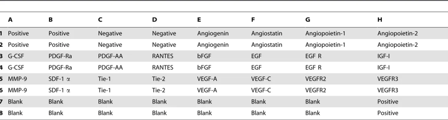
Custom RayBio Human Cytokine Array kit (RayBiotech, Norcross, GA, USA) (Table 1) was used to analyze 20 protein expression in mammary tumors. All sample measurements were performed in duplicate, containing positive and negative controls. Membranes were incubated in 8-well plates with 2 ml of RayBio 1X Blocking Buffer solution for 30 minutes. 500 ug of protein was added to each sample and the membranes were incubated overnight at 4uC. The solution was discarded and the membranes washed three times with 1X RayBio Wash Buffer I, and twice with 1X RayBio Wash Buffer II for 5 minutes each. BiotinConjugated Anti-Cytokines was added and samples were incubated overnight at 4uC. Membranes were washed with Wash Buffer I and II and incubated with 1000X HRP-Conjugated Streptavidin overnight at 4uC. After that, it was washed again with Wash Buffer I and II and incubated for 2 minutes with Detection Buffer. Membranes were exposed in a Multispectral In-Vivo Imaging System (KodakTM Multispectral system, Carestream Health Inc., NY, USA).
Immunohistochemistry
Animals were euthanatized, perfused by intracardiac injection of 10 ml PBS followed by 10 ml 3% paraformaldehyde and tumors were removed and fixed in 3% paraformaldehyde containing 3% sucrose. Tumor sections were prepared for paraffin blocking and sectioning. Standard histochemical staining proce-dures were performed as recommended by the suppliers of primary antibodies. The following antibodies were used to delineate the expression of corresponding antigens: VEGFR2 (Millipore, Billerica, MA, USA), anti-VEGFR3 antibody (Milli-pore, Billerica, MA, USA), anti- von Willebrand Factor (vWF) (Millipore, Billerica, MA, USA) andanti-Ki67 (Millipore, Billerica, MA, USA).
Evaluation of immunohistochemical staining
At least two sections from each tumor underwent staining for different markers and used for analysis. Multiple fields were examined from each slide, especially demarcated areas with brown staining. There were signs of necrosis in the central part of both melatonin and vehicle treated tumors.
For the analysis of immunohistochemistry slides, five areas were photographed at 406magnifications (center, bottom, top, left and right regions) and saved in tiff format. VEGFR2 and VEGFR3 were analyzed based on the intensity of the staining by ImageJ software (NIH, Bethesda MD, USA).Each photograph was divided into four quadrants and 20 spots (small circular ROI) were randomly selected (avoiding the nucleus) in each quadrant, analyzing the intensity from 80 spots from each photographed area. The intensity was determined from a total of 400 spots marked on each slide. A negative control section of the corresponding staining was used for measuring background activity.
Evaluation of micro-vessel density (MVD) was detected by immunohistochemical staining with vWF. Each cell stained positive for vWF was considered to be a micro vessel. Five ‘‘hot spots’’ (area with highest vessel concentration) from each slide were identified and vWF positive areas were counted by two independent observers. The total histological area (mm2) was noted, and MVD was calculated as previously described [44].
Tumors were categorized in relation to cellular proliferation according to the staining of Ki-67.All Ki-67 positive cells were counted from each photographed area. The number of Ki-67 positive cells was normalized to the area of photomicrography.
Statistical analysis
All data are expressed as mean 6 standard error of mean (SEM). Comparison between melatonin and vehicle administra-tion groups was done by Student’s t-test or ANOVA followed by Bonferroni test with GraphPad Prism 4.0 software (La Jolla, CA, USA). Any p-value of 0.05 was considered significant.
Results
MTT assay
We performed the MTT assay with different concentrations of melatonin to assess whether melatonin acts on thein vitroviability of MDA-MB-231 cells. As shown in Figure 1, treatment with 0.0001 mM to 0.1 mM of melatonin did not affect the cell viability after 24 hours (p.0.05). Only a pharmacological concentration of 1 mM of melatonin significantly decreased the cell viability
compared to control cells (*p,0.05 vs control) and compared to all concentrations of melatonin evaluated (#p,0.05) (Figure 1).
Tumor size
To evaluate whether melatonin treatment reduces the breast tumor growthin vivo, we implanted MDA-MB-231 cells in athymic nude mice and treated then with melatonin (40 mg/kg) or vehicle for 21 days.
None of the treated mice showed any loss of weight and lethargy during the treatment for 21 days. On the contrary, the treated animals showed excessive movement but no irritability or aggressive behavior.
The site of implantation of tumor cells (mammary gland or flank) did not alter the rate of tumor growth in the vehicle treated animals (p = 0.88) or in the melatonin-treated animals (p = 0.76),
showing that there was no significant difference in tumor development and response to melatonin treatment between the two models used. Thus, the two models were grouped together into one group for each treatment (melatonin or vehicle) for subsequent analyses.
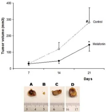
Treated animals showed significantly smaller tumors after 21 days (p,0.05;Figure 2). The mean tumor volume of control and treated animals were 282.0688.5 mm3 and 144.9638.4 mm3, respectively. The mean tumor volume in control animals increased significantly from day 14 (118.9640.2 mm3) today 21 (282.0688.5 mm3), while this was not observed in the treated group (p,0.05, Figure 2). Furthermore, there was tumor regression in an animal treated with melatonin (Day 7 = 27.38 mm3; Day 14 = 8.79 mm3, Day 21 = 4.8 mm3) (Figure 2B). No similar pattern was seen in any of the control mice (Figure 2C, 2D).
In vivo expression of VEGFRs
To determine whether there were any changes in the expression of VEGFRs in the melatonin and vehicle administrated tumors, animals underwent SPECT scanning with Tc-99m tagged VEGF-c. Animals treated with melatonin showed lower activity of Tc-99-HYNIC-VEGF-c in the tumors compared to vehicle treated animals (Figure 3A, 3B).
Semi-quantitative analysis of total radioactivity normalized to contralateral muscles showed that the intensity of radioactivity in the control animals was 183.6620.9%, while the intensity of radioactivity in animals treated with melatonin was 150.5617.1%. Although there was difference in the radioactivity in the tumors between the groups, statistically significant difference was not achieved (p.0.05;Figure 3C).
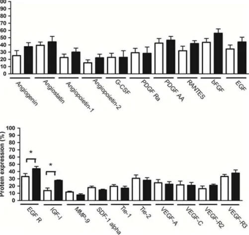
Membrane antibody array
Semi-quantitative densitometry analysis of membranes showed that melatonin did not alter the pattern of expression of the majority of evaluated angiogenic proteins (p.0.05; Figure 4). However, melatonin increased the expression of epidermal growth factor receptor (EGFR) and insulin-like growth factor 1 (IGF-I) proteins (p,0.05;Figure 4).
1 Positive Positive Negative Negative Angiogenin Angiostatin Angiopoietin-1 Angiopoietin-2
2 Positive Positive Negative Negative Angiogenin Angiostatin Angiopoietin-1 Angiopoietin-2
3 G-CSF PDGF-Ra PDGF-AA RANTES bFGF EGF EGF R IGF-I
4 G-CSF PDGF-Ra PDGF-AA RANTES bFGF EGF EGF R IGF-I
5 MMP-9 SDF-1a Tie-1 Tie-2 VEGF-A VEGF-C VEGFR2 VEGFR3
6 MMP-9 SDF-1a Tie-1 Tie-2 VEGF-A VEGF-C VEGFR2 VEGFR3
7 Blank Blank Blank Blank Blank Blank Blank Positive
8 Blank Blank Blank Blank Blank Blank Blank Positive
G-CSF = colony stimulating factor; PDGF-Ra = platelet-derived growth factor receptor, alpha polypeptide; PDGF-AA = platelet-derived growth factor alpha polypeptide; bFGF = fibroblast growth factor (basic); EGF = epidermal growth factor; EGF R = epidermal growth factor receptor; IGF-I = insulin-like growth factor 1 (somatomedin C); MMP-9 = matrix metallopeptidase 9; SDF-1a= Stromal cell-derived factor 1 alpha; Tie-1 = receptor tyrosine kinase of angiopoietin-1, tyrosine
kinase with immunoglobulin-like and EGF-like domains 1; Tie-2 = receptor tyrosine kinase of angiopoietin-1; VEGF-A = vascular endothelial growth factor A;VEGF-C = vascular endothelial growth factor C; VEGFR2 = Vascular endothelial growth factor receptor 2; VEGFR3 = Vascular endothelial growth factor receptor 3. All antibodies are prepared in duplicate.
doi:10.1371/journal.pone.0085311.t001
Figure 1. Inhibitory effect of melatonin on viability of the MDA-MB-231 cell line.The MDA-MB-231 cells were treated with five concentrations of melatonin for 24 h and cell viability was measured by MTT assay. Data are shown as mean 6 S.D. *p,0.05, 1 mM of melatonin vs. Control; #p,0.05, 1 mM of melatonin vs other melatonin’s concentrations.
doi:10.1371/journal.pone.0085311.g001
Immunohistochemistry
The expression of VEGFR2, VEGFR3, vWF and Ki-67 was evaluated for the tumors after 21 days of treatment with melatonin or vehicle by immunohistochemistry.
Lower expression of VEGFR2 was observed in the melatonin treated tumors compared to vehicle treated tumors (p,0.05;
Figure 5). Furthermore, melatonin treated tumors exhibited lower expression of VEGFR3 compared with vehicle treated tumors, but statistically significant difference was not achieved (p.
0.05;Figure 6).
Tumor neovascularization was assessed by quantification of MVD in mammary tumors by vWF immunohistochemistry. Melatonin treatment resulted in a decrease of MVD compared to the vehicle treated tumors (p,0.05;Figure 7).
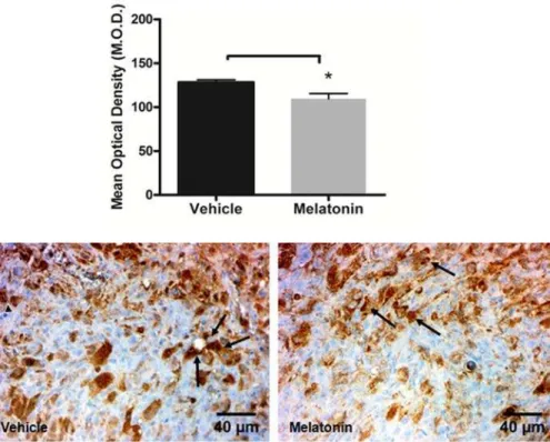
Numerous Ki-67 positive cells were observed in the vehicle treated tumors, whereas none to very few positive cells were observed in the melatonin treated tumors (p,0.05;Figure 8).
Discussion
Results from this study showed that melatonin at pharmacologic concentration (1 mM) was able to reduce ER-negative breast cancer cell viability in vitro. The direct effects of melatonin on mammary cancer have been studied in vitro, basically using the ER-positive human breast cancer cell line (MCF-7) as a model [39]. In physiological concentrations (1 nM corresponding to peak nighttime and 10 pM corresponding to day time serum values in
estrogen-responsive cell lines (MCF-7, T47D, ZR-75-1) more effectively than estrogen negative breast cancer cells such as BT-20, MDA-MB-231, MDA-MB-364, Hs587t, T47Dco, suggesting that the antiproliferative effects of melatonin are mediated through the estrogen-response pathway [39,46].
However, agreeing with our results about ER-negative cells, Leman et al.[47] showed that melatonin at pharmacological level (1 mM) decreases the proliferation of MDA-MB-231 and MCF-7 cells significantly. Proliferation of MDA-MB-435 cells, which are highly metastatic and ER-negative, was not significantly affected by melatonin [47]. Jung et al. [48] showed that melatonin showed weak cytotoxicity only at pharmacologically high concentrations of 8 mM and 16 mM in MDA-MB-231 cells. Melatonin induced apoptosis and inhibited the proliferation via the inhibition of anti-apoptotic genes such as BCL-xL, Mcl-1, cyclin D1, cyclin E, p-STAT3, p-mTOR, and p-AKT at high concentration (12 mM) in ER-negative MDA-MB-231 cells [48].
Melatonin has been reported to bind and activate two distinct receptor types, membrane-bound G protein-coupled receptors, MT1 and MT2, and the nuclear orphan RZR/RORa receptors, members of the steroid/thyroid hormone receptor superfamily [49]. MT1 and MT2 are expressed in human cells in various organs and in neoplastic cells, including breast cancer cells [50].
Through the mediation of a subunit of G protein, MT1 receptors inhibit the activity of adenylcyclase, and thereby decrease the production of adenosine 39, 5-cyclic monophosphate (cAMP). This relationship makes it possible to control the activity of selected protein kinases (PKC, PKA, MAPK) and to influence the levels of transcription factor phosphorylation, that is, cAMP response element-binding, as well as the expression of specific genes, which code proteins involved in the proliferation, angio-genesis, cell differentiation and migration processes [50].
Both MCF-7 and MDA-MB-231 human breast cancer cell lines express low levels of the MT1 receptor [46]; However, overex-pression of the MT1 receptor enhanced the growth-inhibitory and gene-modulatory effects of melatonin in positive but not ER-negative human breast cancer cells [49].
Recent studies have demonstrated higher levels of MT1A mRNA in the MCF-7 cell line compared to the MDA-MB-231 cell line [50,51]. Jablonska et al. [50] showed that there is lower expression of MT1 receptors in breast cancer phenotype of triple negative (ER-, PR -, HER2 -) compared to ER-positive cases and the lower expression of MT1 is correlated with poor prognosis.
However, through activation of the MT1 receptor, melatonin can suppress the development of cancer via a broad spectrum of mechanisms with and without involvement of ER [45]. Oprea-Ilies et al. [52] showed that the MT1 positivity was associated with a lower stage and a smaller tumor size at time of diagnosis in triple-negative breast tumors patients.
Furthermore, melatonin can exert antitumoral properties by a set of complex mechanisms of action, not necessary involving the receptor pathway [26]. Melatonin may act directly, independently of its receptors or via them, making it difficult to understand the action of melatonin at the cellular level [26,50].
Since melatonin decreased ER-negative breast cancer cell viabilityin vitro, we implanted cells in athymic nude mice. Our results showed that melatonin treatment (40 mg/kg) reduced the tumor growth with concomitant decreasing of cell proliferation (Ki-67).
Figure 2. Antitumor effects of melatonin on mammary tumor growth.Melatonin reduced the tumor growth in breast cancer nude mice. Each point in the curves represents the mean6SD (control n = 8; melatonin n = 5). The melatonin inhibited tumor growth, *p,0.05 vs Control.#Significant increase in tumor volume on control group at 14 and 21 after tumor implantation and initiation of treatment with vehicle (p,0.05). Detail: Representative samples of mammary tumors devel-oped by MDA-MB-231 cells implantation on the right flank of mice. A, B. Melatonin treated mammary tumors, B. Mammary tumor which regressed with melatonin treatment. C, D. Vehicle treated mammary tumors.
doi:10.1371/journal.pone.0085311.g002
There are no studies about melatonin treatment in ER-negative breast cancerin vivo, but investigators showed the effectiveness of melatonin in treating rats with breast cancer induced by a carcinogen DMBA [32]. In a study by Cos et al. [53] nude mice bearing ER-positive breast cancer (MCF-7) (inoculated directly into the inguinal mammary fat pad of the mouse) treated with melatonin also showed smaller tumor size compared to control animals. Rao et al. [54] showed that melatonin at 50 mg/kg and 200 mg/kg reduced the incidence of mammary cancer of TG.NK (c-neu) transgenic mice and reduced the number of tumors per mouse and tumor weights as compared with the control group. Although effective, the high-dose of 200 mg/kg caused signifi-cantly lower body weight with high mortality. On the contrary, in our study none of the treated mice showed any adverse effect, loss of weight and lethargy during the treatment with 40 mg/kg for 21 days. Liu et al. [55] performing a study with mice with gastric
tumor (foregastric murine carcinoma cell line subcutaneously inoculated under the right axilla) showed that melatonin treatment in doses of 25 mg/kg, 50 mg/kg and 100 mg/kg reduced tumor volume when correlated with control mice.
It is accepted that melatonin exerts an anti-tumor effect and multiple mechanisms have been suggested for the biological effects of melatonin but are not yet fully established [56]. It has been shown that melatonin has direct anticancer mechanisms in several types of cancer, as pro-apoptotic, anti-proliferative, anti cell-differentiation and anti-angiogenic actions. Melatonin also has indirect anticancer mechanisms such as antioxidative effects and immune system regulation [26,57]. Little is known about the anti-angiogenic effect of melatonin. Some authors have demonstrated that a pharmacological concentration of melatonin may inhibit angiogenesis directly or indirectly, by inhibiting the proliferation of
Figure 3. SPECT analysis ofin vivoaccumulation of Tc-99m-HYNIC-VEGF-c.VEGF-c (which targets both VEGFR2 and VEGFR3) was tagged with HYNIC chelators and then labeled with Tc-99m and injected intravenously in melatonin and vehicle treated mice. One hour after injection, SPECT images were obtained using dedicated animal scanner. Vehicle treated mice showed increased accumulation of Tc-99m-HYNIC-VEGF-c in the mammary tumor (A, Intersection of lines indicate the tumor, with a volume of 865.69 mm3at the 21th day) compared to that of melatonin treated
mammary tumors (B, Intersection of lines indicate the tumor, with a volume of 130.69 mm3at the 21th day) C. Semi-quantitative analysis of total
radioactivity normalized to contralateral muscles showing the intensity of radioactivity in the vehicle and melatonin treated animals. doi:10.1371/journal.pone.0085311.g003
vascular endothelial cells [33] and acting in the inhibition of pro-angiogenic factors [34–37].
In this study, we evaluated the action of melatonin on angiogenesis in ER-negative breast cancer in vivo. Our findings were able to show that melatonin treatment was effective in inhibiting ER-negative mammary tumor angiogenesis, indicated by the reduction of VEGFR2 expression and MVD. Despite the semi-quantitative analysis of Tc-99m-HYNIC-VEGF-c showing lower VEGFR2 expression in the melatonin treated tumors, a statistically significant difference was not achieved. The non-significant differences of Tc-99m-HYNIC-VEGF-c activity be-tween control and treated tumors could be due to the reduction of tumor size in treated groups and the relative expression of VEGFR2 remained unchanged since our analysis used contralat-eral ROI identical to tumor size. However, a decrease of VEGFR2 in melatonin treated mice was confirmed by immunohistochem-istry.
It is noteworthy that the efficacy of anti-angiogenic therapies can be evaluated by nuclear medicine imaging techniques using the tagging of radioactive isotopes to the specific proteins. Previously, Tc-99m was successfully tagged with different proteins and peptides using the new generation chelator, HYNIC (Hydrazine Nicotinamide) [43]. VEGF receptors have been used as imaging agents mediators of angiogenesis, since they are highly expressed in the vascular endothelium, and when injected into the bloodstream, are internalized by binding to VEGF, allowing the accumulation of contrast agents, such as Tc-99m [43,58]. VEGFR2 is the principal receptor transmitting VEGF signals
and is overexpressed in the tumor vasculature compared with normal vasculature [59]. One common mechanism for increased survival and growth of cancer cells is the deregulation of tyrosine kinase receptors like VEGFR2 [60].
In this study, mammary tumors showed necrosis in the central part, both in vehicle and melatonin treated animals, which can be a result of insufficient and defective vasculature in a highly proliferative tumor mass. Hypoxia in tumor tissues is a leading cause of angiogenesis [61]. Low O2 tension of a growing tumor allows stabilization of HIF-1a, leading to increased VEGF-A transcription, which binds to VEGFR2. Activation of intracellular signaling of VEGFR2 stimulates an angiogenic response, leading to cell proliferation, migration, permeability, survival and ultimately resulting in tumor growth [62].
There are no studies evaluating the expression of VEGFRs in response to treatment with melatonin. We also evaluated 20 angiogenic proteins in breast tumor tissue and showed that melatonin treatment did not alter the pattern of expression of the majority of the proteins evaluated. Previous reports have described a decrease in the production of VEGF protein induced by melatonin in human pancreatic carcinoma cells (PANC-1) and human alveolar adenocarcinoma cells (A549) [36,56].
Melatonin suppresses tumor angiogenesis by inhibiting HIF-1a expression and stabilization in prostate cancer [37] and HCT116 colon cancer cells under hypoxic condition in vitro [5]. We evaluated the expression of HIF-1a in tumor tissue of animals treated with melatonin or vehicle, but there was no statistically significant difference (p.0.05, data not shown).
Figure 4. Comparison between proteins expression in mammary tumor in animals treated with melatonin or vehicle.White column = vehicle treatment; Black column = melatonin treatment. Data are shown as mean6S.D. *p,0.05, vs. Control.
doi:10.1371/journal.pone.0085311.g004
Figure 5. Immunohistochemistry staining with VEGFR2 (arrows) in vehicle treated and melatonin treated tumors.Images were taken with 406magnification. A significant decrease was observed at the tumor in melatonin treated tumors compared to vehicle treated tumors (*p, 0.05). Error bars:6standard error.
doi:10.1371/journal.pone.0085311.g005
Figure 6. Immunohistochemistry staining with VEGFR3 (arrows) in vehicle treated and melatonin treated tumors.Images were taken with 406magnification. Melatonin do not decreased significantly the expression of VEGFR3 (p.0.05). Error bars:6standard error.
doi:10.1371/journal.pone.0085311.g006
Figure 7. Immunohistochemistry staining with vWF in vehicle treated and melatonin treated tumors.Images were taken with 406
magnification. Quantitative estimation of micro-vessel density (MVD) by counting positive vessels (arrows) revealed a decrease in MVD after melatonin treatment compared to the vehicle treated tumor (*p,0.05). Error bars:6standard error.
doi:10.1371/journal.pone.0085311.g007
Figure 8. Immunohistochemistry staining with Ki-67 in vehicle treated and melatonin treated tumors.Images were taken with 406
magnification. There was a decreased cell proliferation in tumors treated with melatonin (*p,0.05). Error bars:6standard error. doi:10.1371/journal.pone.0085311.g008