Glucose Sensor MdHXK1 Phosphorylates and
Stabilizes MdbHLH3 to Promote Anthocyanin
Biosynthesis in Apple
Da-Gang Hu, Cui-Hui Sun, Quan-Yan Zhang, Jian-Ping An, Chun-Xiang You, Yu-Jin Hao*
National Key Laboratory of Crop Biology, National Research Center for Apple Engineering and Technology, College of Horticulture Science and Engineering, Shandong Agricultural University, Tai-An, Shandong, China
Abstract
Glucose induces anthocyanin accumulation in many plant species; however, the molecular mechanism involved in this process remains largely unknown. Here, we found that apple hexokinase MdHXK1, a glucose sensor, was involved in sensing exogenous glucose and regulating anthocyanin biosynthesis.In vitroandin vivoassays suggested that MdHXK1 interacted directly with and phosphorylated an anthocyanin-associated bHLH transcription factor (TF) MdbHLH3 at its Ser361site in response to glucose. Furthermore, both the hexoki-nase_2 domain and signal peptide are crucial for the MdHXK1-mediated phosphorylation of MdbHLH3. Moreover, phosphorylation modification stabilized MdbHLH3 protein and enhanced its transcription of the anthocyanin biosynthesis genes, thereby increasing antho-cyanin biosynthesis. Finally, a series of transgenic analyses in apple calli and fruits demon-strated that MdHXK1 controlled glucose-induced anthocyanin accumulation at least partially, if not completely, via regulating MdbHLH3. Overall, our findings provide new insights into the mechanism of the glucose sensor HXK1 modulation of anthocyanin accu-mulation, which occur by directly regulating the anthocyanin-related bHLH TFs in response to a glucose signal in plants.
Author Summary
Glucose is considered as a major regulatory molecule in addition to being essential meta-bolic nutrients and structural components in higher plants. As is well known, hexokinase1 (HXK1) is a glucose sensor that integrates diverse signals to govern gene expression and plant growth in response to environmental cues. Previously, it is reported that the nuclear HXK1 forms a glucose signaling complex core with the vacuolar H+-ATPase B1 (VHA-B1) and the 19S regulatory particle of proteasome subunit (RPT5B), which influences the transcription of target genes. However, it is yet unknown if and how HXK1 directly targets TFs to modulate their function in the nucleus in plants. Our results reveal the important roles of MdHXK1 protein kinase in phosphorylating MdbHLH3 TF to modulate anthocy-anins accumulation in response to glucose in apple.
a11111
OPEN ACCESS
Citation:Hu D-G, Sun C-H, Zhang Q-Y, An J-P, You C-X, Hao Y-J (2016) Glucose Sensor MdHXK1 Phosphorylates and Stabilizes MdbHLH3 to Promote Anthocyanin Biosynthesis in Apple. PLoS Genet 12 (8): e1006273. doi:10.1371/journal.pgen.1006273
Editor:Li-Jia Qu, Peking University, CHINA
Received:January 23, 2016
Accepted:August 2, 2016
Published:August 25, 2016
Copyright:© 2016 Hu et al. This is an open access article distributed under the terms of theCreative Commons Attribution License, which permits unrestricted use, distribution, and reproduction in any medium, provided the original author and source are credited.
Data Availability Statement:All relevant data are within the paper and its Supporting Information files.
Funding:This work was supported by grants from NSFC (31325024, 31272142, 31471854), Ministry of Education (IRT15R42) and Shandong Province (SDAIT-06-03). The funders had no role in study design, data collection and analysis, decision to publish, or preparation of the manuscript.
Introduction
In higher plants, sugars function as major regulatory molecules in addition to being essential metabolic nutrients and structural components. Sugars control gene expression to affect devel-opmental and metabolic processes during the entire plant life cycle and function in response to biotic and abiotic stresses [1–3]. Therefore, rigorous sugar-sensing and sugar-signaling systems are critical for coordinating photosynthesis and carbon metabolism and for adapting to changes in environmental conditions to sustain normal plant growth and development.
Among the myriad of sugars in photosynthesis, glucose is the preferred carbon and energy source. Glucose is involved in many metabolic pathways, including the glycolytic process, in organisms ranging from unicellular microbes to plants and animals [4,5]. In addition to its metabolic function, glucose is the most intensively studied sugar molecule and functions in specific regulatory pathways to modulate plant growth and development [6,7]. Glucose signal-ing modulates the gene expression of enzymes in the glyoxylate cycle and photosynthesis path-way, and is also involved in the decision of whether to initiate the normal seedling
establishment after seed germination [8,9].
Hexokinase 1 (HXK1) is the first plant sugar sensor identified [9,10]. The genetic evidence for HXK1 as a sugar sensor is the isolation of twoArabidopsis gin2(glucose insensitive 2) mutants, both of which are mapped to theHXK1gene [11]. In theArabidopsisgenome, there are threeHXKsand threeHXK-like(HKLs) genes, which execute a variety of physiological functions, including controlling subcellular localization, protein complex formation and tissue-specific expression patterns [12–14]. Moreover, five orthologousHXKshave been identified in the apple genome. Among them, MdHXK1, a well-known apple hexokinase, is highly homolo-gous withArabidopsisAtHXK1 [15]. Generally, HXKs are located on the outer mitochondrial membrane, plastids and even in the nucleus [13,14,16].
The regulatory role of HXK1 in sugar signaling has been identified and characterized in plants in the past two decades. InArabidopsis, HXK1 forms a high-molecular-weight complex together with the V-ATPase subunit VHA-B1 and the proteasome 19S regulatory subunit RPT5B in the nucleus. This complex directly binds to the promoters ofCAB2(chlorophyll a/b binding protein 2) andCAB3genes to confer glucose-mediated transcriptional regulation inde-pendent of glucose metabolism in the cytosol [17]. Both seedlings and adult plants ofvha-b1
andrpt5bmutants display similar phenotypes as thegin2mutant, demonstrating the crucial role of the interaction with HXK1 in glucose signaling [11,17]. In addition, glucose signaling mediated by HXK1 shows crosstalk with ABA, ethylene, auxin, cytokinin and brassinosteroid signaling [18–20]. However, whether HXK1-mediated signaling is involved in the regulation of anthocyanin biosynthesis in plants remains unclear.
Anthocyanins are ubiquitously present in various tissues and organs of plants, especially in the fruit, leaf and flower of ornamental crops. They are responsible for the red, purple and blue coloration of tissues and organs depending on the cellular conditions, such as pH value [21]. Colored organs, such as flowers and fruits, attract pollinators and seed-dispersing animals [22]. Anthocyanins are also antioxidant molecules that protect plants from damage by reactive oxy-gen species (ROS) [23–25]. These properties also make them interesting as food ingredients for human and animal nutrition. Anthocyanins are biosynthesized via the flavonoid pathway in the cytosol and are transported into the vacuole by vacuolar transporters, including ABC and MATE-type transporters [26,27].
biosynthesis by directly binding to the promoters of not only anthocyanin structural genes, such asDFRandUFGT, but also anthocyanin-associatedMYBTF genes to activate their expression [31–34]. Interestingly, MdbHLH3 protein promotes anthocyanin accumulation partially through a putative phosphorylation modification in response to low temperature in apple [32]. However, the protein kinase that mediates the phosphorylation of MdbHLH3 pro-tein is unknown.
Sugars induce anthocyanin biosynthesis in various plant species [35–37]. First, they provide carbon sources, skeletons and glucosides for anthocyanin biosynthesis [38,39]. Second, they increase the expression levels of biosynthetic structural genes and regulatoryMYBgenes [37,40]; however, the precise mechanism by which sugars modulate these genes remains unknown. The present study found that a protein kinase, MdHXK1, is involved in the regula-tion of anthocyanin biosynthesis in response to glucose by interacting with the phosphorylat-ing and stabilizphosphorylat-ing MdbHLH3 protein. Subsequently, the function of MdHXK1 in the modulation of anthocyanin accumulation was characterized in apple calli and fruits. Finally, the potential application of HXK1-mediated glucose signaling in the genetic improvement of horticultural traits is discussed.
Results
MdHXK1 modulates anthocyanin accumulation mainly through glucose
signaling, but not through the catalytic pathway, under the high-glucose
condition
Previous studies have verified that glucose significantly induces anthocyanin biosynthesis in
Arabidopsisseedlings [36]. Similarly, the effect of different concentrations of glucose (0–6%, w/ v) and the HXK inhibitor glucosamine on anthocyanins accumulation was tested inin vitro
shoot cultures of the‘Gala’apple cultivar. The results showed that glucose promotes anthocya-nin accumulation in an HXK-dependent manner in apple (S1 Fig;S5 Text).
Because glucose controls anthocyanin accumulation in an HXK-dependent pathway in apple, it is reasonable to propose that this process is regulated by the catalytic or signaling func-tion of HXK. We isolated theMdHXK1gene from apple to investigate this possibility. The pre-dicted MdHXK1 protein is highly homologous with AtHXK1, which functions as not only a catalytically active kinase but also a glucose sensor inArabidopsis[11,41]. Two catalytically inactive HXK1 mutants have been identified inArabidopsis, namely,HXK1S177Aand
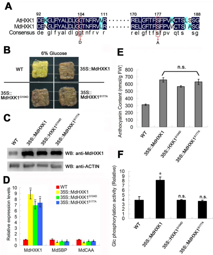
HXK1G104D; these mutants retain signaling functions but not catalytic activities [11]. To inves-tigate whether Ser and Gly at positions 177 and 104 of HXK1, respectively, are conserved between apple andArabidopsis, an alignment of the amino acid sequence of MdHXK1 with AtHXK1 was performed. The result showed that apple MdHXK1 protein exhibited a high sequence similarity (77.31% identity) toArabidopsisAtHXK1 (S2 Fig). The positions of 177 and 104 of apple MdHXK1 are the Ser and Gly residues, respectively, which were the same as those ofArabidopsisAtHXK1 (Fig 1A). These results suggest that the catalytically inactive apple MdHXK1 proteins MdHXK1S177Aand MdHXK1G104Dalso exercised their functions in signaling but not catalytic activities, similar toArabidopsis.
Fig 1. MdHXK1 modulates anthocyanin accumulation mainly through the glucose signaling pathway. (A)Partial amino acid sequences of HXK1 fromArabidopsisand apple orthologs are aligned. The highly conserved amino acids are highlighted with a black background. Conserved Gly (position 104 in AtHXK1 and MdHXK1) and Ser (position 177 in AtHXK1 and MdHXK1) residues are labeled with red boxes.(B)Phenotype of anthocyanin accumulation in the WT control and the35S::MdHXK1,35S::MdHXK1G104Dand35S::MdHXK1S177A
increased by 4.2-, 3.9- and 4.0-fold in the35S::MdHXK1,35S::MdHXK1G104Dand35S::
MdHXK1S177Atransgenic apple calli, respectively, compared with the WT control (Fig 1C), indicating that the target genes were successfully transformed into and expressed in the trans-genic apple calli. In addition, qPCR assays showed thatMdHXK1repressed two classes of pho-tosynthesis genes includingMdSBPandMdCAAin35S::MdHXK1,35S::MdHXK1G104Dand
35S::MdHXK1S177Atransgenic apple calli but not WT apple calli (Fig 1D), suggesting that
Gly104andSer177mutations have similar effect on MdHXK1 asArabidopsisAtHXK1. As a result, the three transgenic apple calli produced nearly the same levels of anthocyanins to each other but at a considerably higher level than in the WT control under 6% glucose con-ditions (Fig 1B and 1E), indicating that MdHXK1 and two point mutants successfully function to promote anthocyanin accumulation in these transgenic apple calli. In addition, the glucose phosphorylation activities were determined for the WT and these transgenic apple calli. The results showed that the35S::MdHXK1transgenic calli exhibited higher glucose phosphoryla-tion activity than the35S::MdHXK1S177Aand35S::MdHXK1G104Dcalli and the WT control (Fig 1F). However, there was no significant difference in glucose phosphorylation activities among the35S::MdHXK1S177Aand35S::MdHXK1G104Dtransgenic calli and the WT control (Fig 1F). Collectively, these results suggest that MdHXK1 modulates anthocyanin accumulation mainly through glucose signaling, but not the catalytic pathway, under the high-glucose (6%)
conditions.
Furthermore, the WT and aforementioned three transgenic calli were also treated with a low glucose concentration (1%) to induce anthocyanin accumulation. The result demonstrated that the35S::MdHXK1S177Aand35S::MdHXK1G104Dtransgenic apple calli produced more anthocyanins than the WT controls but less anthocyanins than the35S::MdHXK1transgenic calli (S3A and S3B Fig). However, the glucose phosphorylation activities of the35S::
MdHXK1G104Dand35S::MdHXK1S177Atransgenic apple calli showed no significant difference compared with the WT control but were considerably lower than the activities for the35S::
MdHXK1transgenic calli (S3C Fig).
Taken together, these results indicate that MdHXK1 induces anthocyanin accumulation depending on both the catalytic activity and signaling under low-glucose conditions but mainly depending on signaling under high-glucose conditions.
MdHXK1 interacts with MdbHLH3 via the conserved hexokinase_2
domain
To screen the target protein of MdHXK1 in its signal pathway, the35S::MdHXK1-Mycvector was constructed and genetically transformed into apple calli (S4A Fig). The35S::MdHXK1-Myc
transgenic calli were used for co-immunoprecipitation (Co-IP) against the monoclonal anti-Myc antibody (S4B Fig). Subsequently, the Co-IPed proteins were analyzed with LC/MS to identify the potential proteins that interact with the MdHXK1 protein. The results showed that the anthocyanin-associated bHLH TF MdbHLH3 is a candidate (S1 Text).
To determine whether MdHXK1 interacts with MdbHLH3 protein, yeast two-hybrid (Y2H) assays were performed. MdHXK1 protein contains two conserved hexokinase domains, i.e., hexokinase_1 and hexokinase_2 (S4C Fig). Therefore, the full-length cDNA ofMdHXK1gene
dark (24 hours dark)-induced glucose starvation to deplete endogenous glucose.(C)Western blotting analysis of MdHXK1 protein abundance in the WT and transgenic apple calli.(D)MdHXK1-mediated glucose-dependent gene repression.MdSBP, sedoheptulose-biphosphatase (accession no. XM_008384867);MdCAA, carbonic anhydrase (accession no. XM_008387117).(E)and(F)Anthocyanin content(E)and glucose phosphorylation activity(F)in the WT and transgenic apple calli. The data are shown as the mean±SE, which were analyzed based on more than 9 replicates. Statistical significance was determined using Student’st-test in different apple calli lines. n.s., P>0.01;*P<0.01.
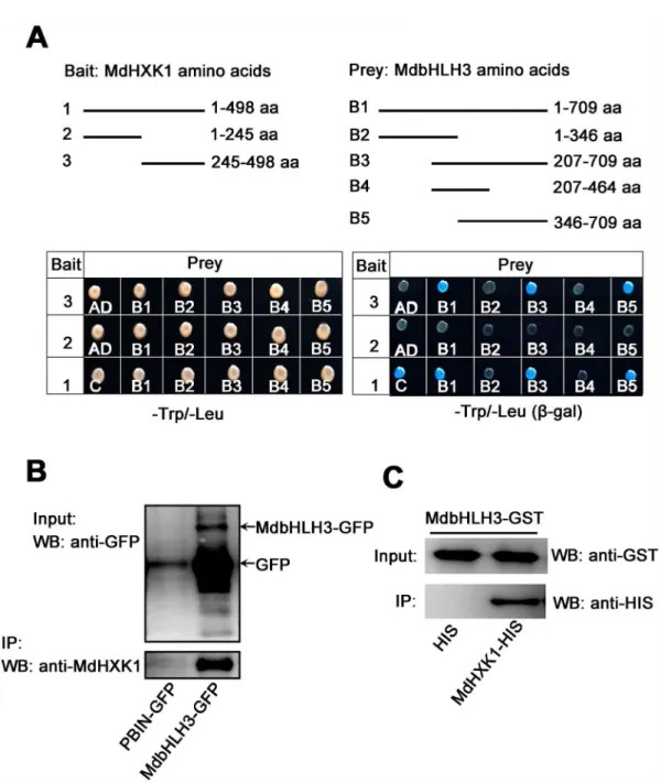
was divided into two fragments, i.e., MdHXK11-245aaand MdHXK1245-498aa. Subsequently, the full-length cDNA and two truncated mutants of theMdHXK1gene were inserted into the pGBT9 vector, independently, as the bait vectors. Moreover, the full-lengthMdbHLH3cDNA and its serially truncated mutants, as previously reported by Xieet al. [32], were inserted into the pGAD424 vector as the prey vectors. The different combinations of bait and prey vectors were transformed into yeast for Y2H assays. The results indicated that the full-length
MdHXK1 strongly interacted with the full-length MdbHLH3 proteins. Furthermore, the trun-cated peptide MdbHLH3346-709aa, i.e., the C-terminus of MdbHLH3, interacted with MdHXK1 proteins at the hexokinase_2 domain MdHXK1245-498aabut not at the hexokinase_1 domain MdHXK11-245aa(Fig 2A).
To further verify the interaction between MdHXK1 and MdbHLH3, anin vivoCo-IP assay using35S::MdbHLH3-GFPtransgenic apple calli was conducted. The result indicated that the MdbHLH3-GFP fusion protein, but not the GFP negative control, interacted with MdHXK1 in apple calli (Fig 2B). In addition, a GST pull-down assay showed that a GST-tagged MdbHLH3 physically interacted with a His-tagged MdHXK1in vitro(Fig 2C). These results indicate that the hexokinase_2 domain of MdHXK1 physically interacts with the C-terminus of the MdbHLH3 protein.
Glucose induces phosphorylation of the MdbHLH3 protein at the Ser
361site
To examine how MdbHLH3 protein responds to glucose, an expression vector35S::
MdbHLH3-Mycwas constructed and genetically transformed into apple calli (S5A Fig). After treatment with or without 6% glucose, the35S::MdbHLH3-Mycoverexpressing calli were used for Western blotting with the anti-Myc antibody. The results showed that the position of the MdbHLH3 proteins shifted from a faster- to a slower-migrating band in the transgenic apple calli treated with 6% glucose compared to those without glucose (Fig 3A), indicating that the MdbHLH3 protein was post-translationally modified in response to glucose. Furthermore, treatment with calf intestine alkaline phosphatase (CIP), which cleaves exposed phosphate resi-dues from ribonucleotides and deoxyribonucleotides, converted the slower-migrating form of MdbHLH3 to the faster-migrating form (Fig 3A), indicating that the glucose-induced post-translational modification for the MdbHLH3 protein in apple calli is predominantly a phosphorylation.
To examine the potential phosphorylation sites of the MdbHLH3 protein, the glucose-induced phosphorylated MdbHLH3 protein in the35S::MdbHLH3-Mycoverexpressing calli was captured with anti-Myc antibody-conjugated agarose beads and separated in an
SDS-PAGE gel. After proteolytic digestion and purification, the protein sample was subjected to liquid chromatography-tandem mass spectrometry (LC-MS/MS) to detect the phosphoryla-tion sites. The serine at residue 361 (Ser361) of the MdbHLH3 protein exhibited a high phos-phopeptide signal intensity (Fig 3B;S2andS4Texts), suggesting that it is a potential phosphorylation site.
Subsequently, a monoclonal antibody specifically against the MdbHLH3 phosphorylation site at residue 361 was prepared and named as the anti-MdbHLH3S361antibody (S5B Fig). This antibody specifically recognized the glucose-induced phosphorylation of MdbHLH3 protein in the WT apple calli (Fig 3C), which was consistent with the results shown inFig 3A. These results indicate that glucose induces the phosphorylation of the MdbHLH3 protein at the Ser361site in apple calli.
0%, 1%, 3% and 6% and then used for immunoblotting with the anti-MdbHLH3S361antibody. The results showed that the MdbHLH3 protein was not phosphorylated when the calli grew in absence of glucose, whereas the phosphorylation intensity of the MdbHLH3 protein increased gradually with glucose concentration (Fig 3D). Moreover, apple calli were treated with 6% glu-cose for different times (0, 10, 20, 30 and 60 min) to examine whether treatment time affects the Fig 2. MdHXK1 physically interacts with MdbHLH3. (A)The C-terminus of MdHXK1 specifically interacts with the C-terminus of MdbHLH3 in a yeast two-hybrid assay. Top panels show the schematic representation of the different MdHXK1 and MdbHLH3 deletions in yeast vectors. Bottom panels show their interaction as indicated by yeast growth andβ-gal staining in a serial of yeast two-hybrid assays.(B)In vivoCo-IP assays of the interaction between MdHXK1 and MdbHLH3 in transgenic apple calli.(C)In vitroGST pull-down assays of the interaction between MdHXK1 and MdbHLH3. Anti-His immunoblot (IB) shows the amount of MdHXK1-His bound by the indicated MdbHLH3-GST protein.
phosphorylation of the MdbHLH3 protein. The results showed that the phosphorylation inten-sity of MdbHLH3 proteins in the calli gradually increased with the treatment duration (Fig 3E). These results indicate that the MdbHLH3 protein is phosphorylated in response to glucose and that this modification is positively associated with glucose concentration and treatment time.
In addition, glucose-induced phosphorylation of the MdbHLH3 protein could be observed in apple leaves (S5C Fig), indicating that the glucose-induced phosphorylation of the
MdbHLH3 protein occurred in different apple tissues and organs.
Glucose-induced MdbHLH3 phosphorylation is required for MdHXK1
Considering the interaction between MdHXK1 and MdbHLH3 proteins, it is reasonable to hypothesize that the MdHXK1 protein kinase mediates the phosphorylation of MdbHLH3 pro-tein in apple calli. To verify this hypothesis, new transgenic apple calli,35S::antiMdHXK1, were obtained, which contained an antisense fragment specific toMdHXK1cDNA and exhibited Fig 3. Glucose induces the phosphorylation of the MdbHLH3 protein at the Ser361site. (A)Glucose inducedthe mobility shift of the MdbHLH3 protein, which was abolished by the phosphorylation inhibitor calf intestine alkaline phosphatase (CIP) in the35S::MdbHLH3-Myctransgenic apple calli. Note: MdbHLH3-P represents phosphorylated MdbHLH3 protein unless noted otherwise in this study.(B)Collision-induced dissociation mass spectrum showing the phosphorylation of Ser-361, a glucose-induced phosphorylation site in MdbHLH3. Top panel: the structural diagram of MdbHLH3 protein and its phosphorylation site. Bottom panel: the phosphorylation sites were identified using LC-MS/MS. MdbHLH3-Myc protein from transgenic apple calli was affinity purified as in (A) before being subjected to in-gel digestion with AspN.(C)Glucose induced the phosphorylation of the MdbHLH3 protein, which was abolished by CIP in WT apple calli. Western blotting was conducted with an anti-MdbHLH3S361antibody specifically against the phosphorylation site.(D)and(E)The glucose-induced
phosphorylation of MdbHLH3 protein depends on glucose concentration(D)and treatment time(E). The WT apple calli was treated with different concentrations of glucose (0, 1%, 3% or 6%) for 30 min(D), or treated with 6% glucose for different times (0, 10, 20, 30, or 60 min)(E). A Western blotting assay was performed with an anti-MdbHLH3S361antibody.
considerably lower transcript and protein levels of MdHXK1 than the WT control (S6A and S6B Fig). Subsequently, immunoblotting assays with the anti-MdbHLH3S361antibody were performed using the WT control and the35S::MdHXK1and35S::antiMdHXK1transgenic apple calli after treatment with or without glucose. The result showed that the35S::MdHXK1overexpressing calli exhibited a considerably higher phosphorylation level of the MdbHLH3 protein, whereas that of the35S::antiMdHXK1-suppressing calli were lower than the WT control in response to glucose treatment (Fig 4A). This result suggests that the MdHXK1 protein kinase is necessary, if not suffi-cient, for the glucose-induced phosphorylation of the MdbHLH3 protein in apple calli.
To further verify that MdHXK1 directly phosphorylates the MdbHLH3 protein, in-gel assays were conducted using prokaryon-expressed and purified MdHXK1-GST and
Fig 4. MdHXK1 mediates the glucose-induced phosphorylation of the MdbHLH3 protein. (A)The glucose-induced MdbHLH3 phosphorylation was enhanced in the35S::MdHXK1overexpressing apple calli but inhibited in
the35S::antiMdHXK1suppressing apple calli.(B)MdHXK1in vitrophosphorylates MdbHLH3 but not
MdbHLH3S361A. The kinase assay was initiated by adding radiolabeled ATP to the mixture of MdHXK1-His kinase and MdbHLH3-GST (or MdbHLH3S361A-GST). SDS-PAGE gel with coomassie blue-stained MdHXK1-His, MdbHLH3-GST and MdbHLH3S361A-GST proteins (bottom panel); autoradiograph showing MdbHLH3 phosphorylation by MdHXK1 (top panel, top band labeled with asterisk) and MdHXK1 autophosphorylation (top panel, bottom bands labeled with triangle).(C)Mutation G104D or S177A of the MdHXK1 protein does not affect its ability to phosphorylate MdbHLH3. The35S::MdHXK1,35S::MdHXK1G104Dand35S::MdHXK1S177Atransgenic
apple calli were used. Protein amounts were normalized based on the protein folds of the35S::MdHXK1transgenic apple calli.(D)Signal peptide and hexokinase_2 domain of MdHXK1 play a crucial role in the ability of MdHXK1 to phosphorylate the MdbHLH3 protein. The Myc-tag recombined vector plasmids of MdHXK135-242aa(hexokinase_1), MdHXK1245-491aa(hexokinase_2), MdHXK11-21aa+35-242aa(Signal peptide + hexokinase_1), MdHXK11-21aa+245-491aa (Signal peptide + hexokinase_2) and MdHXK11-499aa(Signal peptide + hexokinase_1 + hexokinase_2) were transformed into the WT apple calli. Protein amounts were normalized based on the protein folds of the WT control.
(E)Co-localization analysis of the full-length or truncated mutants of MdHXK1-GFP and MdbHLH3-RFPin vivo. The full-length and truncated mutants of MdHXK1 as mentioned in(D)were fused to the green fluorescent protein (GFP) tag. The full-length MdbHLH3 was fused to the red fluorescent protein (RFP) tag. For each image, two constructs, as indicated, were transferred into protoplasts of apple calli cells and then analyzed using confocal microscopy. Yellow colors in the merged images indicate the co-localization of the two signals. Bars = 20μm. Note: In(C)and
(D), protein bands were quantified by scanning densitometry using a Hewlett Packard Scanjet scanner and Scanplot software. All of the protein amounts were normalized based on the protein folds of band 1.
MdbHLH3-His fusion proteins. As a result, MdbHLH3 protein was phosphorylated by the recombined MdHXK1 (Fig 4B). Furthermore, thisin vitrophosphorylation assays were per-formed with anti-MdbHLH3S361antibody. The result showed that MdbHLH3-His proteins were phosphorylated by MdHXK1, while MdbHLH3 mutation MdbHLH3S361A-His were not (S7 Fig). These results demonstrated that the MdbHLH3 protein is a direct substrate of the MdHXK1 protein kinase.
In addition, the phosphorylation status of the MdbHLH3 protein was determined in the glu-cose-treated35S::MdHXK1-,35S::MdHXK1G104D- and35S::MdHXK1S177A-overexpressing apple calli lines. Interestingly, there was no visible difference in the phosphorylation levels of these three transgenic calli (Fig 4C), indicating that the abolishment of MdHXK1 catalytic function as indicated by the phosphorylation activity is unable to affect the phosphorylation level of the MdbHLH3 protein.
Both the hexokinase_2 domain and signal peptide are crucial for the
MdHXK1-mediated phosphorylation of the MdbHLH3 protein
To further examine which kinase domain functions to phosphorylate the MdbHLH3 protein, vectors were constructed to contain the truncatedMdHXK1cDNA fragmentsMdHXK135-242aa
andMdHXK1245-491aa, which encode hexokinase_1 and hexokinase_2 domains, respectively. The resulting vectors35S::MdHXK135-242aa-Mycand35S::MdHXK1245-491aa-Mycwere genetically transformed into the WT apple calli, independently. Subsequently, the35S::MdHXK135-242aa -Mycand35S::MdHXK1245-491aa-Myctransgenic apple calli were used for immunoblotting assays with anti-Myc and anti-MdbHLH3S361antibodies, respectively. The results showed that the trun-cated proteins MdHXK135-242aaand MdHXK1245-491aawere successfully expressed in the 2 trans-genic calli. However, there was no visible difference in the phosphorylation level of MdbHLH3 between the WT control and 2 transgenic calli, i.e.,35S::MdHXK135-242aa-Mycand35S::
MdHXK1245-491aa-Myc(Fig 4D).
In addition to the hexokinase_1 and hexokinase_2 domains, MdHXK1 also contains a sig-nal peptide ranging from 1 to 22 amino acid residues at the N-terminus (S4C Fig). Given that signal peptides are polypeptide chains that are used as‘address labels’for sorting proteins to their correct subcellular destinations, it was hypothesized that the signal peptide of MdHXK1 is involved in the MdbHLH3 phosphorylation process. To verify this hypothesis, three vectors of MdHXK1 cDNA including the signal peptide domain, i.e.,35S::MdHXK11-21aa+35-242aa-Myc,
35S::MdHXK11-21aa+245-491aa-Mycand35S::MdHXK11-499aa-Myc, were constructed and suc-cessfully transformed into the WT apple calli (Fig 4D). The resulting transgenic calli were used for immunoblotting assays with anti-Myc and anti-MdbHLH3S361antibodies. The results showed that the phosphorylation intensities of MdbHLH3 proteins were considerably higher in the35S::MdHXK11-21aa+245-491aa-Mycand35S::MdHXK11-499aa-Myctransgenic calli than in the WT control. However, the level of MdbHLH3 phosphorylation was highly similar in the
35S::MdHXK11-21aa+35-242aa-Myctransgenic calli as in the WT control (Fig 4D). Therefore, the signal peptide and hexokinase_2 domain are crucial for MdHXK1-mediated phosphorylation of the MdbHLH3 protein.
protoplasts were observed in a subcellular localization assay using a laser confocal microscope. The results showed that MdHXK11-499aa-GFP was co-localized with MdbHLH3-RFP in the nucleus (Fig 4E). Moreover, similar to MdHXK11-499aa-GFP, MdHXK11-21aa+245-491aa-GFP together with MdbHLH3-RFP resided in the nucleus, whereas other truncated peptides, includ-ing MdHXK135-242aa-GFP, MdHXK1245-491aa-GFP and MdHXK11-21aa+35-242aa-GFP, were not co-localized with MdbHLH3-RFP in the nucleus (Fig 4E).
Taken together, the signal peptide and hexokinase_2 domain of the MdHXK1 protein are essential for its nuclear co-localization together with the MdbHLH3 protein, which is crucial for MdHXK1-mediated phosphorylation of the MdbHLH3 protein.
Phosphorylation modification stabilizes the MdbHLH3 protein and
enhances its transcriptional activation of downstream genes
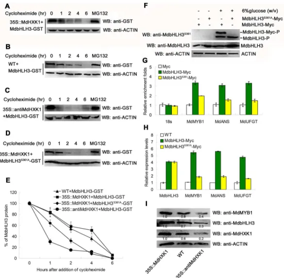
To examine whether MdHXK1 influences the stability of MdbHLH3 proteins, the prokaryon-expressed and purified MdbHLH3-GST fusion proteins were incubated with plant total pro-teins that were extracted from the WT control and the35S::MdHXK1and35S::antiMdHXK1
transgenic apple calli. Subsequently, protein degradation assays were performed. The results showed that MdbHLH3-GST proteins were more stable in the protein extracts of the35S::
MdHXK1transgenic calli than in those of the WT control (Fig 5A, 5B and 5E), whereas they were degraded at a more rapid speed in the protein extracts of35S::antiMdHXK1transgenic calli compared to those of the WT control (Fig 5C and 5E). These results suggest that MdHXK1-mediated phosphorylation of the MdbHLH3 protein may increase its stability.
To further verify that phosphorylation influences the stability of the MdbHLH3 protein, a site-directed S361A mutation was introduced into the MdbHLH3 protein. The mutated cDNA
MdbHLH3S361Awas inserted into the expression vector for prokaryon-expression and purifica-tion of MdbHLH3S361A-GST fusion proteins, which were then incubated with the total proteins extracted from the WT calli. The protein sample was used for Western blotting with the anti-GST antibody. The results showed that the MdbHLH3S361A-GST proteins degraded at a rapid speed compared with the wild-type MdbHLH3-GST proteins (Figs4B,5D and 5E), indicating that the inhibition of phosphorylation promoted the degradation of MdbHLH3 proteins. In addition, MdHXK1 also enhanced the stability of the endogenous MdbHLH3 proteins (S8A–S8D Fig). To examine whether phosphorylation of the MdbHLH3 protein influences its binding capacity to the downstream genes, such asMdMYB1,MdANSandMdUFGT, the35S::
MdbHLH3-Mycand35S::MdbHLH3S361A-Myctransgenic apple calli were used for ChIP-PCR analysis (Fig 5F;S9 Fig). The results showed that the phosphorylated MdbHLH3-Myc protein exhibited a higher enrichment in the promoters ofMdMYB1and anthocyanins biosyn-thetic structural genes than the non-phosphorylated MdbHLH3S361A-Myc (Fig 5G). As a result, those genes showed higher expression levels in the35S::MdbHLH3-Myctransgenic apple calli than theMdbHLH3S361A-Mycapple calli (Fig 5H). Furthermore, the abundance of the endoge-nous MdbHLH3 and MdMYB1 proteins were higher in35S::MdHXK1overexpressing calli but lower in35S::antiMdHXK1suppressing calli than in the WT control (Fig 5I).
Therefore, phosphorylation modification stabilizes the MdbHLH3 protein and enhances its transcriptional activation of downstream genes.
MdHXK1 promotes anthocyanin accumulation in an
MdbHLH3-dependent manner
Fig 5. Phosphorylation modification stabilizes the MdbHLH3 protein and enhances its binding capacity to the promoters of downstream genes. (A-C)Cell-free degradation assays demonstrate that MdHXK1 stabilizes the MdbHLH3 protein. The recombined MdbHLH3-GST protein was co-incubated with the isolated total proteins extracted from the WT control (B) and the35S::MdHXK1(A)and35S::antiMdHXK1(C)transgenic apple calli. In addition, the mixed proteins were treated with 20μg /mL cycloheximide for 0, 1, 2, 4 or 6 h. The degradation of MdbHLH3 protein was measured using Western blotting with an anti-GST antibody. MG132 was used as a positive control to stabilize the MdbHLH3 protein unless noted otherwise.(D)Ser361 is important for the
MdHXK1-mediated stabilization of MdbHLH3.(E)The graph shows the quantitation of the Western blot data in(A),
(B),(C)and(D).(F)Western blotting assay of the phosphorylation intensity for MdbHLH3 and MdbHLH3S361A proteins inMdbHLH3-MycandMdbHLH3S361A-Myctransgenic calli. The anti-MdbHLH3S361
antibody was used.
(G)ChIP-qPCR assays of the enrichments of the target gene promoters in the35S::MdbHLH3-Mycand35S::
MdbHLH3S361A-Myctransgenic calli compared to the35S::Myctransgenic calli.(H)Relative expression levels of
MdMYB1,MdANSandMdUFGTin the WT control and in the35S::MdbHLH3-Mycand35S::MdbHLH3S361A-Myc
transgenic calli.(I)Protein abundance of MdHXK1, MdbHLH3 and MdMYB1 in the WT control and in the35S::
MdHXK1and35S::antiMdHXK1transgenic apple calli. Protein amounts were normalized based on the protein
folds of the35S::MdHXK1transgenic apple calli. In(G)and(H), the data are shown as the mean±SE, which were analyzed based on more than 9 replicates. Statistical significance was determined using Student’st-test in different apple calli lines. n.s., P>0.01;*P<0.01;**P<0 .001.
resulting vectors were then transformed into apple calli. In the present study, we obtained nine types of transgenic apple calli, namely,35S::MdHXK1,35S::MdbHLH3,35S::MdbHLH3S361A,
35S::MdHXK1+35S::MdbHLH3,35S::MdHXK1+35S::MdbHLH3S361A,35S::MdHXK1+35S::
antiMdbHLH3,35S::antiMdHXK1+35S::MdbHLH3,35S::antiMdHXK1+35S::MdbHLH3S361A
and35S::antiMdHXK1+35S::antiMdbHLH3(Fig 6A). TheMdHXK1andMdbHLH3genes were successfully overexpressed or suppressed in the corresponding calli compared with the WT control (Fig 6B), indicating that the genetic transformation was successful in apple calli. As downstream genes, the transcript levels ofMdANSandMdUFGTgenes were positively cor-related with that of theMdbHLH3gene; however,MdANSandMdUFGTwere considerably lower in the35S::MdbHLH3S361Atransgenic calli than in the35S::MdbHLH3calli (Fig 6B). In addition, the transcription activity of theMdANSpromoter was positively associated with the transcript level ofMdHXK1genes (S10 Fig).
These transgenic apple calli were used to determine the anthocyanin content. The results showed that overexpression ofMdHXK1andMdbHLH3, either alone or together, noticeably enhanced the anthocyanin content in the corresponding transgenic calli compared with the WT control (Fig 6C). Moreover, the35S::MdbHLH3S361Atransgenic calli produced less antho-cyanins than the35S::MdbHLH3calli (Fig 6C), indicating that the phosphorylation site Ser361 is crucial for MdbHLH3 to regulate the biosynthesis of anthocyanins.
Furthermore, the35S::MdHXK1transgenic calli produced more anthocyanins, but the35S::
MdHXK1+35S::antiMdbHLH3calli produced less than the WT control (Fig 6A and 6C), indi-cating that the suppression of theMdbHLH3gene inhibited the MdHXK1-mediated increase of anthocyanin biosynthesis. Therefore, MdHXK1 regulates anthocyanin accumulation at least partially, if not completely, depending on the presence of MdbHLH3.
MdHXK1 works together with MdbHLH3 to modulate anthocyanin
accumulation in apple fruits
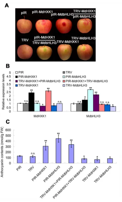
To investigate whether MdHXK1 and MdbHLH3 regulate anthocyanin accumulation in apple fruits in a similar manner as in calli, a viral vector-based method was applied to alter their expression using vector pRI for overexpression and vector TRV for suppression. Four viral constructs, including pRI-MdHXK1, TRV-MdHXK1, pRI-MdbHLH3 and TRV-MdbHLH3, were obtained. Each construct and two combinations, i.e., TRV-MdHXK1+pRI-MdbHLH3 and pRI-MdHXK1+TRV-MdbHLH3, were used for fruit infiltration, with the empty vectors as controls (Fig 7A). The results showed that the transcript levels ofMdHXK1andMdbHLH3
genes were enhanced after being infiltrated with pRI-MdHXK1 and pRI-MdbHLH3 but decreased with TRV-MdHXK1 and TRV-MdbHLH3, respectively (Fig 7B).
Subsequently, anthocyanin levels were measured in apple peel tissues around the sites infiltrated with the different viral constructs. The results showed that both MdHXK1 and MdbHLH3 positively regulate anthocyanin accumulation and that the MdHXK1-mediated anthocyanin accumulation required MdbHLH3 in apple fruits (Fig 7C), similar to the apple calli (Fig 6C).
Discussion
Fig 6. MdHXK1 controls anthocyanin accumulation viaMdbHLH3in apple calli. (A)Anthocyanin accumulation in WT and transgenic apple calli grown on MS medium supplement with 6% glucose under 17°C plus UVB light. The numbers 1–10 represent the WT and transgenic apple calli containing different combinations of constructs, as indicated.(B)qRT-PCR analysis of the relative expression levels ofMdHXK1,
calli.(C)Anthocyanin content of the WT and transgenic apple calli in(A). In(B)and(C), the data are shown as the mean±SE, which were analyzed based on more than 9 replicates. Statistical significance was
determined using Student’st-test in different apple calli lines. n.s., P>0.01;*P<0.01;**P<0 .001.
doi:10.1371/journal.pgen.1006273.g006
Fig 7. Transient expression ofMdHXK1andMdbHLH3via the viral vector-based transformation alters anthocyanin levels in apple fruits. (A)Apple fruit peel coloration around the injection sites. The full-length cDNA ofMdHXK1andMdbHLH3genes were cloned into the pIR vector for overexpression, whereas their antisense cDNA fragments were inserted into the TRV vector for suppression. The empty vectors were used as controls.(B)The qRT-PCR analysis of the relative expression levels ofMdHXK1andMdbHLH3 genes around the injection sites.(C)Anthocyanin content of the injected apple fruit peel in(A). In(B)and(C), the data are shown as the mean±SE, which were analyzed based on more than 9 replicates. Statistical significance was determined using Student’st-test in different apple calli lines. n.s., P>0.01;*P<0.01;
**P<0 .001.
Glucose-induced anthocyanin accumulation results from
MdHXK1-dependent glucose signaling and metabolic functions
Sugars are the major sources of carbon and energy metabolites and play key roles in plant growth and development. Sugars also act as effective signaling molecules throughout plant life [43,44]. InArabidopsis, HXK1 is a crucial enzyme in glucose catabolism; HXK1 senses glucose and initiates its signaling pathway [11]. Glucose-promoted aliphatic glucosinolate biosynthesis is regulated by HXK1-mediated signaling via the MYB TFs MYB28 and MYB29 [45]. Most recently, it was reported that glucose treatment greatly enhances anthocyanin content and induces the expression ofPsWD40-2,PsMYB2,PsCHS1,PsCHI1andPsF3’H1through glucose signaling inPaeonia suffruticosacut flowers [37]. Among the WBM genes,MYBandWD40genes, but notbHLHgenes, are induced at the transcriptional level by glucose. The present study found that a glucose-dependent signaling pathway is involved in the regulation of antho-cyanin accumulation in apple. This process depended on functional MdHXK1, which directly phosphorylated and stabilized the WBM component MdbHLH3 protein at the post-transla-tional level (Figs6A–6Cand7A–7C). MdbHLH3 modulates both anthocyanin biosynthetic structural genes and the regulatoryMdMYB1gene, thereby promoting anthocyanin accumula-tion [32].
Furthermore, anthocyanin accumulation is induced by glucose, which is not due to the osmotic effects of glucose (S1C and S1D Fig) [46]. MdHXK1 promoted anthocyanins accumu-lation mainly via the glucose signaling pathway under the high-glucose condition (Fig 1B–1F) and via both glucose metabolism and the signaling pathway under the low-glucose condition (S3 Fig). Given that sugars are the major sources of carbon and energy, higher plants require sugars for normal metabolism [9]. Glucose provides carbon skeletons for anthocyanin biosyn-thesis via its HXK1-dependent catalytic metabolism pathway, especially under low-glucose conditions (S3 Fig) [47]. Moreover, the MdHXK1-dependent glucose signaling pathway also plays a vital role in anthocyanin biosynthesis (Fig 1B–1E;S3 Fig). Therefore, glucose promotes anthocyanin biosynthesis depending on both signaling and metabolism under low-glucose conditions in apple. However, when carbohydrates are derived from glucose to meet the needs of anthocyanin synthesis under high-glucose conditions (e.g., 6% glucose), the glucose mainly served as signaling molecules to initiate anthocyanin biosynthesis (Fig 1B–1E). Taken together, glucose-induced anthocyanin accumulation is the result of MdHXK1-dependent glucose sig-naling together with catalytic metabolism pathways.
In the present study, the catalytic and signaling functions of MdHXK1 were characterized using its two catalytically inactive mutants,MdHXK1S177AandMdHXK1G104D, in apple (Fig 1), both of which retain signaling functions but not catalytic activities, similar to their activities in
Arabidopsis[11]. Most recently, Fenget al. [48] successfully resolved the crystal structures of two AtHXK1 inactive forms, AtHXK1S177Aand AtHXK1G104D, and analyzed the biochemical properties of AtHXK1 inArabidopsis. These findings provide biochemical and structural insights into how HXK1 functions at the atomic level, thereby providing a structural explana-tion for the dual funcexplana-tions of HXK1 in plants.
The nuclear HXK1 complex regulates glucose-mediated transcription
activation
nuclear HXK1 complexes [49]. A large number of putative TFs identified in the nuclear HXK1 complexes interact directly with VHA-B1 and/or RPT5B but not directly with HXK1 [49]. In addition, nuclear-localized HXK1 has also been implicated in the control of the transcriptional activity and proteasome-mediated degradation of EIN3 (ethylene-insensitive3), a key tran-scriptional regulator in ethylene signaling [50]. The present study found that HXK1 directly interacted with MdbHLH3 (Fig 2B–2D), a key bHLH transcriptional regulator in anthocyanin biosynthesis [32]. However, it is unclear whether the HXK1/VHA-B1/RPT5B nuclear complex is also involved in these processes.
Furthermore, a R2R3 MYB regulator MdMYB1 interacts with the N-terminus of MdbHLH3 to regulate anthocyanin biosynthesis [32]. The present study found that the hexokinase_2 domain of MdHXK1 strongly interacted with the C-terminus of MdbHLH3 to modulate antho-cyanin accumulation (Fig 2B). Therefore, there is no competition for the interaction of the MdbHLH3 protein with MdMYB1 and MdHXK1 in the regulation of anthocyanin biosynthesis.
MdbHLH3 phosphorylation may be an MdHXK1-mediated single-site
phosphorylation event
In apples, a putative phosphorylation modification is involved in the MdbHLH3-mediated anthocyanin accumulation in response to low temperature [32]. However, the protein kinase that mediates the phosphorylation of MdbHLH3 protein is not yet identified. The present study found that the MdHXK1 protein kinase is directly involved in the glucose-induced phos-phorylation of MdbHLH3 protein, thereby modulating anthocyanin biosynthesis (Fig 4A and 4B). In addition, the hexokinase_2 domain of MdHXK1, which may be required for signal pep-tide cleavage based on its functions in protein secretion and subcellular localization [51,52], plays a key role in the phosphorylation of the MdbHLH3 protein (Fig 4D and 4E).
Additionally, several bHLH TFs are phosphorylated by external environmental stimuli. For example, multiple light-induced Ser/Thr phosphorylation sites are found in the phyB-interact-ing bHLH TF PIF3 inArabidopsis[53]. Multisite light-induced phosphorylation of the bHLH TFs PIF1 and PIF5 has been confirmed using photobiological and genetic approaches [54,55]. In addition to PIFs, another bHLH TF, TWIST1, is phosphorylated at Thr125 and Ser127 to control pro-metastatic functions in prostate cancer cells [56]. In contrast to the aforemen-tioned bHLH TFs, the bHLH TF speechless is phosphorylated to promote stomatal develop-ment at a single serine 186 site inArabidopsis[57]. Similarly to the bHLH TF speechless, only a single phosphorylation site in the bHLH TF MdbHLH3 protein was detected in apple (Fig 3B and 3C;S2 Text), suggesting that MdbHLH3 phosphorylation may be a single-site phosphory-lation event in apple or at least that its Serine 361 plays a crucial role in anthocyanin biosynthe-sis (Figs5D–5H,6and7).
MdHXK1 protein kinase stabilizes MdbHLH3 to regulate the expression
of anthocyanin biosynthesis genes
As is well known, the MYB-bHLH-WDR (MBW) complex plays an important role in regulat-ing anthocyanin and proanthocyanidin biosynthesis. In apple, MdbHLH3 physically interacts with MdMYB1 and specifically binds to the promoters of anthocyanin structural genes, such as
calli accumulated more malate than the WT control (S11 Fig), possibly due to the MdHXK1-mediated stabilization of MdbHLH3.
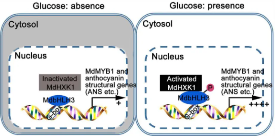
The glucose supply promotes anthocyanin biosynthesis and organ coloration in different plant species, such asArabidopsis, grape, andPaeonia suffruticosa[36,37,40]. However, the mechanism underlying the glucose signaling-mediated regulation ofMYBTFs,WDRand anthocyanin structural genes remains unclear [37,40]. Here, a working model is proposed to illuminate how glucose regulates anthocyanin accumulation in apple (Fig 8). Under glucose deprivation conditions, the kinase activity of MdHXK1 is inactivated and fails to phosphorylate MdbHLH3 protein (Fig 3A and 3C). As a result, a small amount of MdbHLH3 protein binds to the promoters of anthocyanin structural genes, leading to reduced anthocyanin accumulation (Figs6and7). When exposed to glucose, the kinase activity of MdHXK1 is activated, and then, MdHXK1 phosphorylates and stabilizes the MdbHLH3 protein, which further regulates the expression of the anthocyanin biosynthetic genes and the regulatoryMYBgenes (Figs4A, 4B,5 and6B), ultimately enhancing anthocyanin biosynthesis in apple (Figs6A, 6C,7A and 7C). In addition, ectopic expression of the appleMdHXK1gene also increased anthocyanin accumula-tion in the transgenicArabidopsis(S12 Fig), suggesting that the mechanism by which HXK1 controls anthocyanin accumulation in response to glucose is conserved in different species.
In summary, the current study provides new insights into the molecular mechanism of MdHXK1 stabilization of the MdbHLH3 protein, which occurs via phosphorylation, thereby promoting the accumulation of anthocyanins in plant cells in response to glucose signals. Because color is one of the most eye-catching traits for fresh fruits and ornamental plants [60,61], there is considerable interest for the organ coloration in the breeding programs for these economically important plants. Taken together, the regulatory mechanism uncovered in the present study is also useful for the development of novel biotechnological strategies for improving the quality of apple fruit and other horticultural crops.
Materials and Methods
Plant materials and growth conditions
Thein vitroshoot cultures of apple were obtained from detoxified buds of‘Gala’apples. They were maintained at 25°C under long-day conditions (16 h light/8 h dark) on Murashige and Skoog (MS) medium supplemented with 0.8 mg L-16-BA and 0.2 mg L-1IAA and subcultured at a 4-week interval before being used for further studies.
Fig 8. Model demonstrating that MdHXK1 protein kinase stabilizes MdbHLH3 via phosphorylation to modulate anthocyanin accumulation in response to glucose in apple.
The apple calli used in this study were induced from the young embryos of the‘Orin’apple (Malus domesticaBorkh.). The calli were grown on MS medium containing 0.5 mg of L-1 indole-3-acetic acid (IAA) and 1.5 mg of L-1 6-benzylaminopurine (6-BA) at 25°C in the dark. The apple calli were subcultured three times at 15-day intervals before being used for genetic transformation and in other assays. Additionally, all the apple calli were suffered from a dark (24 hours dark)-induced glucose starvation before being treated with exogenous glucose in this study, unless noted otherwise.
The apple fruits used for the injection of viral vectors were collected from mature trees of the cultivar‘Red Delicious’that were grown in a commercial orchard near Tai-An City. Fruits were bagged at 35 DAB (days after blooming); the bagged fruits were harvested at 140 DAB and de-bagged before injection.
The present study used theArabidopsis(Arabidopsis thaliana) ecotype‘Columbia,’the
MdHXK1overexpression lineMdHXK1-OVX1, the glucose-insensitive mutantgin2, and the function-complementary lineMdHXK1-R1(overexpression ofMdHXK1in agin2mutant background). Seeds were surface sterilized with 70% (v/v) ethanol and sown on 0.8% (w/v) agar plates containing half-strength MS medium and different glucose concentrations. The seeds were stratified for three days at 4°C and transferred into constant light
(100μmol m2s-1) at 20°C for 2 weeks of growth. Before being used for exogenous glucose
treatment, 2-weeks-oldArabidopsisplants were suffered from a dark (24 hours dark)-induced glucose starvation.
Construction of the expression vectors and genetic transformation
To constructMdHXK1andMdbHLH3sense overexpressing and antisense suppressing vectors, the full-length cDNA ofMdHXK1andMdbHLH3, a specific fragment ofMdHXK1and a con-served fragment ofMdbHLH3were isolated from‘Gala’apple using RT-PCR. Furthermore, truncated sense overexpression vectors, includingMdHXK135-242aa,MdHXK1245-491aa,
MdHXK11-21aa+35-242aaandMdHXK11-21aa+245-491aa, were also isolated from‘Gala’apple using RT-PCR. All of the cDNA were digested with EcoRI/BamHI and cloned into the pRI plant transformation vector downstream of the CaMV35Spromoter. All of the primers used in this study are listed inS3 Text.
In addition, two point mutants ofMdHXK1, namely, G104D (altering Glycine to Aspartate at position 104) and S177A (altering Serine to Alanine at position 177), and theMdbHLH3
point mutant Ser361A(mutation of Serine to Alanine at position 361), were obtainedusing site-directed mutagenesismethods. The resulting cDNA were digested with EcoRI/BamHI and cloned into the pRI plant transformation vector downstream of the CaMV35Spromoter. The primers used in this study are listed inS3 Text.
In addition, the full-length cDNA ofMdHXK1andMdbHLH3were also cloned into the PRI plant transformation vector with a Myc tag downstream of the CaMV35Spromoter, and subsequently, the recombined expression vectorsMdHXK1-MycandMdbHLH3-Mycwere used for genetic transformation.
For apple calli transformation, the constructs, including35S::MdHXK1,35S::MdbHLH3,
35S::MdbHLH3S361A,35S::MdHXK1+35S::MdbHLH3,35S::MdHXK1+35S::MdbHLH3S361A,
35S::MdHXK1+35S::antiMdbHLH3,35S::antiMdHXK1+35S::MdbHLH3,35S::antiMdHXK1
+35S::MdbHLH3S361A,35S::antiMdHXK1+35S::antiMdbHLH3, and35S::MdbHLH3-Myc, were introduced into‘Orin’apple calli using anAgrobacterium-mediated method as described by Huet al. [59].
floral dip method [59]. The seeds of the transgenic plants were individually harvested and sub-sequently selfed. Homozygous transgenic lines were used for further investigation.
RNA extraction and quantitative RT-PCR assays
RNA extraction and quantitative RT-PCR (qRT-PCR) assays were performed with the meth-ods described by Huet al. [59]. All of the primers used for qRT-PCR are listed inS3 Text.
Protein extraction and Western blotting
Protein extraction and Western blotting assays were conducted as described by Huet al. [59]. The monoclonal antibody of anti-MdHXK1, anti-MdbHLH3S361(specifically against the MdbHLH3 phosphorylation site at residue 361) and anti-GST antibody were prepared by the Abmart Company (Shanghai, China).
Yeast two-hybrid assays
Yeast two-hybrid assays were performed using the Matchmaker GAL4-based two-hybrid sys-tem (Clontech, Palo Alto, CA, USA). Full-length cDNA and truncated mutants of MdHXK1, including MdHXK11-245 aaand MdHXK1245-498aa, were inserted into the pGBT9 vector. The associated yeast two-hybrid vectors of MdbHLH3, which were inserted into vector pGAD424, are detailed in Xieet al. [32]. All of the constructs were transformed into yeast strain AH109 using a lithium acetate method. Yeast cells were cultured on minimal medium -Leu/-Trp according to the manufacturer’s instructions. Transformed colonies were plated onto minimal medium -Leu/-Trp/-His/-Ade with or withoutβ-galactosidase to test for possible interactions.
Co-immunoprecipitation (Co-IP) procedures
The WT and35S::MdbHLH3-GFPtransgenic apple calli were treated with 50μM MG132 for
16 h to stabilize the MdbHLH3-GFP and MdHXK1 proteins. The Co-IP was carried out as described by Ohet al. [62]. The eluted samples were immunoblotted using GFP and anti-MdHXK1 antibodies.
GST pull-down assays
For the GST pull-down assays, full-length cDNA ofMdbHLH3were inserted into the pGEX-4T-1 vector, whereas that ofMdHXK1was inserted into pET-32a. All of the recombinant pro-teins were used to perform GST pull-down assays as described by Ohet al. [62].
Identification of phosphorylation sites using LC-MS/MS
MdbHLH3 proteins were immunoprecipitated with anti-MdbHLH3 antibody-conjugated aga-rose beads and then separated on an SDS-PAGE gel and stained with Coomassie brilliant blue (CBB). The band containing phosphorylated MdbHLH3 protein was cut from the stained SDS-PAGE gel. The protein digestion, phosphopeptide enrichment, mass spectrometry data acquisition, data analysis, and label-free quantitation were carried out as described by Wang
et al. [63].
Detection of phosphoproteins
proteins extraction was performed for Western blotting assays with an anti-Myc antibody. Actin served as a protein-loading control.
In vitro
kinase assay
A total of 0.2μg of recombinant His-tagged protein kinase MdHXK1 and 1μg of
MdbHLH3S361A-GST and normal MdbHLH3-GST proteins were incubated in 25μL of
reac-tion buffer [20 mM Tris (pH 7.5), 5 mM MgCl2, 10 mM NaCl and 2 mM DTT] with 100μM
ATP and [λ-32P]ATP (0.2 mCi per reaction) at room temperature for 30 min. Recombinant MdbHLH3S361A-GST was served as a negative control in thein vitrokinase assay. The phos-phorylated proteins were visualized using autoradiography after separation on a 12% SDS-PAGE gel.
Construction of the viral vectors and transient expression in apple fruits
To construct antisense expression viral vectors, a specific fragment ofMdHXK1and a con-served fragment ofMdbHLH3were amplified with RT-PCR using apple fruit cDNA as the template. The resulting products were cloned into the tobacco rattle virus (TRV) vector in the antisense orientation under the control of the dual35Spromoter. The vectors were named TRV-MdHXK1 and TRV-MdbHLH3. To generate overexpression viral vectors, full-length cDNA ofMdHXK1andMdbHLH3were inserted into the IL-60 vector under the control of the35Spromoter. The resulting vectors were named MdHXK1-IL and MdbHLH3-IL.
The antisense expression viral vectors were transformed intoAgrobacterium tumefaciens
strain GV3101 for inoculations. Fruit infiltrations were performed as previously described [59]. The injected apple fruits were kept in the dark at room temperature for two days and sub-sequently placed in the highlight at 10°C for one week. The peel of the injected part was then harvested for gene expression analysis and anthocyanin content determination.
Analysis of glucose phosphorylation activity
Glucose phosphorylation activity was measured using an enzyme-linked assay according to Schaffer and Petreikov [64]. The assays contained a total volume of 1 mL of 30 mM HEPES--NaOH, pH 7.5, 2 mM MgCl2, 0.6 mM EDTA, 9 mM KCl, 1 mM NAD, 1 mM ATP, and 1 unit of NAD-dependent glucose-6-phosphate dehydrogenase (G6PDH). To assay glucose phos-phorylation, 25μL of the desalted extract was added to start the reaction under 25°C.
Reduc-tion of NAD within 5 min was monitored by the increase in absorpReduc-tion at 340 nm. Activity was calculated in terms ofμmol of NAD reduced per minute.
Transient expression in protoplasts of apple calli cells and fluorescence
detection
Determination of the total anthocyanin content
The total anthocyanins were extracted using a methanol-HCl method and detected as described by Huet al. [59].
Statistical analysis
Samples were analyzed in triplicates, and the data are expressed as the mean ± standard deviation unless noted otherwise. Statistical significance was determined using Student’st-test. A difference at P0.01 was considered significant, and P0.001 was considered extremely significant.
Supporting Information
S1 Fig. Glucose promotes anthocyanin accumulation in an HXK-dependent manner in apple. (A)Different concentration of glucose was tested for their ability to induce anthocyanin accumulation inin vitroshoot cultures of the‘Gala’apple cultivar. The shoot cultures of apple were plated on Murashige and Skoog (MS) agar containing 0.6 mg L-16-BA and 0.2 mg L-1 IAA plus different concentration of glucose (contains 1%, 2%, 3% and 6%) as indicated. Antho-cyanin accumulation in apple leaves was measured after 7 days of growth under 17°C low tem-perature induction and continuous light.(B)Anthocyanin content of apple leaves in(A).(C) The phenotype as indicated by a red color for anthocyanin accumulation inin vitroshoot cul-tures of the‘Gala’apple cultivar treated with 6% glucose and 6% mannitol or 6% glucose plus glucosamine.(D)The anthocyanin content of apple shoot cultures in(C). The data are shown as the mean ± SE, which were analyzed based on more than 9 replicates. Statistical significance was determined using Student’st-test in apple shoot cultures. n.s., p>0.01;p<0.001.
(TIF)
S2 Fig. Amino acid sequence alignment of apple MdHXK1 andArabidopsisAtHXK1 pro-teins.The conserved amino acid residues were labeled with black boxes. The alignment of sequences was generated using a‘‘multiple sequence alignment”method with DNAMAN soft-ware.
(TIF)
S3 Fig. Glucose modulates anthocyanin accumulation by the signaling and catalytic path-way under 1% glucose condition. (A)WT,35S::MdHXK1(WT background),35S::
MdHXK1G104D(WT background), and35S::MdHXK1S177A(WT background) transgenic apple calli showed anthocyanin accumulation phenotype on MS agar media containing 1% glucose. The apple calli were placed at 10°C under long-day conditions (16 h light/8 h dark) for 10 days. (B)and(C)Anthocyanin content(B)and glucose phosphorylation activity(C)in WT and transgenic apple calli in(A). In(B)and(C), data are shown as mean±SE, which were analyzed based on more than 9 replicates. Statistical significance was determined using Student’sttest in different apple calli lines. n.s., P>0.01;P<0.01;P<0.001.
(TIF)
http://smart.embl-heidelberg.de/. The pink rectangle indicates the signal peptide, while the gray rectangle shows the hexokinase_1 and hexokinase_2 domains respectively. The numbers below domains indicated the predicted starting and ending numbers of amino acid.
(TIF)
S5 Fig. Western blotting assay of the MdbHLH3 protein with Myc and anti-MdbHLH3S361antibodies. (A)Western blotting assay of MdbHLH3 protein abundance by using anti-Myc antibody in wild type (WT) and35S::MdbHLH3-Myctransgenic apple calli. The ACTIN was served as a protein-loading control.(B)Western blotting assay of the specific-ity of the anti-MdbHLH3S361antibody. TheMdbHLH3-Myctransgenic apple calli was pre-incubated in MS medium plus 6% glucose for 3 hours. Subsequently, the proteins extraction was used for Western blotting assays with an antibody of MdbHLH3S361phosphorylation site. (C)Glucose induced the phosphorylation of MdbHLH3 protein and was abolished by CIP in WT apple plants. The apple plants were pre-incubated in MS medium plus glucose (0 or 6%) and 5 U of CIP for 1 or 3 hours. Subsequently, the proteins extraction was used for Western blotting assays with an antibody of MdbHLH3S361phosphorylation site.
(TIF)
S6 Fig. The transcript and protein level of MdHXK1 in wild-type (WT) and35S:: antiMdHXK1transgenic apple calli. (A)Relative expression level ofMdHXK1in WT and
35S::antiMdHXK1transgenic apple calli. Data are shown as mean±SE, which were analyzed based on more than 9 replicates. Statistical significance was determined using Student’sttest in different apple calli lines.P<0.01.(B)The protein abundance of MdHXK1 in WT and35S:: antiMdHXK1transgenic apple calli.
(TIF)
S7 Fig. MdHXK1in vitrophosphorylates MdbHLH3 but not MdbHLH3S361A.The kinase assay was initiated by adding radiolabeled ATP to the mixture of MdHXK1-His kinase and MdbHLH3-GST (or MdbHLH3S361A-GST). The phosphorylated MdbHLH3-GST protein were detected with anti-MdbHLH3S361antibody. Note: MdbHLH3-GST-P represent the phos-phorylated MdbHLH3-GST protein.
(TIF)
S8 Fig. MdHXK1in vivokinase activity stabilizes the MdbHLH3 protein. (A-C)MdHXK1
in vivokinase activity stabilizes MdbHLH3 protein. The isolated total proteins from35S::
MdHXK1(A), WT(B)and35S::antiMdHXK1(C)apple calli were treated with 20μg /ml
cyclo-heximide for 0, 1, 3 and 6 h. The degradation of MdbHLH3 protein was followed by western blotting with anti-MdbHLH3 antibody.(D)The graph shows the quantitation of the western blot data in(A),(B), and(C).
(TIF)
S9 Fig. Western blotting assay of MdbHLH3S361Aprotein abundance using a Myc antibody in wild-type (WT) and35S::MdbHLH3S361A-Myctransgenic apple calli.The ACTIN was served as a protein-loading control.
(TIF)
S10 Fig. MdHXK1 activates theMdANRpromoter as detected by GUS assays. (A)The effec-tors and reporter constructs in the binary veceffec-tors were introduced into apple calli for GUS activity assays.pMdANR::GUStransgenic apple calli were transformation with35S::MdHXK1
calculated from the results of three independent experiments. (TIF)
S11 Fig. Malate content in the WT control as well as the35S::MdHXK1overexpressing and 35S::antiMdHXK1suppressing transgenic apple calli.
(TIF)
S12 Fig. The mechanism through which glucose-mediated HXK1 controls anthocyanin accumulation is conserved in different species. (A)Glucose-mediated HXK1 promotes anthocyanin biosynthesis inArabidopsis. The WT (Ler),AtHXK1mutantgin2,MdHXK1 over-expressor line HXK1-OVX1 and function complementary line HXK1-R1 were grown on one-half-strength MS medium without sugar (Control) or with 6% glucose (w/v), and 6% mannitol (w/v) at 10°C under long-day conditions (16 h light/8 h dark) for 10 days.(B)Anthocyanin content of WT and transgenicarabidopsisas indicated in(A).(C)Amino acid alignment of the conserved Ser361of HXK1 proteins in apple and other species. The conserved serine residue at position 361 were indicated with red pentagram. The alignment of all the sequences was gener-ated using a‘‘multiple sequence alignment”method with DNAMAN software.
(TIF)
S1 Text. Identification of MdHXK1-interacting proteins in co-immunoprecipitation using an LC/MS assay.
(XLS)
S2 Text. Identification of potential phosphorylation sites in MdbHLH3 with an LC/MS assay.
(XLS)
S3 Text. List of primers used in this study. (XLS)
S4 Text. Map of potential phosphorylation sites in MdbHLH3 with an LC/MS assay. (PDF)
S5 Text. Glucose promotes anthocyanin accumulation in an HXK-dependent manner in apple.
(DOCX)
Acknowledgments
We thank Prof. Ilan Sela of Hebrew University of Jerusalem, Israel, for IL-60-BS binary vectors, Prof. Takaya Moriguchi of National Institute of Fruit Tree Science, Japan, for‘Orin’apple calli and Shanghai Applied Protein Technology Co. Ltd for the technical assistance in LC/MS prote-omic and phosphorylation sites assays.
Author Contributions
Conceived and designed the experiments:YJH DGH.
Performed the experiments:DGH CHS QYZ JPA CXY.
Analyzed the data:DGH.
Contributed reagents/materials/analysis tools:DGH YJH JPA.
References
1. Biemelt S, Sonnewald U. Plant-microbe interactions to probe regulation of plant carbon metabolism. J Plant Physiol. 2006; 163: 307–318. PMID:16368160
2. Leon P, Sheen J. Sugar and hormone connections. Trends Plant Sci. 2003; 8: 110–116. PMID: 12663220
3. Seo YS, Cho JI, Lee SK, et al. Current insights into the primary carbon metabolic flux that occurs in plants undergoing a defense response. Plant Stress. 2007; 1: 42–49.
4. Plaxton WC. The organization and regulation of plant glycolysis. Annu Rev Plant Biol. 1996; 47(1): 185–214.
5. Moreno F, Ahuatzi D, Riera A, Palomino CA, Herrero P. Glucose sensing through the Hxk2-dependent signaling pathway. Biochem Soc T. 2005; 33(1): 265–268.
6. Ho SL, Chao YC, Tong WF, Yu SM. Sugar coordinately and differentially regulates growth and stress related gene expression via a complex signal transduction network and multiple control mechanisms. Plant Physiol. 2001; 125: 877–890. PMID:11161045
7. Chen JG. Sweet sensor, surprising partners. Sci Signal. 2007; 2007: pe7.
8. Graham IA, Denby KJ, Leaver CJ. Carbon catabolite repression regulates glyoxylate cycle gene expression in cucumber. Plant Cell. 1994; 6: 761–772. PMID:12244257
9. Jang JC, Leon P, Zhou L, Sheen J. Hexokinase as a sugar sensor in higher plants. Plant Cell. 1997; 9: 5–19. PMID:9014361
10. Jang JC, Sheen J. Sugar sensing in higher plants. Plant Cell. 1994; 6: 1665–1679. PMID:7827498
11. Moore B, Zhou L, Rolland F, et al. Role of theArabidopsisglucose sensor HXK1 in nutrient, light, and hormonal signaling. Science. 2003; 300: 332–336. PMID:12690200
12. Claeyssen E, Rivoal J. Isozymes of plant hexokinase: occurrence, properties and functions. Phyto-chemistry. 2007; 68: 709–731. PMID:17234224
13. Granot D. Role of tomato hexose kinases. Funct Plant Biol. 2007; 34: 564–570.
14. Karve A, Rauh BL, Xiaoxia X, et al. Expression and evolutionary features of the hexokinase gene family
inArabidopsis. Planta. 2008; 228: 411–425. doi:10.1007/s00425-008-0746-9PMID:18481082
15. Li M, Feng F, Cheng L. Expression patterns of genes involved in sugar metabolism and accumulation during apple fruit development. PLoS One. 2012; 7(3): e33055. doi:10.1371/journal.pone.0033055 PMID:22412983
16. Kandel-Kfir M, Damari-Weissler H, German MA, et al. Two newly identified membrane-associated and plastidic tomato HXKs: characteristics, predicted structure and intracellular localization. Planta. 2006; 224: 1341–1352. PMID:16761134
17. Cho YH, Yoo SD, Sheen J. Regulatory functions of nuclear hexokinase1 complex in glucose signaling. Cell. 2006; 127: 579–589. PMID:17081979
18. Kushwah S, Jones AM, Laxmi A. Cytokinin interplay with ethylene, auxin and glucose signaling controls
Arabidopsisseedling root directional growth. Plant Physiol. 2011: pp-111.
19. Kushwah S, Laxmi A. The interaction between glucose and cytokinin signal transduction pathway in
Ara-bidopsis thaliana. Plant Cell Environ. 2014; 37(1): 235–253. doi:10.1111/pce.12149PMID:23763631
20. Gupta A, Singh M, Laxmi A. Interaction between glucose and brassinosteroid during the regulation of lateral root development inArabidopsis. Plant Physiol. 2015; 168(1): 307–320. doi:10.1104/pp.114. 256313PMID:25810094
21. Yoshida K, Mori M, Kondo T. Blue flower color development by anthocyanins: from chemical structure to cell physiology. Nat Prod Rep. 2009; 26: 884–915. doi:10.1039/b800165kPMID:19554240
22. Winkel-Shirley B. Flavonoid biosynthesis: a colorful model for genetics, biochemistry, cell biology, and biotechnology. Plant Physiol. 2001; 126: 485–493. PMID:11402179
23. Gould KS, McKelvie J, Markham KR. Do anthocyanins function as antioxidants in leaves? Imaging of H2O2in red and green leaves after mechanical in jury. Plant Cell Environ. 2002; 25: 1261–1269.
24. Nagata T, Todoriki S, Masumizu T, et al. Levels of active oxygen species are controlled by ascorbic acid and anthocyanin inArabidopsis. J Agric Food Chem. 2003; 51: 2992–2999. PMID:12720382
25. Hu DG, Ma QJ, Sun CH, et al. Overexpression ofMdSOS2L1, an CIPK protein kinase, improves the antioxidant metabolites to enhance salt tolerance in apple and tomato. Physiol Plantarum. 2015; doi: 10.1111/ppl.12354
26. Debeaujon I, Peeters AJ, Léon-Kloosterziel KM, Koornneef M. TheTRANSPARENT TESTA12gene of
Arabidopsisencodes a multidrug secondary transporter-like protein required for flavonoid
27. Gomez C, Terrier N, Torregrosa L, Ageorges A. Grapevine MATE-Type proteins act as vacuolar H+ -dependent acylated anthocyanin transporters. Plant Physiol. 2009; 150: 402–415. doi:10.1104/pp. 109.135624PMID:19297587
28. Koes R, Verweij W, Quattrocchio F. Flavonoids, a colorful model for the regulation and evolution of bio-chemical pathways. Trends Plant Sci. 2005; 10: 236–242. PMID:15882656
29. Ballester AR, Molthoff J, de Vos R. Biochemical molecular analysis of pink tomatoes, deregulated expression of the gene encoding transcription factorSlMYB12leads to pink tomato fruit color. Plant Physiol. 2010; 152: 71–84. doi:10.1104/pp.109.147322PMID:19906891
30. Albert NW, Lewis DH, Zhang H, et al. Members of an R2R3-MYB transcription factor family inPetunia are developmentally and environmentally regulated to control complex floral and vegetative pigmen-tation patterning. Plant J. 2011; 65: 771–784. doi:10.1111/j.1365-313X.2010.04465.xPMID: 21235651
31. Xu W, Dubos C, Lepiniec L. Transcriptional control of flavonoid biosynthesis by MYB-bHLH-WDR com-plexes. Trends Plant Sci. 2015; 20(3): 176–185. doi:10.1016/j.tplants.2014.12.001PMID:25577424
32. Xie XB, Li S, Zhang RF, et al. The bHLH transcription factor MdbHLH3 promotes anthocyanin accumu-lation and fruit colouration in response to low temperature in apple. Plant Cell Environ. 2012; 35: 1884– 1897. doi:10.1111/j.1365-3040.2012.02523.xPMID:22519753
33. Figueroa P, Browse J. Male sterility inArabidopsisinduced by overexpression of aMYC5-SRDX chime-ric repressor. Plant J. 2015; 81(6): 849–860. doi:10.1111/tpj.12776PMID:25627909
34. Liu Z, Zhang Y, Wang J, et al. Phytochrome-interacting factors PIF4 and PIF5 negatively regulate anthocyanins biosynthesis under red light inArabidopsisseedlings. Plant Sci. 2015; 238: 64–72. doi: 10.1016/j.plantsci.2015.06.001PMID:26259175
35. Hara M, Oki K, Hoshino K, Kuboi T. Enhancement of anthocyanins biosynthesis by sugar in radish (Raphanus sativus) hypocotyl. Plant Sci. 2003; 164: 259–265.
36. Teng S, Keurentjes J, Bentsink L, Koornneef M, Smeekens S. Sucrose-specific induction of anthocya-nins biosynthesis inArabidopsisrequires theMYB75/PAP1gene. Plant Physiol. 2005; 139: 1840– 1852. PMID:16299184
37. Zhang C, Fu J, Wang Y, et al. Glucose supply improves petal coloration and anthocyanins biosynthesis
inPaeonia suffruticosa‘Luoyang Hong’cut flowers. Postharvest Biol Technol. 2015; 101: 73–81.
38. Hribar U, Poklar UN. The metabolism of anthocyanins. Curr Drug Metab. 2014; 15(1): 3–13. PMID: 24329109
39. Hedrich R, Sauer N, Neuhaus HE. Sugar transport across the plant vacuolar membrane: nature and regulation of carrier proteins. Curr Opin Plant Biol. 2015; 25: 63–70. doi:10.1016/j.pbi.2015.04.008 PMID:26000864
40. Dai ZW, Meddar M, Renaud C, et al. Long-termin vitroculture of grape berries and its application to assess the effects of sugar supply on anthocyanin accumulation. J Exp Bot. 2014; 65(16): 4665–4677. doi:10.1093/jxb/ert489PMID:24477640
41. Zhao J, Sun MH, Hu DG, Hao YJ. Molecular cloning and expression analysis of apple hexokinase gene
MdHXK1. Hortic Plant J. 2015; 42(8): 1437–1447.
42. Zheng Y, Tian L, Liu H, et al. Sugars induce anthocyanin accumulation and flavanone 3-hydroxylase expression in grape berries. Plant Growth Regul. 2009; 58(3): 251–260.
43. Ramon M, Rolland F, Sheen J. Sugar sensing and signaling. TheArabidopsisBook. 2008; 6: e0117. doi:10.1199/tab.0117PMID:22303242
44. Smeekens S, Ma J, Hanson J, Rolland F. Sugar signals and molecular networks controlling plant growth. Curr Opin Plant Biol. 2010; 13: 274–279. doi:10.1016/j.pbi.2009.12.002PMID:20056477
45. Miao H, Wei J, Zhao Y, et al. Glucose signaling positively regulates aliphatic glucosinolate biosynthesis. J Exp Bot. 2013; ers399.
46. Martin T, Oswald O, Graham IA.Arabidopsisseedling growth, storage lipid mobilization, and photosyn-thetic gene expression are regulated by carbon: nitrogen availability. Plant Physiol. 2002; 128: 472– 481. PMID:11842151
47. Lukaszewicz M, Matysiak-Kata I, Skala J, et al. Antioxidant capacity manipulation in transgenic potato tuber by changes in phenolic compounds content. J Agric Food Chem. 2004; 52(6): 1526–1533. PMID: 15030206
48. Feng J, Zhao S, Chen X, et al. Biochemical and structural study ofArabidopsishexokinase 1. Acta Crystallographica Section D: Biological Crystallography. 2015; 71(2): 0–0.