Assessment of Point-of-Care Diagnostics for
G6PD Deficiency in Malaria Endemic Rural
Eastern Indonesia
Ari W. Satyagraha1*, Arkasha Sadhewa1, Rosalie Elvira1, Iqbal Elyazar2,
Denny Feriandika1, Ungke Antonjaya2, Damian Oyong2, Decy Subekti2, Ismail E. Rozi1, Gonzalo J. Domingo3, Alida R. Harahap1, J. Kevin Baird2,4
1Eijkman Institute for Molecular Biology, Jakarta, Indonesia,2Eijkman-Oxford Clinical Research Unit, Jakarta, Indonesia,3PATH, Seattle, Washington, United States of America,4Centre for Tropical Medicine, Nuffield Department of Medicine, University of Oxford, Oxford, United Kingdom
Abstract
Background
Patients infected byPlasmodium vivaxorPlasmodium ovalesuffer repeated clinical attacks without primaquine therapy against latent stages in liver. Primaquine causes seriously threatening acute hemolytic anemia in patients having inherited glucose-6-phosphate dehy-drogenase (G6PD) deficiency. Access to safe primaquine therapy hinges upon the ability to confirm G6PD normal status. CareStart G6PD, a qualitative G6PD rapid diagnostic test (G6PD RDT) intended for use at point-of-care in impoverished rural settings where most malaria patients live, was evaluated.
Methodology/Principal Findings
This device and the standard qualitative fluorescent spot test (FST) were each compared against the quantitative spectrophotometric assay for G6PD activity as the diagnostic gold standard. The assessment occurred at meso-endemic Panenggo Ede in western Sumba Island in eastern Indonesia, where 610 residents provided venous blood. The G6PD RDT and FST qualitative assessments were performed in the field, whereas the quantitative assay was performed in a research laboratory at Jakarta. The median G6PD activity5 U/ gHb was 9.7 U/gHb and was considered 100% of normal activity. The prevalence of G6PD deficiency by quantitative assessment (<5 U/gHb) was 7.2%. Applying 30% of normal G6PD activity as the cut-off for qualitative testing, the sensitivity, specificity, positive predic-tive value, and negapredic-tive predicpredic-tive value for G6PD RDT versus FST among males were as follows: 100%, 98.7%, 89%, and 100% versus 91.7%, 92%, 55%, and 99%; P = 0.49, 0.001, 0.004, and 0.24, respectively. These values among females were: 83%, 92.7%, 17%, and 99.7% versus 100%, 92%, 18%, and 100%; P = 1.0, 0.89, 1.0 and 1.0, respectively.
a11111
OPEN ACCESS
Citation:Satyagraha AW, Sadhewa A, Elvira R, Elyazar I, Feriandika D, Antonjaya U, et al. (2016) Assessment of Point-of-Care Diagnostics for G6PD Deficiency in Malaria Endemic Rural Eastern Indonesia. PLoS Negl Trop Dis 10(2): e0004457. doi:10.1371/journal.pntd.0004457
Editor:Photini Sinnis, Johns Hopkins Bloomberg School of Public Health, UNITED STATES
Received:November 12, 2015
Accepted:January 22, 2016
Published:February 19, 2016
Copyright:© 2016 Satyagraha et al. This is an open access article distributed under the terms of the
Creative Commons Attribution License, which permits unrestricted use, distribution, and reproduction in any medium, provided the original author and source are credited.
Data Availability Statement:All relevant data are within the paper and its Supporting Information files.
Conclusions/Significance
The overall performance of G6PD RDT, especially 100% negative predictive value, demon-strates suitable safety for G6PD screening prior to administering hemolytic drugs like prima-quine and many others. Relatively poor diagnostic performance among females due to mosaic G6PD phenotype is an inherent limitation of any current practical screening methodology.
Author Summary
G6PD is an enzyme that chemically protects us from otherwise toxic substances, like some chemotherapeutic agents. About 8% of people exposed to malaria have an inherited disor-der that impairs G6PD activity, leaving them vulnerable to harm by an important therapy against malaria, primaquine. This drug alone prevents repeated clinical attacks stemming from dormant parasites residing in the human liver. Absent certain knowledge of patient G6PD status, healthcare providers managing patients infected byPlasmodium vivaxor
Plasmodium ovalemalaria must choose between risk of harm caused by hemolytic toxicity of primaquine and that caused by the parasite after withholding therapy. Resolving that therapeutic dilemma requires assessment of patient G6PD status at the point-of-care in the impoverished rural tropics, where the vast majority of malaria patients live. Current technology for such screening is impractical in that setting. In this study we evaluated screening designed for practicality at the endemic tropical point-of-care: a rapid diagnostic test for G6PD (G6PD RDT; CareStart G6PD, AccessBio, USA). We found the G6PD RDT to be effective in screening volunteers living in rural eastern Indonesia. This G6PD RDT kit costs relatively little ($1.50), was simple to execute and interpret, required no special-ized equipment or skills, performed well at ambient tropical temperatures (>30°C), and
required no cold chain storage. This and similar kits may permit safe universal access to primaquine therapy against relapse ofP.vivax, a vitally important step forward in mitigat-ing the global burden of morbidity and mortality imposed by this pernicious parasite.
Introduction
Glucose-6-phosphate dehydrogenase deficiency (G6PDd) is the most common inherited disor-der, affecting about 400 million people [1–3]. G6PD enzyme catalyzes the first and rate-limit-ing reaction of the pentose phosphate pathway, the only means of reducrate-limit-ing nicotinamide adenine dinucleotide phosphate (NADPH) in red blood cell cytosol. In turn, NADPH is the sole source of electrons for reducing glutathione, the principal means of maintaining healthy reduction-oxidation (redox) equilibrium in cytosol. Oxidative stress upon red blood cells with impaired G6PD activity leads to threatening redox imbalance. Most people with G6PDd none-theless lead healthy lives of normal longevity, and it is only exposure to certain drugs, chemi-cals, foods or infections that impose hemolytic crisis and risk of serious harm. In the malaria endemic rural tropics, the most threatening scenario is becoming infected by the parasite Plas-modium vivaxand being prescribed the drug primaquine to prevent the repeated clinical attacks (called relapses) deriving from latent liver stages called hypnozoites [2].
Primaquine is an 8-aminoquinoline drug licensed as a hypnozoitocide in 1952 and remains the only therapy available for preventing relapse [4]. During its clinical development in and after World War II, investigators observed hemolytic sensitivity in some subjects. Only later, in 1956, did those investigators identify deficiency in G6PD as the cause of that sensitivity [5]. study design, data collection and analysis, decision to
publish, or preparation of the manuscript.
Those early studies, conducted in prisoner volunteers in the USA, characterized primaquine sensitivity in African-Americans expressing the A- variant of G6PDd typically expressing 10–
20% of normal G6PD activity. Primaquine hemolyzed only older red blood cell populations in those subjects and hemolysis ceased despite continued exposure to high doses of primaquine [6,7]. These findings led to the view of primaquine-induced hemolysis as relatively mild and self-limiting. However, studies in the 1960s revealed other variants of G6PDd, including the exquisitely primaquine-sensitive Mediterranean variant [8–12]. Mediterranean variant typi-cally exhibited<5% of normal activity and primaquine-induced hemolysis occurred even
among the youngest red blood cells—severe and unlimited hemolysis without cessation of dos-ing. Over the decades that followed, confirmed severe hemolytic crises and deaths due to pri-maquine toxicity in G6PD deficient patients accumulated [13–17]. In South and Southeast Asia, where more than 80% of vivax malaria attacks occur, the extraordinary diversity of G6PDd is dominated by Mediterranean-like, severely deficient variants [18,19].
Recent recognition ofPlasmodium vivaxas a pernicious infection and its multiple relapses a serious clinical and public health threat [20–22] elevated awareness of the problem of G6PDd as a very significant barrier to safe primaquine therapy [23,24]. Absent the ability to identify G6PDd patients among those infected byP.vivax, providers must choose between risk of harm caused by the drug and that caused by the repeated clinical attacks allowed by withholding the drug. Resolving this therapeutic dilemma requires identifying those at risk of harm with prima-quine therapy and thus ensuring those not at risk obtain the enormous therapeutic benefit of primaquine.
The most widely used and recommended procedure for screening patients for G6PD defi-ciency, excluding newborn screening, is the fluorescent spot test (FST), described by G6PD pio-neer Ernest Beutler in 1966 [25]. In a laboratory setting the test is relatively simple and
inexpensive. However, in the setting of the impoverished rural tropics, the requirements for laboratory skills, refrigeration, specialized equipment, and high costs have excluded its avail-ability to the vast majority of patients suffering malaria. Expert consensus defined practicality criteria for point-of-care G6PD diagnostics that included simplicity of use, ease of interpreta-tion, no specialized equipment or cold chain, and relatively low cost [26–28]. Expert consensus also acknowledged that the availability of such robust devices where most malaria patients live is a key to the control and elimination of endemicP.vivaxmalaria [28].
In the current study, the performance of the CareStart G6PD (G6PD RDT, AccessBio, USA) device against the FST using quantitative spectrophotometric G6PD assay as diagnostic gold standard was compared among residents in a malaria-endemic area of rural eastern Indonesia. The G6PD RDT performed as well as the FST.
Methods
Ethics Statement
This study has been ethically approved by the Eijkman Institute Research Ethics Commission (EIREC) (project No. 69, February 13th, 2014). After obtaining the informed consent from 610 healthy subjects at least 6 years old, recruitment ceased at targeted full enrollment. Written informed consent was obtained from all study participants. Parents or guardians signed the informed consents for minors under 18 years of age.
Population and Study Site
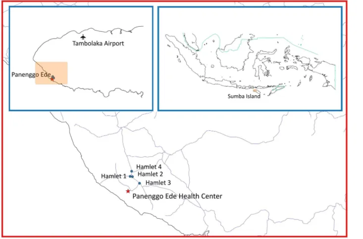
The village Panenggo Ede is located in the western coastal region of Southwest Sumba regency (Fig 1), where G6PDd prevalence was known to be>5% [29]. A total of 1117 people resided in
gathered at churches or other social functions, and explained the study procedures and intent. Residents were then invited to a study center established in the village at designated times and dates between April and May 2014. The inclusion criteria were people6 years old, healthy and willing to sign informed consent. A total of 350 females and 260 males provided a 3mL sample of whole venous blood collected into tubes containing EDTA anticoagulant. Samples were held at 4°C prior to processing and analysis on-site (G6PD RDT) or nearby temporary laboratory (FST) within 3 hours on the same day, or within 3 days at the laboratory in Jakarta (quantitative G6PD).
G6PD RDT
The principle of the CareStart G6PDTscreening test is reduction of a colorless nitro-blue tetra-zolium dye to purple colored formazan. Thus, whereas a colorless test outcome indicates G6PD deficiency, a purple color reflects G6PD activity (Fig 3). Readers of the test were instructed to consider only a diagnosis of deficient or normal, with the demand to classify as deficient any test strip exhibiting a colorless to distinctly lighter hue of purple compared to that of most other tests. This approach would ensure safety when primaquine therapy would follow the diagnosis of G6PD normal.
Two microliters of whole blood was removed from the EDTA tube by a stick device included in the RDT kit and placed into the sample window, immediately followed by two drops of a provided buffer solution into the assay window according to the manufacturer’s Fig 1. Geographic location of the study site.
instructions. After ten minutes at the ambient temperature of approximately 30°C, the RDT was visually read and classified as deficient or normal.
Fluorescent Spot Test
At the end of each day of work in the village, venous blood was transferred on ice packs to a field laboratory in Weetabula to conduct the fluorescent spot test (FST, Trinity Biotech, Ire-land; Cat. No. 203-A) using deficient (Cat. No. G5888), intermediate (Cat. No. G5029) and normal (Cat. No. G6888) G6PD controls from the same company. This qualitative test is a modification of Beutler’s test in which glucose-6-phosphate and NADP+reagents (substrate solution) in the presence of G6PD sample produce fluorescent NADPH and 6-phosphogluco-nate. Progress of the reaction was observed in the dark under long-wave ultraviolet illumina-tion of sample filter paper (Whatman No. 1 filter paper, Cat. No. 1001–150) at intervals of zero, 5 and 10 minutes. Briefly, 200μl of substrate solution and 10μl of gently mixed venous blood was put into a 5 ml tube and mixed by manual swirling. A single drop of this solution Fig 2. Flow-chart of the study in Panenggo Ede where those tests performed in field or field laboratory were confirmed by DNA analysis at the Eijkman Institute in Jakarta, Indonesia.
Fig 3. Photographs illustrating visual test outcomes for the G6PD RDT (top) and FST (bottom).For FST, samples were spotted at time 0, 5 and 10 minutes interval and the dark spots were considered deficient (D) and the bright ones were considered normal (N). RDT with purple color was considered normal (N) and no color was considered deficient (D).
was transferred onto filter paper marked as time zero. The tube was then placed into a 37°C water bath for 5 min, when another drop was placed onto filter paper marked as time 5 min. This was repeated for the final sample at 10 min. The filter papers were allowed to dry at room temperature (25°-29°C) before visual inspection under UV light in an otherwise dark room. Deficient (no fluorescence), intermediate (weak fluorescence) and normal (strong fluores-cence) controls were done for every set of 10 samples from the subjects. Readers were instructed to classify intermediate test outcomes as deficient.
G6PD Quantitative Test
The principle of the G6PD quantitative assay from Trinity Biotech (Cat. No. 345-B) is similar to the FST. Fluorescence from NADPH produced in the same substrate solution mixture was read at 340nm using a high-grade, temperature-controlled spectrophotometer (UV-1800 UV-VIS Shimadzu). The same G6PD controls from Trinity Biotech were conducted for each set of 25 samples from the subjects. The assay was performed in an air-conditioned (~25°C) laboratory at the Eijkman Institute in Jakarta within 3 days of blood withdrawal. The venous blood tubes were kept at 4°C at all times prior to use in Jakarta. Hemoglobin level was deter-mined using 10μl of blood into a micro-cuvette supplied by the manufacturer of the HemoCue system (HemoCue AB, Sweden) and immediately read in the instrument (Hb201+) of that sys-tem for hemoglobin measurement prior to the G6PD quantitative assay. The manufacturer’s instructions were strictly followed for measuring absorbance at 340nm and deriving an esti-mate of G6PD activity in U/g Hb at 30°C using the incubated spectrophotometer. Although the manufacturer recommended a cut off value<4.6 U/g Hb for deficient activity, we selected <5U/g Hb on the basis of prior survey in the same area showing a median G6PD activity of 10
U/g Hb [29]. We aimed for a 50% cut off value, that being the limit of relative safety with respect to potential hemolytic loss of red blood cell populations, knowing this value correlated with the proportion of deficient red blood cells in a laboratory model of the female heterozy-gous state [30]. The assay was performed in triplicates where a mean was derived to be used for downstream analyses.
G6PD Variant Genotyping
Samples for G6PD genotyping were selected on the basis of a deficient classification by G6PD quantitative assay (<5U/gHb), or by having Hb<8g/dL (Fig 2). DNA from the buffy coat of
Table 1. PCR primers and RFLP conditions for G6PD variants common in Southwest Sumba Regency and PCR primers for detecting SAO, HbE andαthalassemia.
Variant Primer Primer Sequence (5’!3’) Expected PCR Product (bp)
RE Expected Result (bp)
References
Deficient Normal
Vanua Lava VL-9F CAG CCT GGG GCA GTG TCT GTG CT
366 EcoNI 366 346 Our design
VL-9R GCG GTT GGC CTG TGA CCC CTG GTG
20
Viangchan VC-9F TGG CTT TCT CTC AGG TCT AG
126 XbaI 106 126 Nuchprayoon
et al, 2002 VC-9R GTC GTC CAG GTA CCC TTT
GGG G
20
Chatham CT-9F CAA GGA GCC CAT TCT CTC
CCT T
208 BstXI 100 130 Gandomaniet al, 2011 CT-9R TTC TCC ACA TAG AGG AGG
ACG GCT GCC AAA GT
78 78
30
Kaiping KP-9F ACG TGA AGC TCC CTG ACG C 227 MnlI 206 227 Laosombatet al,
2006 KP-9R GTG CAG CAG TGG GGT GAA
CAT A
21
SAO OVF
1098
GGG CCC AGA TGA CCC TCT TGC
175 (148 for SAO) - - - Jarolimet al, 1991
OVR 1272
GCC GAA GGT GAT GGC GGG TG
HbE Com C ACC TCA CCC TGT GGA GCC
AC
293 MnlI 122 106 Pramoodjago
et al, 1999 TLR
62320
CTA TTG GTC TCC TTA AAC CTG TCT TGT AAC CTT GCT A
106 60
alpha Thalassemia (Multiplex PCR—2 gene deletion)
SEA-alpha F
CTC TGT GTT CTC AGT ATT GGA GGG AAG GAG
1110 660 - - - Liuet al, 2000
SEA-alpha R
ATA TAT GGG TCT GGA AGT GTA ACC CTC CCA
alpha R TGA AGA GCC TGC AGG ACC AGG TCA GTG ACC G
FILL-alpha F
AAG AGA ATA AAC CAC CCA ATT TTT AAA TGG GCA
550 - -
- FILL-alpha R
GAG ATA ATA ACC TTT ATC TGC CAC ATG TAG CAA
THAI-alpha F
CAC GAG TAA AAC ATC AAG TAC ACT CCA GCC
411 - -
- THAI-alpha R
TGG ATC TGC ACC TCT GGG TAG GTT CTG TAC C
αThalassemia (Multiplex
PCR—1 gene deletion)
2/3P TGT TGG CAC ATT CCG GGA CAG
1940 (normal) - - - Setianingsihet al, 2003 XY1 GCG CCG AGC CTG GCC AAA
CCA TCA CTT TTC
2220 (-3.7 kb deletion) 3R1 TGC ATC CTC AAA GCA CTC
TAG GGT CCA GCG T
1673 (-4.2 kb deletion) SA3P TAA GCT AGA GCA TTG GTG
GTC ATG C
XYHA GAA GTA CGT CCG ACC AGC TTA GCC A
RE is restriction enzyme; bp is base pair.
Red Cell Disorder Genotyping
DNA extracted from venous blood was also genotyped for Southeast Asian ovalocytosis (SAO), alpha thalassemia, and hemoglobin E (HbE).Table 1lists the primers for SAO, one and two gene deletions for alpha thalassemia and HbE. The PCR conditions for each mutation were as reported elsewhere [32]. PCR conditions for one-gene deletions were as previously reported [33].
Analytical Rationale and Statistics
In the current study the diagnostic objective was not G6PD deficiencyper se, but a diagnostic outcome indicating either hazard or safety with administration of a potentially hemolytic drug. As such, diagnostic performance of the G6PD screening techniques was linked to the perceived primaquine safety margin of 30% of normal activity per WHO recommendation [26]. We aimed to classify all male hemizygotes and female heterozygotes having less than variable thresholds of normal G6PD activity (<10%,<30%, or<60%) as deficient. The median G6PD
activity among subjects having5U/g Hb was considered 100% of normal. These thresholds represented an examination of variance in diagnostic performance representing poor, good, and complete safety, respectively, with respect to exposure to primaquine. Poor safety at 10% would likely include patients at risk of hemolysis, whereas complete safety at a 60% would unnecessarily deny some patients primaquine treatment. The 30% cut off value represents a compromising balance of those problems.
Diagnostic performance of the qualitative G6PD RDT and FST were assessed against the quantitative G6PD classification as“deficient”or“normal”at G6PD activity thresholds. Fur-ther, the analyses were segregated by sex for the simple reason that hemizygosity versus hetero-zygosity (males and females, respectively) profoundly impacts diagnostic performance for G6PD deficiency [34]. Males tend to be wholly deficient or normal, whereas females will pres-ent the full spectrum of G6PD activity levels due to mosaicism of this X-linked trait [35].
Standard methods for calculation of sensitivity, specificity, positive predictive value, and negative predictive value were applied to the G6PD RDT and FST for each threshold of percent of normal G6PD activity. The meaning of these parameters in the context of a diagnosis guid-ing primaquine therapy bears explanation here. Sensitivity and specificity are easily grasped, i.e., rate of true positives and rate of true negatives, respectively. The terms“positive”and“ neg-ative”refer to what is defined here as“deficient”and“normal”G6PD phenotype, respectively. A test negative for G6PD activity is positive for G6PD deficiency, and vice versa for a positive test for G6PD activity. The terminology“deficient”(positive for deficiency) versus“normal”
(negative for deficiency) recommended by WHO [26], avoids confusion and was adopted here. Further, the terms“deficient predictive value”(DPV) and“normal predictive value”(NPV) were applied for consistency and clarity, but using precisely the same standard mathematical methods for all of these statistics.
Results
Inherited Blood Disorders in the Community
The overall prevalence of G6PDd at Panenggo Ede by quantitative assay (<5U/g Hb) was 7.2%
(44/610), 9.2% for males (24/260) and 5.7% (20/350) for females. Southeast Asian ovalocytosis (SAO) occurred in 12.7% (78/610), alpha thalassemia (alpha Thal) in 15.1% (92/610), and hemoglobin E (HbE) in 16.4% (100/610) of residents. Double mutations occurred among 13 residents having G6PDd (5 with SAO, 3 with alphaThal, 5 with HbE), and one subject had G6PDd, SAO and HbE. SAO occurred in 21 subjects also having alpha Thal, and in 7 people also having HbE. In total, 44.3% (270/610) of the population had one or more of these four blood disorders.
Malaria and Anemia
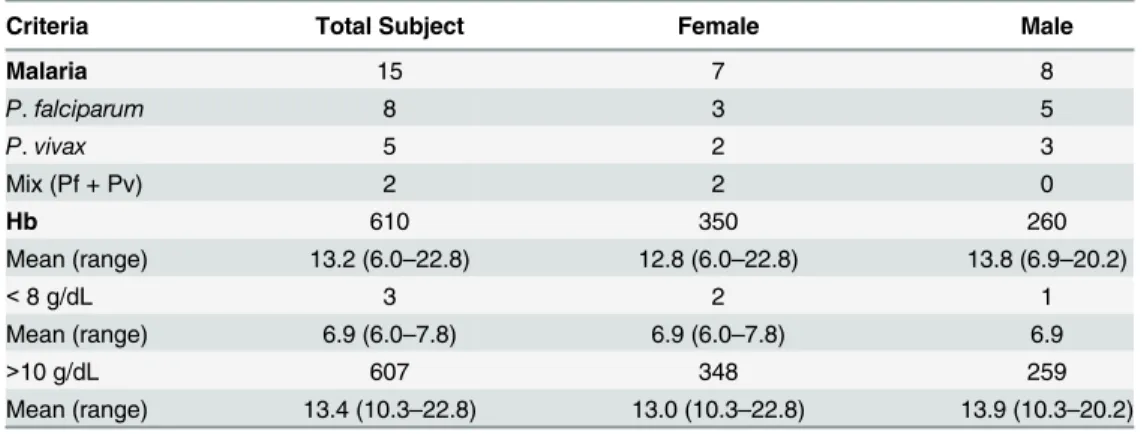
Table 2summarizes findings of malaria and anemia in the community. The overall prevalence of microscopically patent parasitemia was 2.5% (15/610); with 53%P.falciparum, 33%P.
vivax, and 14% mixed by these species. The mean level (and range) of Hb in the study popula-tion was 13.2(6.0–22.8) g/dL. Only 3 subjects had levels<8.0g/dL, and the majority had
10.0g/dL (607; 99.5%). Males and females had similar but statistically distinct levels of Hb: 13.8 (6.9–20.2), and 12.8 (6.0–22.8), respectively (P<0.0001). Among the three severely anemic
subjects (<8.0g/dL), genotyping for G6PD variants revealed one as a female (Hb 6.0g/dL)
het-erozygous for Vanua Lava variant with a quantitative G6PD value of 13.55 U/gHb. The other Fig 4. Chart illustrating the rationale for assessing diagnostic performance of qualitative G6PD screening devices in the context of a clinical decision to offer or withhold primaquine therapy in patients withP.vivaxmalaria.Each classification (bold font top), clinical outcome (normal font middle), and risk or benefit (italics bottom) of diagnostic performance appears in each box of classification.
two were G6PD normal genotype and phenotype. Hemoglobin level did not appear to be sig-nificantly different between subjects with or without any particular inherited blood disorder evaluated.
G6PDd Characteristics
Fig 5Aillustrates the results of genotyping of the 44 subjects deemed G6PDd by quantitative assay (<5.0 U/g Hb). Vanua Lava dominated at 50% (22/44), followed by Viangchan at 30%
(13/44), Coimbra Shunde at 11% (5/44), Chatham at 7% (3/44), and 1 subject was not success-fully genotyped (2%).Fig 5Billustrates G6PD activity values for subjects classified as normal by G6PD activity, as well as with those classified as deficient and successfully genotyped. Het-erozygous females having5.0 U/gHb would have been excluded from the genotyping survey and would be included among normals in the figure. The values illustrated for heterozygotes inform only the diagnostic assessment rather than as a survey of their G6PD activity ranges. Among hemizygous males, however, the G6PD activity mean and range may be considered estimates of residual enzyme activity among the specific variants: 0.8 U/g Hb (0.27–2.5 U/g Hb) for Vanua Lava; 0.97 U/g Hb (0.52–1.62 U/g Hb) for Viangchan; and 0.09 U/g Hb (0.03–
0.16 U/g Hb) for Coimbra Shunde. Remarkably low G6PD activity was also observed in the two females expressing Coimbra Shunde variant (0.57 U/g Hb; the mean of 0.11 and 1.04 U/g Hb). Chatham variant was found only in 3 females. G6PD activity did not vary with age in this study, as reported in another study from the same region [29].
Diagnostic Assessment
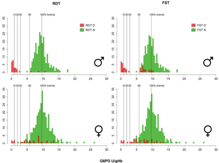
Fig 6illustrates diagnostic outcomes for the G6PD RDT and FST across quantitative G6PD activity values in males and females. The tests performed similarly, with each discerning G6PD deficiency at a threshold of 10% of normal activity among males and females. However, 2 FST tests in males were read as normal at<10% of activity. The tests also performed similarly at a
60% activity threshold for both tests. The FST in males showed a propensity for false deficient reads, even at or above 100% of normal activity, but was especially frequent between 65% and 85% of normal activity. Three false deficient reads occurred among males with the G6PD RDT at 65%, 90%, and 115% of normal G6PD activity. Although both tests properly identified all female heterozygotes below a 30% threshold (with a single exception for the G6PD RDT at 22% of normal activity), each also exhibited a profound propensity for false deficient reads all across the range of G6PD activity values.
Table 2. Malaria and anemia in the community.
Criteria Total Subject Female Male
Malaria 15 7 8
P.falciparum 8 3 5
P.vivax 5 2 3
Mix (Pf + Pv) 2 2 0
Hb 610 350 260
Mean (range) 13.2 (6.0–22.8) 12.8 (6.0–22.8) 13.8 (6.9–20.2)
<8 g/dL 3 2 1
Mean (range) 6.9 (6.0–7.8) 6.9 (6.0–7.8) 6.9
>10 g/dL 607 348 259
Mean (range) 13.4 (10.3–22.8) 13.0 (10.3–22.8) 13.9 (10.3–20.2)
Fig 5. Variants of G6PD found in Panenggo Ede.(A) Bar graph showing different variants found in males (blue) and females (red) and (B) showed the boxplot showing the activity of these variants in comparison to normal. VL, VC, CT and CO stands for Vanua Lava, Viangchan, Chatham and Coimbra Shunde variant of G6PD respectively. Black line across each box plot is the median for each group.*Indicated that the group contained homozygous females as well.
Table 3summarizes the statistical analyses of these diagnostic outcomes among males and females diagnostic thresholds of 10%, 30%, and 60% for both of the qualitative G6PD screening kits. At the 30% threshold the G6PD RDT showed superior sensitivity and specificity in males compared to the same for the FST: 100% and 98.7% versus 91.7% and 92.4%, respectively (P = 0.48 and P<0.001 for sensitivity and specificity respectively). Among females at the 30%
threshold, no statistically significant differences occurred between the sensitivities and specific-ities of the two kits: 83.3% and 100% vs. 92.7% and 92.2% (P = 1 and P = 0.89) for G6PD RDT vs. FST, respectively. Deficient predictive value (DPV) for the G6PD RDT for males at 30% of normal G6PD activity was superior to the same with FST: 63.0% versus 37.5% (P = 0.05), respectively. Among females at the same threshold, DPV was 10.0% and 9.1% (P = 1). Among males for both G6PD RDT and FST at 30% threshold, normal predictive value (NPV) was 100% and 99.1% respectively (P = 0.23), and for females 100% and 100% (P = 1).
Fig 6. Graphs illustrate diagnostic outcomes for the G6PD RDT (left) and FST (right) among males (top) and females (bottom) across quantitative G6PD activity values for each subject.A qualitative test classification as deficient is shown in red, and green for normal. Vertical lines within each identify specific diagnostic thresholds (10%, 20%, 30%, 60% and 100%) employed to calculate diagnostic performance characteristics.
Discussion
This assessment of a new RDT for G6PDd (CareStart G6PD) revealed performance character-istics essentially similar to the current screening standard, the FST. Whereas the G6PD RDT meets essential performance characteristics defined by expert consensus [26–28], the FST meets almost none of those. The availability of practical G6PD diagnostic devices at the periph-ery of healthcare delivperiph-ery in the endemic tropics would meet an urgent need to provide prima-quine therapy to the G6PD-normal majority infected by the relapsing malarias [23,24,28,36]. Consistency in satisfactory diagnostic performance of the G6PD RDT should impel making it broadly available in order to resolve the therapeutic dilemma of primaquine, G6PD deficiency andP.vivaxorP.ovalemalarias.
A study of a Cambodian population (n = 938) having 7.9% G6PD deficiency dominated by the Viangchan variant (92%) reported good performance of the G6PD RDT relative to the FST [37]. Investigators in Ghana also reported satisfactory performance of the G6PD RDT in a pop-ulation (n = 206) dominated by the A- variant [38], as well as a study (n = 456) in Haiti [39]. All of these studies applied a quantitative diagnostic threshold of<30% of normal G6PD.
Con-cordance among these studies offers assurance of satisfactory diagnostic performance of the G6PD RDT among settings of distinct G6PDd variant composition, malaria endemicity, and teams managing the evaluation. Taken together, these real world assessments of the G6PD RDT indicate suitability for intended use in guiding safe access to primaquine therapy against relapse.
In the current study, the G6PD RDT provided a superb margin of safety in the context of G6PD screening for the purpose of reaching a clinical decision on primaquine therapy (i.e., NPV = 100%). The FST resulted in two male subjects being falsely classified as normal despite enzyme activity below 10% of normal (NPV = 99.1%). Deviation from 100% NPV means vul-nerable patients will be in danger of exposure to primaquine (Fig 4), and this did occur in the current study with female G6PD RDT testing with NPV = 99.7% (at<30% analysis in one
Table 3. G6PD diagnostic tests analyzed in 610 subjects living in Panenggo Ede village, Southwest Sumba, tested against G6PD gold standard test.
Cut off Performance indicator1 Male P-value Female P-value
RDT2 FST3 RDT FST
10% Sensitivity 100.0 (80.5–100) 88.2 (63.6–98.5) 0.48 100.0 (29.2–100) 100.0 (29.2–100) 1.0
Specificity 95.9 (92.6–98.0) 89.7 (85.2–93.2) 0.01 92.2 (88.9–94.8) 91.4 (87.9–94.1) 0.78
DPV 63.0 (42.4–80.6) 37.5 (22.7–54.2) 0.05 10.0 (2.1–26.5) 9.1 (1.9–24.3) 1.0
NPV 100.0 (98.4–100) 99.1 (96.8–99.9) 0.23 100.0 (98.9–100) 100.0 (98.8–100) 1.0
30% Sensitivity 100.0 (85.8–100) 91.7 (73–99) 0.49 83.3 (35.9–99.6) 100.0 (54.1–100) 1.0
Specificity 98.7 (96.3–99.7) 92.4 (88.2–95.4) 0.001 92.7 (89.5–95.2) 92.2 (88.8–94.8) 0.89
DPV 88.9 (70.8–97.6) 55.0 (38.5–70.7) 0.004 16.7 (5.6–34.7) 18.2 (7–35.5) 1.0
NPV 100.0 (98.4–100) 99.1 (96.8–99.9) 0.24 99.7 (98.3–100) 100.0 (98.8–100) 1.0
60% Sensitivity 96.0 (79.6–99.9) 88.0 (68.8–97.5) 0.61 44.0 (24.4–65.1) 60.0 (38.7–78.9) 0.39
Specificity 98.7 (96.3–99.7) 92.3 (88.2–95.4) 0.001 94.2 (91–96.4) 94.5 (91.4–96.7) 1.0
DPV 88.9 (70.8–97.6) 55.0 (38.5–70.7) 0.004 36.7 (19.9–56.1) 45.5 (28.1–63.6) 0.61
NPV 99.6 (97.6–100) 98.6 (96.1–99.7) 0.36 95.6 (92.8–97.6) 96.8 (94.3–98.5) 0.53
1
Proportion and 95% confidence interval.
2CareStart RDT (Access Bio) and tested infield setting with temperature 28
–34°C and humidity between 55–76%.
3
FST (Trinity Biotech) and tested infield laboratory with temperature 26–29°C.
females subject with 22% of normal G6PD), as well as in two of the studies cited above: 97.7% [38], 98.2% [39].
G6PD RDT and FST each showed a propensity for false deficient reads across the spectrum of G6PD activity among subjects, especially females. Test failure to chemically develop may result in falsely deficient outcomes. This apparently occurred at a relatively high rate in this study and others. The DPV of G6PD RDT and FST at a 30% enzyme activity threshold for females, 17% and 18%, reflected this diagnostic problem. In other words, 83% and 82% of female subjects screening as deficient were actually“normal”(>30% G6PD activity). Partial
development of color or fluorescence would have prompted test readers to classify lesser color intensity as“deficient”. We viewed this approach as clinically appropriate with respect to pre-venting exposure to primaquine in patients at risk, i.e. protecting NPV with compromise of DPV. That compromise results in patients who could safely consume primaquine therapy being denied it (Fig 4).
Female heterozygotes present a serious diagnostic problem. In the current study all subjects were evaluated for quantitative G6PD activity, using<5.0 U/gHb to classify each as deficient
(with Hb level>8.0 g/dL).Fig 6clearly illustrates females almost exclusively occurring in the
range of 30% to 60% of normal G6PD activity. They screened as both deficient and normal in that range, largely depending upon placement within that range, precisely as observed in a lab-oratory-based study of G6PD RDT and FST [30]. Consequently, any normal classification of females by screening may not be considered assurance of safety with primaquine therapy. As expressed by WHO [28], females cleared for primaquine therapy by a normal G6PD screen may nonetheless require clinical monitoring for assurance of safety.
This study employed well-trained laboratorians as readers of the qualitative G6PD diagnos-tic kits evaluated. This had no impact on the primary objective of this study—examining the performance of the new G6PD RDT relative to the FST standard. Each test likely benefitted equally from the relatively high level of skill of the readers. Thus, while the tests were per-formed in the setting of a village in the endemic rural tropics, those performing the tests were imported from the setting of a sophisticated modern medical research laboratory. An evalua-tion of the suitability of the G6PD RDT should be done employing the intended end-users, i.e., paramedics or specially trained residents who today conduct malaria RDT diagnostics and dis-pense antimalarial therapy at the village level. Proper training on analysis and documentation as well as standard operating procedure must be implemented with use of G6PD RDT by less well trained staff. The use of venipuncture rather than fingerstick blood sample represents another limitation of the study. However, others have demonstrated no difference in G6PD activity estimates from venous versus capillary blood samples [40].
Screening for G6PD deficiency by qualitative point-of-care kits like the first-generation one evaluated here will likely be improved. Nonetheless, in the meantime the present version of G6PD RDT certainly offers an option that is conspicuously better than the current standard of care for most patients with vivax malaria—no G6PD screening and the raw choices of risk of harm by the drug or by the parasite in withholding it. The broad availability of practical and effective kits would vastly mitigate G6PD deficiency as a serious barrier to access to primaquine therapy against relapse.
Conclusions
harm with primaquine therapy. Both screening kits often misclassified G6PD normal subjects as deficient, which would result in withholding primaquine therapy from patients who could safely consume it. All qualitative tests for G6PD suffer the drawback of classifying many female heterozygotes as G6PD normal despite significantly impaired G6PD activity (i.e., 30% to 60% of normal), exposing them to risk of harm with primaquine therapy. The degree of that risk is poorly understood and requires a great deal more work, both in terms of assessing it and miti-gating it with improved diagnostics. There is also a need to evaluate the stability of the RDT during storage in the field.
Supporting Information
S1 Checklist. STARD Checklist.
(DOCX)
Acknowledgments
The authors express their gratitude to the residents of Panenggo Ede, those who volunteered as subjects and others who guided and assisted our efforts there. We thank Fitri Wulandari and Agus at EOCRU who rendered technical and administrative assistance, as did Dedi Sudiana in the field. We would like to thank Saraswati, Mewahyu Dewi, Lia Waslia and Jeni for laboratory assistance.
Author Contributions
Conceived and designed the experiments: AWS GJD JKB. Performed the experiments: AWS AS RE DF UA DO DS ARH. Analyzed the data: AWS AS RE IE GJD ARH JKB. Contributed reagents/materials/analysis tools: AWS GJD IE IER. Wrote the paper: AWS AS IE GJD JKB. Prepare field logistics: UA DS DO DF.
References
1. Cappellini MD, Fiorelli G. Glucose-6-phosphate dehydrogenase deficiency. Lancet 2008; 371: 64–74.
doi:10.1016/S0140-6736(08)60073-2PMID:18177777
2. Youngster I, Arcavi L, Schechmaster R, Akayzen Y, Popliski H, Shimonov J, Beig S, Berkovitch M. Medications and glucose-6-phosphate dehydrogenase deficiency: and evidence-based review. Drug Saf 2010; 33: 713–26. doi:10.2165/11536520-000000000-00000PMID:20701405
3. Howes RE, Battle KE, Satyagraha AW, Baird JK, Hay SI. G6PD deficiency: global distribution, genetic variants and primaquine therapy. Adv Parasitol 2013; 81: 133–201. doi:10.1016/B978-0-12-407826-0. 00004-7PMID:23384623
4. Baird JK, Hoffman SL. Primaquine therapy for malaria. Clin Infect Dis 2004; 37: 1659–67.
5. Alving AS, Carson PE, Flanagan CL, Ickes CE. Enzymatic deficiency in primaquine-sensitive erythro-cytes. Science 1956; 124: 484–5.
6. Dern R, Beutler E, Alving AS. The hemolytic effect of primaquine. II. The natural course of hemolytic anemia and mechanism of its self-limited character. J Lab Clin Med 1954; 44: 171–6. PMID:13184224 7. Alving AS, Johnson CF, Tarlov AR, Brewer GJ, Kellermeyer RW, Carson PE. Mitigation of the
haemoly-tic effect of primaquine and enhancement of its action against exoerythrocyhaemoly-tic forms of the Chesson strain of Plasmodium vivax by intermittent regimens of drug administration. Bull WHO 1960; 22: 621–
31. PMID:13793053
8. Pannacciulli I, Tizianello A, Ajmar F, Salvidio E. The course of experimentally induced hemolytic ane-mia in a primaquine-sensitive Caucasian: a case study. Blood 1965; 25: 92–5. PMID:14255977 9. George JN, Sears DA, McCurdy PR, Conrad ME. Primaquine sensitivity in Caucasians: hemolytic
10. Bonsignore A, Foraini G, Fantoni A, Leoncini G, Segni P. Relationship between age and enzymatic activities in human erythrocytes from normal and fava bean-sensitive subject. J Clin Invest 1964; 43: 834. PMID:14169512
11. Piomelli S, Corash LM. Davenport DD, Miraglia J, Amorosi EL. In vivo liability of glucose-6-phosphate dehydrogenase in GdA-and GdMediterraneandeficiency. J Clin Invest 1968; 47: 940
–8. PMID:5641629 12. Salvidio E, Pannacciulli, Tizianello A, Ajmar F. Nature of hemolytic crises and the fate of G6PD
defi-cient, drug-damaged erythrocytes in Sardinians. New Engl J Med 1967; 276: 1339–44. PMID: 6024564
13. Ebisawa I, Muto T. Malaria in Laos. Primaquine sensitivity of the Laotians and Japanese. Jpn J Exp Med 1972; 42: 415–7. PMID:4538385
14. Chopra SA. Hemolytic crisis in a Zanzibari Arab girl with G6PD deficiency and sickle cell trait. East Afr Med J 1968; 45: 726–7. PMID:5721119
15. Ziai M, Amirhakimi GH, Reinhold JG, Tabatabee M, Gettner MS, Bowman JE. Malaria prophylaxis and treatment in G6PD deficiency: an observation on the toxicity of chloroquine and primaquine. Clin Pediatr 1967; 6: 242–3.
16. Abeyarantne KP, Halpe NL. Sensitivity to primaquine in Ceylonese children due to deficiency of eryth-rocytic glucose-6-phosphate dehydrogenase deficiency. Ceylon Med J 1968; 13: 134–8. PMID: 5721557
17. Lacerda MV, Fragoso SC, Alecrim MG, Alexandre MA, Magalhaes BM, et al. Postmortem characteriza-tion of patients with a clinical diagnosis of Plasmodium vivax: to what extent does this parasite kill? Clin Infect Dis 2012; 55: e67–74. PMID:22772803
18. Clyde DF. Clinical problems associated with the use of primaquine as a tissue schizontocidal and gametocytocidal drug. Bull WHO 1981; 59: 391–5. PMID:6976846
19. Howes RE, Piel FB, Patil AP, Nyangiri OA, Gething PW, et al. G6PD deficiency prevalence and esti-mates of affected populations in malaria endemic countries: a geostatistical-based map. PLoS Med 2012; 9: e1001339. doi:10.1371/journal.pmed.1001339PMID:23152723
20. Anstey NM, Douglas NM, Poespoprodjo JR, Price RN. Plasmodium vivax: clinical spectrum, risk factors and pathogenesis. Adv Parasitol 2012; 80: 151–201. doi:10.1016/B978-0-12-397900-1.00003-7
PMID:23199488
21. Baird JK. Evidence and implications of mortality associated with acute Plasmodium vivax malaria. Clin Microbiol Rev 2013; 26: 36–57. doi:10.1128/CMR.00074-12PMID:23297258
22. Naing C, Whittaker MA, Nyunt Wai V, Mak JW. Is Plasmodium vivax a severe malaria?: a systematic review and meta-analysis. PLoS Negl Trop Dis 2014; 8: e3071. doi:10.1371/journal.pntd.0003071 PMID:25121491
23. World Health Organization. Global Technical Strategy for Malaria 2016–2030. 2015. Geneva. 35pp. 24. World Health Organization. 2015. Control and Elimination of Plasmodium vivax Malaria. A Technical
Brief. Geneva. 64pp.
25. Beutler E. A series of new screening procedures for pyruvate kinase deficiency, glucose-6-phosphate dehydrogenase deficiency, and glutathione reductase deficiency. Blood 1966; 28: 553–62. PMID: 5923607
26. Domingo GJ, Satyagraha AW, Anvikar A, Baird JK, Bancone G, et al. G6PD testing in support of treat-ment and elimination of malaria: recommendations for evaluation of G6PD tests. Malaria J 2013; 12: 391.
27. von Seidlein L, Auburn S, Espino F, Shanks D, Cheng Q, et al. Review of key knowledge gaps in glu-cose-6-phosphate dehydrogenase deficiency detection with regard to the safe clinical deployment of 8-aminoquinoline treatment regimens: a workshop report. Malaria J 2013; 12: 112.
28. World Health Organization. 2015. Point-of-care G6PD testing to support safe use of primaquine for the treatment of vivax malaria. WHO Evidence Review Group meeting report 8–9 October 2014, WHO/
UNAIDS Building, Geneva, Switzerland (http://www.who.int/malaria/mpac/mpac-march2015-erg-g6pd. pdfaccessed 26 October 2015).
29. Satyagraha AW, Sadhewa A, Baramuli V, Elvira R, Ridenour C, et al. G6PD deficiency at Sumba in eastern Indonesia is prevalent, diverse and severe: implications for primaquine therapy against relaps-ing vivax malaria. PLOS Negl Trop Dis 2015; 9: e0003602. doi:10.1371/journal.pntd.0003602PMID: 25746733
30. Baird JK, Dewi M, Subekti D, Elyazar I, Satyagraha AW. Non-inferiority of glucose-6-phosphate dehy-drogenase deficiency diagnosis by a point-of-care rapid test vs the laboratory fluorescent spot test demonstrated by copper inhibition in normal human red blood cells. Transl Res 2015; 165: 677–688.
31. Saunders MA, Hammer MF, Nachman MW. Nucleotide variability at G6PD and the signature of malaria selection in humans. Genetics 2002; 162: 1849–61. PMID:12524354
32. Liu YT, Old JM, Miles K, Fisher CA, Weatherall DJ, Clegg JB. Rapid detection of a-thalassaemia dele-tions and a-globin gene triplication by multiplex polymerase chain reacdele-tions. Brit J Haematol 2000; 108:295–299.
33. Setianingsih I, Harahap A, Nainggolan IM. Alpha thalassemia in Indonesia: phenotypes and molecular defects. Adv Exp Med Biol 2003; 531: 47–56. PMID:12916780
34. Ainoon O, Alawiyah A, Yu YH, Cheong SK, Hamidah NH, Boo NY, Zaleha M. Semiquantitative screen-ing test for G6PD deficiency detects severe deficiency but misses a substantial proportion of partially-deficient females. Southeast Asian J Trop Med Public Health 2003; 34: 405–14. PMID:12971572 35. Baird JK, Satyagraha AW, Bancone G. Glucose-6-phosphate dehydrogenase deficiency and
prima-quine hemolytic toxicity. In: Encyclopedia of Malaria (Hommel M, Kremsner PG, eds). Springer. 2014.
36. Baird JK. Point-of-care G6PD diagnostics for Plasmodium vivax malaria is clinical and public health urgency. BMC Med,in press.
37. Roca-Feltrer A, Khim N, Kim S, Chy S, Canier L, et al. Field trial evaluation of the performances of point-of-care tests for screening G6PD deficiency in Cambodia. PLoS One 2014; 9: e116143. doi:10. 1371/journal.pone.0116143PMID:25541721
38. Adu-Gyasi D, Asante KP, Newton S, Dosoo D, Amoako S, et al. Evaluation of the diagnostic accuracy of the CareStart G6PD deficiency rapid diagnostic test (RDT) in a malaria endemic area in Ghana, Africa. PLoS One 2015; 10: e0125796. doi:10.1371/journal.pone.0125796PMID:25885097
39. von Fricken ME, Weppelmann TA, Eaton WT, Masse R, Beau de Rochars MV, Okech BA. Performance of the CareStart glucose-6-phosphate dehydrogenase (G6PD) rapid diagnostic test in Gressier, Haiti. Am J Trop Med Hyg 2014; 91: 77–80. doi:10.4269/ajtmh.14-0100PMID:24778197