http://www.uem.br/acta ISSN printed: 1679-9275 ISSN on-line: 1807-8621
Doi: 10.4025/actasciagron.v38i2.27689
Biochemical characterization of systemic bacteria in bananas,
sensitivity to antibiotics and plant phytotoxicity during shoot
proliferation
Janiffe Peres de Oliveira1 and Jonny Everson Scherwinski-Pereira2*
1
Programa de Pós-graduação em Biotecnologia, Centro de Apoio Multidisciplinar, Universidade Federal do Amazonas, Manaus, Amazonas, Brazil. 2Parque Estação Biológica, Empresa Brasileira de Pesquisa Agropecuária, Recursos Genéticos e Biotecnologia, Avenida W5 Norte, Cx. Postal 02372, 70770-917, Brasília, Distrito Federal, Brazil. *Author for correspondence. E-mail: jonny.pereira@embrapa.br
ABSTRACT. The objective of this work was to characterize the biochemically systemic bacterial isolated from banana plants, to evaluate the bacterial sensitivity to antibiotics, and to determine the phytotoxicity of banana shoots during in vitro proliferation. Systemic bacteria belonging to the Klebsiella and Aeromonas
genera were isolated from the “Maravilha” (FHIA 01 AAAB), “Preciosa” (PV 4285 AAAB) and “Thap Maeo” (AAB) varieties and were then characterized. Tests of shoot sensitivity to antibiotics were performed, and the minimum inhibitory concentration (MIC) and phytotoxic effects of selected antibiotics to plants were determined. Among the 20 antibiotics evaluated, the strains showed sensitivity to cefaclor, cefalexin, cefalotin, nalidixic acid, chloramphenicol, and vancomycin. However, during MIC determination, the best results were obtained with cefaclor, vancomycin or nalidixic acid alone in concentrations ranging from 512 to 1,024 mg L-1.
In culture medium, cefaclor at 1,024 mg L-1 was the only antibiotic to affect the multiplication and the shoot
survival in culture.
Keywords:Musa spp., micropropagation, contamination, endophytic microorganisms, antimicrobial control.
Caracterização bioquímica de bactérias sistêmicas em bananeiras, sensibilidade a
antibióticos e fitotoxicidade de plantas durante a proliferação de brotos
RESUMO. O objetivo do trabalho foi caracterizar bioquimicamente bactérias sistêmicas isoladas de plantas de bananeiras, avaliar a sensibilidade das bactérias a antibióticos e determinar a fitotoxicidade de brotos de bananeiras durante a proliferação in vitro. Bactérias sistêmicas pertencentes aos gêneros Klebsiella e
Aeromonas foram isoladas a partir das variedades “Maravilha” (FHIA 01 AAAB), “Preciosa” (PV 4285 AAAB) e “Thap Maeo” (AAB), sendo em seguida caracterizadas. Testes de sensibilidade das brotações aos antibióticos foram desenvolvidos e a mínima concentração inibitória (MIC) e os efeitos fitotóxicos dos antibióticos selecionados em relação aos brotos foram determinados. Entre os 20 antibióticos avaliados, verificou-se que as bactérias mostraram sensibilidade para o cefaclor, cefalexina, cefalotina, ácido nalidíxico, cloranfenicol e vancomicina. Entretanto, durante a determinação da MIC os melhores resultados foram obtidos com cefaclor, vancomicina e ácido nalidixico em concentrações entre 512 a 1.024 mg L-1. Em meio de cultura, o cefaclor na
concentração de 1.024 mg L-1 foi o único a afetar a multiplicação e a sobrevivência de brotos em cultivo.
Palavras-chave: Musa spp., micropropagação, contaminação, microrganismos endofíticos, controle microbiano.
Introduction
In the culture of plant cells, tissues, and organs, the main reasons for the loss of plant material are contaminations caused by fungi, bacteria, and yeasts. These microbial contaminations in growth media may be attributed to ineffectiveness in the process of explant disinfection or to inefficient aseptic practices in the handling of the culture. However, the greatest evidence of the source of microbial contamination in the multiplication stage relates to endophytic organisms, as these contaminants are not generally eliminated by disinfectant agents because they are
Acta Scientiarum. Agronomy Maringá, v. 38, n. 2, p. 193-200, Apr.-June, 2016
explants can present high levels of contamination during the establishment and multiplication phases, constituting the major cause of loss of material during micropropagation of the species (Thomas, Swarna, Patil, Prakash, & Rawal, 2008; Thomas & Soly, 2009).
The most practical measure to be taken to prevent the spread of these contaminations, which can lead to total loss of the material under cultivation, is the autoclaving and disposal of the contaminated material. However, in cases where maintenance of the contaminated plant material is necessary, complete control of the contamination is essential (Scherwinski-Pereira, Mattos, & Fortes, 2003).
One alternative for the reduction of contamination problems is the application of curative treatments, and several experiments using antimicrobial substances to complement the action of disinfectants, thereby improving the efficiency during disinfection of the material, have been published (Kulkarni, Kelkar, Watve, & Krishnamurthy, 2007; Mbah & Wakil, 2012; Msogoya, Kanyagha, Mutigitu, & Mamiro, 2012). However, the success of efforts to control microorganisms using substances, in particular those intended for controlling bacteria, the main sources of contamination during large-scale micropropagation, depends on the isolation, identification, and testing of the sensitivity of the bacteria to antibiotics. This is because control of these contaminants is only possible through the use of substances that are within the spectrum of effectiveness against these microorganisms (Thomas et al., 2008; Donnarumma et al., 2011).
During in vitro culture, it is also important that the selected antibiotic is effective against the contaminating bacteria without compromising the normal development of the plants. For this reason, it is essential to conduct tests to evaluate the phytotoxicity of the antibiotic on the explants (Mittal, Gosal, Senger, & Kumar, 2009; Grzebelus & Skop, 2014).
Although contaminations during in vitro culture are considered by researchers in the field to be the main cause of loss of material, studies in this area have made very few advances in recent years, and much remains to be done, particularly in banana culture, due to the growing demand for micropropagated plantlets.
The objective of this work was to characterize the biochemically systemic bacteria isolated from banana plants, to evaluate the bacterial sensitivity to antibiotics, and to determine the phytotoxicity of banana shoots during in vitro proliferation.
Material and methods
Systemic bacteria were isolated from the propagative material of the Maravilha (FHIA 01 AAAB), Preciosa (PV 4285 AAAB) and Thap Maeo (AAB) varieties, previously established in vitro by Oliveira, Costa, and Scherwinski-Pereira (2008). Contaminated material was selected approximately thirty days following establishment, and using a flame-sterilized platinum loop, the contaminants were then transferred individually to Petri dishes containing Nutrient Agar medium (NA) (peptone, 5 g L-1; meat extract 3 g L-1; glucose, 5 g L-1; agar,
15 g L-1; pH 7.0 ± 0.2) for the purpose of
purification, based on the morphological characteristics of the bacteria, particularly pigmentation, texture, surface, and border. Once inoculated on the NA medium, the bacterial material was incubated at 28 ± 1°C for five days until complete growth of the colonies was observed. The sowing of bacterial material was repeated in new media using the cross-streak method until purification was observed.
Once isolated and purified, the bacteria were initially evaluated in terms of shape, pigmentation, surface, and texture and were then assessed by Gram staining. They were sent to the Fundação André Tosselo (André Tosselo Foundation, Campinas, São Paulo State) to be identified at the level of family, genus, and species by standard biochemical tests (Krieg & Holt, 1994).
Following identification, susceptibility testing of the isolated bacteria was performed using the disk diffusion sensitivity method, by means of paper disks impregnated with twenty different types of antibiotics: cefalexin (30 μg mL-1), chloramphenicol
(30 μg mL-1), streptomycin (10 μg mL-1), amikacin
(30 μg mL-1), ampicillin (10 μg mL-1), penicillin
(10 μg mL-1), rifampicin (5 μg mL-1), sulfonamide
(300 μg mL-1), cefaclor (30 μg mL-1), cefotaxime
(30 μg mL-1), cefoxitin (30 μg mL-1), nalidixic acid
(30 μg mL-1), oxacillin (1 μg mL-1), cefalotin
(30 μg mL-1), vancomycin (30 μg mL-1), tetracycline
(30 μg mL-1), amoxicillin (10 μg mL-1), gentamicin
(10 μg mL-1), erythromycin (15 μg mL-1), and
novobiocin (5 μg mL-1).
To perform the test, a portion of each culture was first transferred to individual Erlenmeyer flasks containing 50 mL of nutrient broth medium (NB) (5 g L-1 of peptone, 3 g L-1 of meat extract, 5 g L-1 of
the distribution of paper disks impregnated with the antibiotics. In this phase, the cultures remained under incubation at 28 ± 1°C without light, in accordance with the methodology described by Scherwinski-Pereira, Mattos, and Fortes (2003).
The sensitivity of the bacterial isolates to the antibiotics was evaluated for up to forty-eight hours of incubation, as determined by measuring the inhibition zone formed in millimeters. The isolates that presented the formation of an inhibition zone of a minimum of eight millimeters were considered sensitive to the antibiotics tested. In total, six observation units, each made up of one disk impregnated with a specific antibiotic, were evaluated for each isolate, to determine the average inhibition zone. The Petri dishes used to evaluate the sensitivity of the bacterial isolates to the antibiotics were completely randomly arranged during the cultivation.
Having determined the most effective antibiotics, the minimum inhibitory concentration (MIC) was determined using the method proposed by Scherwinski-Pereira et al. (2003). To this end, the isolates were transferred to new NA media and were incubated at 28°C for a period of eighteen to twenty-four hours. Using a platinum loop, aliquots of the incubated isolates were transferred individually to Erlenmeyer flasks containing 50 mL of a solution of NB medium and were maintained under agitation at 100 rpm for twenty-four hours at a temperature of 28±1°C. After this period, 1 mL of the bacterial suspension was removed and transferred to test tubes containing 9 mL of saline solution to perform serial dilutions to obtain the most probable number of cells in a 10-5 suspension.
Eleven test tubes with saline solution were prepared in duplicate, and the six antibiotics previously selected in the disk diffusion sensitivity test were added: cefaclor, vancomycin, nalidixic acid, cefalotin, chloramphenicol, and cefalexin. The antibiotics were cold-sterilized by means of filtration (Millipore® 0,22 μm) and were individually added to
test tubes containing 2 mL of solution to obtain a dilution of from 1/
2 to 1/1,024, corresponding to
concentrations of the test antibiotic ranging from 2 to 1,024 mg L-1. For each treatment, an aliquot of 100 μL
of a bacterial suspension (10-5) was added, using tubes
containing only saline solution as controls.
Subsequently, the tubes were maintained at 28±1°C in the dark and under agitation (100 rpm). The turbidity of the media was tested for up to ninety-six hours of incubation. To confirm the results of the inhibition of bacterial growth, aliquots of 100 μL of the clear dilutions of the trial agents were smeared onto Petri dishes containing NA medium, in accordance with the methodology developed by
Scherwinski-Pereira et al. (2003). This process was performed in triplicate, evaluating the growth of colonies for up to seventy-two hours of incubation.
Having determined the MIC, the phytotoxicity of the antibiotics to cultivation was then assessed. Banana shoots of the Preciosa variety, obtained during the in vitro multiplication phase and measuring approximately 1.2 cm, were cultivated in MS (Murashige & Skoog, 1962) medium with the addition of 4 mg L-1 N6-benzylaminopurine (BAP).
The three most effective antibiotics for bacterial control (nalidixic acid, cefaclor, and vancomycin) were individually added to this growth medium at concentrations of 0 (control), 512, and 1,024 mg L-1.
As described above, the antibiotics were cold-sterilized using 0.22 μm filters (Millipore®) and were
then added to growth medium during the cooling process (40 to 50°C). After adding the antibiotics to the medium, the explants were inoculated and kept in a growth room at 25 ± 2°C, with a photoperiod of 16 hours and radiation of 30 μmol m-2 s-1. Explant
survival percentage, shoot height, and multiplication rate were evaluated in two successive subcultures of thirty days each.
The statistical design used for the test was totally randomized with five replications. The treatments were arranged in a 3 x 3 factorial scheme, with three types of antibiotics tested in three concentrations (0, 512, and 1,024 mg L-1) for a total of nine
treatments with four explants per batch. The data obtained were submitted for variance analysis, and the averages were compared using Tukey’s test at a 5% probability.
Results and discussion
Although species of the genus Aeromonas are often involved in studies focusing on their pathogenicity in humans, and although they most frequently occur in water and fish, there are studies in the literature that claim that soil is the most important reservoir of these species because the soil is where the strains survive for the longest periods of time and are capable of being transmitted through plant material (Janda & Abott, 2010), which may explain their presence in banana culture explants. Bacteria of the species Klebsiella are more commonly found in association with banana culture. Braga, Sá, and Mustafá (2001) reported losses of up to 75% of banana explants of the Caipira variety during the establishment phase caused by four different species of bacteria – among them, Klebsiella.
Acta Scientiarum. Agronomy Maringá, v. 38, n. 2, p. 193-200, Apr.-June, 2016
endophytic microorganisms have a beneficial symbiotic relationship with the host plant, which may be related to the production of growth hormones or even biological nitrogen fixation (Ryan, Germaine, Franks, Ryan, & Dowling, 2008). Among these endophytic organisms, bacteria of the genus Klebsiella have been routinely used as model organisms for genetic and biochemical studies of biological nitrogen fixation (BNF). Therefore, the presence of a nitrogen-fixing species associated with different banana varieties may be a strong indicator that the banana is a valuable endophytic flora that needs to be further studied, both for its BNF potential and for other possible symbiotic activities.
In terms of morphological and biochemical characteristics, Aeromonas colonies are light cream in color, with irregular borders, smooth, shiny surfaces, creamy texture, and characteristic odor (Table 1). The cells are in the form of short rods with convex circular form and are facultative anaerobic, mobile, nonspore-forming, catalase and oxidase positive, urease and Gram negative (Table 2). In plant tissue culture, eradication of this genus is considered to be problematic because it is very resistant to and difficult to eliminate with chlorine-based agents (Sisti, Alabano, & Brandi, 1998), such as hypochlorite, a substance commonly used in the disinfection of explants cultivated in vitro.
Table 1. Morphological characterization of the systemic bacteria
Aeromonas hydrophila and Klebsiela pneumoniae isolated from banana plants.
Morphological characteristics Aeromonas hydrophila Klebsiela pneumoniae
Color1
Light cream Cream
Shape1 Circular and convex Circular and elevated
Texture Creamy Creamy
Motility + -
Cellular morphology rod Short rod
1After 24 hours in nutrient agar at 28°C, pH 7,0 ± 0,2.
Among the morphological and biochemical characteristics of Klebsiella colonies are their cream color, elevated circular form, smooth, shiny, regular surface, and creamy texture (Table 1). The cells are
short rods, Gram negative, facultative anaerobic and immotile, without spores, catalase positive, and oxidase negative (Table 3).
According to Scherwinski-Pereira et al. (2003), this genus belongs to the Enterobacteriaceae family and is directly linked to high rates of loss of materials during in vitro cultivation.
The two genera isolated and identified in this study both presented negative Gram stain test results, corroborating with the results obtained by Nietsche et al. (2006), who reported that the highest percentages
of contaminating bacteria found in banana explants from the varieties Prata Anã and SH36-40 were Gram negative, with values of 62 and 57%, respectively.
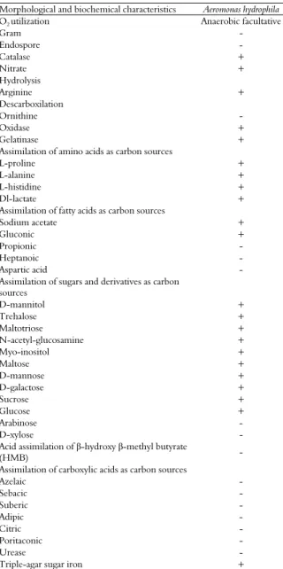
Table 2. Morphological and biochemical characterization of the systemic bacteria Aeromonas hydrophila isolated from banana plants.
Morphological and biochemical characteristics Aeromonas hydrophila
O2 utilization Anaerobic facultative
Gram - Endospore - Catalase + Nitrate + Hydrolysis Arginine + Descarboxilation Ornithine - Oxidase + Gelatinase + Assimilation of amino acids as carbon sources
L-proline + L-alanine + L-histidine + Dl-lactate + Assimilation of fatty acids as carbon sources
Sodium acetate +
Gluconic + Propionic - Heptanoic -
Aspartic acid -
Assimilation of sugars and derivatives as carbon
sources
D-mannitol + Trehalose + Maltotriose + N-acetyl-glucosamine + Myo-inositol + Maltose + D-mannose + D-galactose + Sucrose + Glucose + Arabinose - D-xylose - Acid assimilation of β-hydroxy β-methyl butyrate
(HMB) -
Assimilation of carboxylic acids as carbon sources
Azelaic - Sebacic - Suberic - Adipic - Citric - Poritaconic -
Urease -
Triple-agar sugar iron +
When the antibiotic sensitivity tests were conducted, the genus Aeromonas showed more pronounced sensitivity to the antibiotics cefalotin, chloramphenicol, cefotaxime, nalidixic acid, erythromycin, cefalexin, tetracycline, and cefaclor. The antibiotics most effective in forming the largest halos of inhibition against the genus
Klebsiella were vancomycin, chloramphenicol,
family, including organisms of the genus
Klebsiella, also found that they were especially
sensitive to cefotaxime.
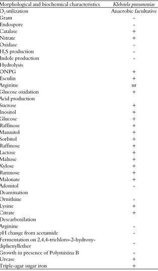
Table 3. Morphological and biochemical characterization of the systemic bacteria Klebsiela pneumoniae isolated from banana plants.
Morphological and biochemical characteristics Klebsiela pneumoniae
O2 utilization Anaerobic facultative
Gram - Endospore - Catalase + Nitrate + Oxidase -
H2S production -
Indole production -
Hydrolysis ONPG + Esculin + Arginine nr
Glucose oxidation +
Acid production
Sucrose + Inositol + Glucose + Raffinose + Mannitol + Sorbitol + Raffinose + Lactose + Maltose + Xylose + Ramnose + Malonate + Adonitol - Deamination Ornithine - Lysine + Citrate + Descarboxilation Arginine -
pH change from acetamide -
Fermentation on
2,4,4-trichloro-2-hydroxy-diphenyllether -
Growth in presence of Polymixina B -
Urease +
Triple-agar sugar iron +
These results enabled the selection of antibiotics for use in a subsequent research to determine the minimum inhibitory concentration (MIC) of the contaminants, a step considered fundamental by Scherwinski-Pereira et al. (2003), who stated that the success of procedures with antibiotics for in vitro culture can only be achieved by isolating and identifying the bacteria and performing tests to determine their sensitivity to antimicrobial substances. According to Scherwinski-Pereira and Costa (2010), due to the high cost of treatment and their phytotoxicity, antibiotics should only be used for culture-specific contaminants because only the bacteria within the spectrum of action of each antibiotic will be controlled.
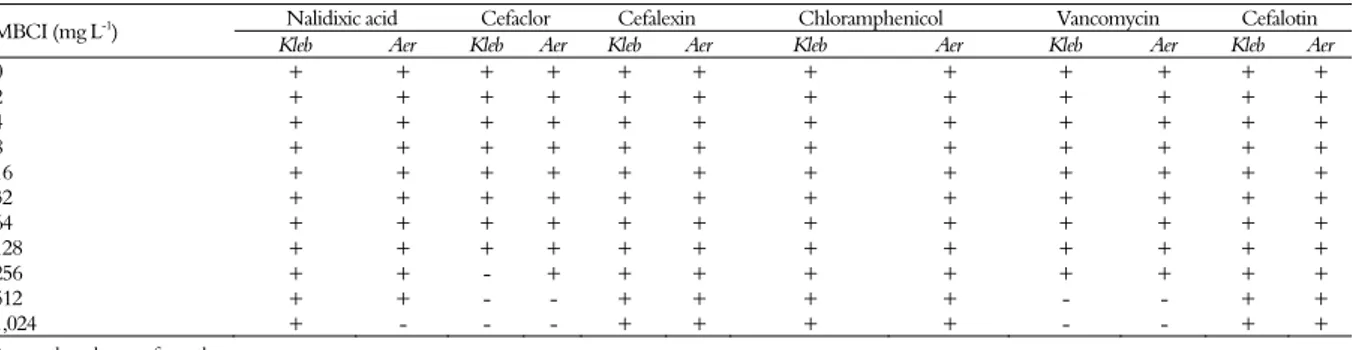
Of the six antibiotics tested, only three inhibited bacterial growth: nalidixic acid, cefaclor, and
vancomycin. However, nalidixic acid was only effective for one of the genera in the study and produced bactericidal effects only in treatments containing the highest concentration tested (1,024 mg L-1). Cefaclor and Vancomycin inhibited
growth of both isolates at half of this concentration (512 mg L-1)(Table 5).
Table 4. Culture susceptibility test of the identified bacterial contaminants to different antibiotics.
Antibiotics Concentration (μg mL-1)
Inhibition halo (mm)
Klebsiella Aeromonas
Cefalotin 30.0 18.0 ± 1.0s 30.0 ± 1.0s
Gentamicin 10.0 11.0 ± 0.0s 15.0 ± 1.0s
Rifampicin 5.0 10.0 ± 1.0s 16.0 ± 2.0s
Vancomycin 30.0 20.0 ± 4.0s 19.0 ± 2.0s
Penicillin 10.0 0.0 ± 0.0r 13.0 ± 2.0s
Chloramphenicol 30.0 21.0 ± 8.0s 23.0 ± 3.0s
Ampicillin 10.0 6.0 ± 4.0r 6.0 ± 1.0r
Cefotaxime 30.0 29.0 ± 3.0s 27.0 ± 8.0s
Streptomycin 10.0 13.0 ± 1.0s 16.0 ± 1.0s
Novobiocin 5.0 12.0 ± 1.0s 13.0 ± 7.0s
Nalidixic acid 30.0 19.0 ± 1.0s 29.0 ± 1.0s
Amoxicillin 10.0 0.0 ± 0.0r 17.0 ± 1.0s
Erythromycin 15.0 2.0 ± 4.0r 20.0 ± 3.0s
Sulfonamide 300.0 0.0 ± 0.0r 2.0 ± 4.0r
Cefalexin 30.0 17.0 ± 1.0s 31.0 ± 4.0s
Cefoxitin 30.0 26.0 ± 3.0s 19.0 ± 10s
Oxacillin 1.0 8.0 ± 1.0s 12.0 ± 6.0s
Amikacin 30.0 13.0 ± 1.0s 17.0 ± 1.0s
Tetracycline 30.0 20.0 ± 1.0s 21.0 ± 2.0s
Cefaclor 30.0 26.0 ± 2.0s 39.0 ± 4.0s
*The sensitivity of contaminants was determined after up to 48 hours incubation, determining
the inhibition halo size (mm). It were considered susceptible to antibiotics the contaminants that showed the formation of a halo of inhibition of 8 mm according to Scherwinski-Pereira et al. (2003); s = susceptible; r = resistant.
In general, in spite of both isolates presenting sensitivity only at the higher concentrations tested (512 and 1,024 mg L-1), the growth of Klebsiella was
also inhibited in a medium containing Cefaclor at a concentration of 256 mg L-1, demonstrating that this
microorganism is more susceptible to this product than the genus Aeromonas.
During phytotoxicity testing, the survival rates of the propagative material cultivated in vitro were 100%, with phytotoxic effects only being reported in the treatment with 1,024 mg L-1 Cefaclor, which led
Acta Scientiarum. Agronomy Maringá, v. 38, n. 2, p. 193-200, Apr.-June, 2016 Table 5. Minimum inhibitory concentration (MIC) of antibiotics for bacterial strains Klebsiella (Kleb) and Aeromonas (Aer) isolated from banana during micropropagation.
MBCI (mg L-1) Nalidixic acid Cefaclor Cefalexin Chloramphenicol Vancomycin Cefalotin
Kleb Aer Kleb Aer Kleb Aer Kleb Aer Kleb Aer Kleb Aer
0 + + + + + + + + + + + +
2 + + + + + + + + + + + +
4 + + + + + + + + + + + +
8 + + + + + + + + + + + +
16 + + + + + + + + + + + +
32 + + + + + + + + + + + +
64 + + + + + + + + + + + +
128 + + + + + + + + + + + +
256 + + - + + + + + + + + +
512 + + - - + + + + - - + +
1,024 + - - - + + + + - - + +
+: growth; -: absence of growth.
Table 6. Multiplication and survival rate of banana shoots in MS (Murashige & Skoog, 1962) medium with the addition of 4 mg L-1 N6
-benzylaminopurine (BAP)(1).
Concentration (mg L-1) Multiplication rate Survival rate (%)
Cefaclor Nalidixic Acid Vancomycin Cefaclor Nalidixic Acid Vancomycin
0,0 2.2 aA 2.2 aA 2.2 aA 100.0 aA 100.0 aA 100.0 aA
512 1.3 bB 2.9 aA 2.5 aA 100.0 aA 100.0 aA 100.0 aA
1,024 0.2 bB 2.1 aA 2.6 aA 35.0 bB 100.0 aA 100.0 aA
CV (%) 24.5 17,8
(1)
Means followed by equal letters, lower case in the columns and upper case in the lines, within each variable, do not differ by Tukey’s test, at 5% probability.
However, the same behavior was not observed in treatments with vancomycin and nalidixic acid. With these antibiotics added to the medium, the multiplication values of cultures reached approximately 2.0 shoots per explant, similar to the results obtained by Costa, Scherwinski-Pereira, Pereira, and Oliveira (2006) and Oliveira et al. (2008) in multiplication experiments of bananas without the addition of any antibacterial agent. Additionally, no symptoms of phytotoxicity were visually observed in the shoots cultivated in medium containing these antibiotics, regardless of the concentrations used.
Although costly, the addition of antibiotics to the growth medium can provide efficient results with respect to the loss of plant material. Lima and Moraes (2006) observed a reduction in bacterial contamination of up to 66.6% when antibiotics were added to MS medium during the cultivation of bananas of the Caipira variety. However, the effectiveness of the use of an antibiotic to control bacteria depends on its form and spectrum of action. Vancomycin, for example, belongs to the group of glycopeptides, which act as inhibitors of bacterial cell wall synthesis, weakening and causing the death of the bacteria (Kohanski, Dwyer, & Collins, 2010). Despite its spectrum of action being limited to Gram positive bacteria and its usage at generally high concentrations in this study, vancomycin was effective in the control of Klebsiella and Aeromonas -
both Gram negative bacteria, which indicates that the spectrum of action of this bactericide can be enhanced when its concentration is increased, as shown by Scherwinski-Pereira et al. (2003), who suggested
increased concentrations of antibiotics in the growth medium when they present low toxic effects on crops.
As with vancomycin, cefaclor also acts as an inhibitor of bacterial cell wall synthesis. This bactericide belongs to the group of cephalosporins and can be effective both for Gram positive and Gram negative bacteria. Despite the positive action in the control of bacteria during in vitro cultivation (El-Shaboury, Saleh, Mohamed, & Rageh, 2007), in this study the bactericidal potential of cefaclor negatively affected the development of banana explants when added to growth medium in high concentrations.
Nalidixic acid belongs to the group of quinolones that hinder bacterial replication by affecting the DNA synthesis of the bacteria. Soon after identification and due to its activity against aerobic Gram-negative bacteria, Lescher, Froelich, Gruett, Bailey, & Brundage (1962) reported that this antibiotic was effective against species of the genus Klebsiella, whereas Jacoby (2005) and Minarini and Darini (2012) reported that several mechanisms make
Klebsiella resistant to quinolones, including nalidixic
acid.
antibiotics are used in MS medium lead us to infer another factor that may influence the rate of phytotoxicity of a product: the genotype. This hypothesis can be confirmed by comparing the work of Lima and Moraes (2006) with that of Carneiro, Silva, Ximenes, Carneiro, and Borges (2000). The former, working with the antibiotic rifampicin, reported no anomalies in banana plants of the Caipira cultivar propagated in MS medium, whereas the latter, using equivalent concentrations of the same antibiotic, detected phytotoxic effects on plants of the Maçã variety, as evidenced by deformation of the aerial part and a reduction in the final size of the shoots.
Conclusion
Bacteria of endophytic origin belonging to the genera Klebsiella and Aeromonas are contaminants of banana plants during micropropagation;
The most effective antibiotics for controlling them are cefaclor, vancomycin, and nalidixic acid, at concentrations between 512 and 1,024 mg L-1;
Among the three antibiotics selected, the rate of multiplication and survival of banana shoots is only affected by cefaclor when added to the multiplication medium at concentrations greater than or equal to 512 mg L-1;
At concentrations between 512 and 1,024 mg L-1,
vancomycin and nalidixic acid do not affect either the multiplication or the survival rate of banana shoots during in vitro multiplication.
Acknowledgements
Authors gratefully acknowledge National Council of Scientific and Technological Development (CNPq) for financial support and scholarships.
References
Braga, M. F., Sá, M. E. L., & Mustafá, P. C. (2001). Avaliação de um protocolo para multiplicação in vitro da bananeira (Musa AAA cv. Caipira). Revista Brasileira de Fruticultura, 23(2), 25-219.
Carneiro, M. F., Silva, G. D., Ximenes, P. A., Carneiro, I. F., & Borges, J. D. (2000). Avaliação de produtos na contaminação de explantes de banana (Musa AAB cv. Maçã). Pesquisa Agropecuária Tropical, 30(1), 29-35. Costa, F. H. S., Scherwinski-Pereira, J. E., Pereira, M. A.
A., & Oliveira, J. P. (2006). Efeito da interação entre carvão ativado e N6-benzilaminopurina na propagação in vitro de bananeira, cv. Grande Naine (AAA). Revista Brasileira de Fruticultura, 28(2), 280-283.
Donnarumma, F., Capuana, M., Vettori, C., Petrini, G., Giannini, R., Indorato, C., & Mastromei, G. (2011). Isolation and characterisation of bacterial colonies
from seeds and in vitro cultures of Fraxinus spp. from Italian sites. Plant Biology, 13(1), 169-76. doi: 10.1111/j.1438-8677.2010.00334.x
El-Shaboury, S. R., Saleh, G. A., Mohamed, F. A., & Rageh, A. H. (2007). Analysis of cephalosporin antibiotics. Journal of Pharmaceutical and Biomedical Analysis, 45(1), 1-19. doi: 10.1016/j.jpba.2007.06.002 Grzebelus, E., & Skop, L. (2014). Effect of β-lactam
antibiotics on plant regeneration in carrot protoplast cultures. In Vitro Cellular and Developmental Biology-Plant, 50(5), 568-575. doi: 10.1007/s11627-014-9626-0 Jacoby, G. A. (2005). Mechanisms of resistance to
quinolones. Clinical Infectious Diseases, 41(2), S120-S126. doi: 10.1086/428052
Janda, J. M., & Abott, S. L. (2010). The genus Aeromonas: Taxonomy, pathogenicity, and infection. Clinical
Microbiology Reviews, 23(1), 35-73. doi:
10.1128/CMR.00039-09
Kohanski, M. A., Dwyer, D. J., & Collins, J. J. (2010). How antibiotics kill bacteria: from targets to networks.
Nature Reviews Microbiology, 8(6), 423-435. doi: 10.1038/nrmicro2333
Krieg, N. R., & Holt, J. (1994). Bergey´s manual of determinative bacteriology. Baltimore, MD: Williams and Wilkins.
Kulkarni, A. A., Kelkar, S. M., Watve, M. G., & Krishnamurthy, K. V. (2007). Characterization and control of endophytic bacterial contaminants in in vitro cultures of Piper spp., Taxus baccata subsp.
wallichiana, and Withania somnifera. Canadian Journal of Microbiology, 53(1), 63-74.
Lescher, G. Y., Froelich, E. J., Gruett, M. D., Bailey, J. H., & Brundage, R. P. (1962). 1,8-Naphthyridine derivatives: a new class of chemotherapy agents.
Journal of Medicinal and Pharmaceutical Chemistry, 5(5), 1063-1068.
Lima, J. D., & Moraes, W. S. (2006). Controle de bactérias contaminantes em explantes de bananeira (Musa AAA cv. Caipira). Pesquisa Agropecuária Tropical, 36(3), 181-186.
Mbah, E. I., & Wakil, S. M. (2012). Elimination of bacteria from in vitro yam tissue cultures using antibiotics.
Journal of Plant Pathology, 94(1), 53-58.
Minarini, L. A. R., & Darini, A. L. C. (2012). Mutations in the quinolone resistance-determining region of gyrA and parC in Enterobacteriaceae isolates from Brazil.
Brazilian Journal of Microbiology, 43(4), 1309-1314. Mittal, P., Gosal, S. S., Senger, A., & Kumar, P. (2009).
Impact of cefotaxime on somatic embryogenesis and shoot regeneration in sugarcane. Physiology and Molecular Biology of Plants, 15(3), 257-265.
Msogoya, T., Kanyagha, H., Mutigitu, J., & Mamiro, M. K. D. (2012). Identification and management of microbial contaminants of banana in vitro cultures.
Journal of Applied Biosciences,55, 3987-994.
Murashige, T., & Skoog, F. (1962). A revised medium for rapid growth and bioassays with tobacco tissue culture.
Acta Scientiarum. Agronomy Maringá, v. 38, n. 2, p. 193-200, Apr.-June, 2016
Nietsche, S., Marques, S. V., Pereira, M. C. T., Salles, B., Xavier, A. A.; França, A. C., & Silva, L. S. (2006). Estabelecimento in vitrode explantes de três cultivares de bananeira. Ciência Rural, 36(3), 989-991.
Nogueira, K. S., Higuti, I. H., Nascimento, A. J., Terasawa, L. B., Oliveira, S., Matos, A. P., ... Costa, L. M. D. (2006). Occurrence of extended-spectrum beta-lactamases in Enterobacteriaceae isolated from hospitalized patients in Curitiba, southern Brazil.
Brazilian Journal of Infectious Diseases, 10(6), 390-395. Oliveira, J. P., Costa, F. H. S., & Scherwinski-Pereira, J. E.
(2008). Micropropagación y estimativa de producción de mudas de bananos para la Amazonia Occidental.
Pesquisa Agropecuária Brasileira, 43(10), 1429-1432. Ryan, R. P., Germaine, K., Franks, A., Ryan, D. J., &
Dowling, D. N. (2008). Bacterial endophytes: Recent developments and applications. FEMS Microbiology Letters,278(1), 1-9.
Scherwinski-Pereira, J. E., & Costa, F. H. S. (2010). Estratégias de seleção e uso de substâncias químicas antimicrobianas para o controle de contaminantes na cultura de tecidos de plantas. In J. E. Scherwinski-Pereira (Ed.), Contaminações microbianas na cultura de células, tecidos e órgãos de plantas (p. 261-292). Brasília, DF: Embrapa Informação Tecnológica.
Scherwinski-Pereira, J. E., Mattos, M. L. T., & Fortes, G. R. L. (2003). Identificação e controle com antibióticos de bactérias endofíticas contaminantes em explantes de
batata micropropagados. Pesquisa Agropecuária Brasileira, 38(7), 827-834.
Sisti, M., Alabano, A., & Brandi, G. (1998). Bactericidal effect of chlorine on motile Aeromonas spp. in drinking supplies and influence of temperature on disinfection efficacy. Letters in Applied Microbiology, 26(5), 347-350. Thomas, P. (2007). Isolation and identification of five
alcohol defying Bacillus spp. covertly associated with in vitro culture of seedless watermelon. Current Science, 92(7), 983-987.
Thomas, P., & Soly, T. A. (2009). Endophytic bacteria associated with growing shoot tips of banana (Musa
sp.) cv. Grand Naine and the affinity of endophytes to the host. Microbial Ecology, 58(4), 952-964.
Thomas, P., Swarna, G. K., Patil, P., Prakash, P., & Rawal, R. D. (2008). Ubiquitous presence of normally non-culturable endophytic bacteria in field shoot-tips of banana and their gradual activation to quiescent cultivable form in tissue cultures. Plant Cell, Tissue and Organ Culture, 93(1), 39-54.
Received on May 6, 2015. Accepted on August 14, 2015.
License information: This is an open-access article distributed under the terms of the Creative Commons Attribution License, which permits unrestricted use, distribution, and reproduction in any medium, provided the original work is properly cited.