http://dx.doi.org/10.1590/s2175-97902017000216070
A
r
*Correspondence: R. B. T. Alves. Departamento de Tecnologia de Alimentos. Universidade Federal de Viçosa. Av. P.H. Rolfs s/n, Campus Universitário, CEP: 36570-000, Viçosa, Minas Gerais, Brasil. E-mail: [email protected]
Physical and chemical quality, biodiversity, and thermodynamic
prediction of adhesion of bacterial isolates from a water
purification system: a case study
Roberta Barbosa Teodoro Alves
1*, Nélio José de Andrade
1, Edimar Aparecida Filomeno Fontes
1,
Patrícia Campos Bernardes
2, Antônio Fernandes de Carvalho
11Food Technology Department, Federal University of Viçosa, Viçosa, MG, Brazil, 2Food Engineering Departmen, Federal University of Espírito Santo, Alegre, ES, Brazil
The objective of this study was to evaluate the quality of water puriication system and identify the bacteria this system, predict bacterial adherence according to the hydrophobicity of these microorganisms and of the polypropylene distribution loop for puriied water.The assessment of drinking water that supplies the puriication system allowed good-quality physical, chemical, and microbiological speciications. The physicochemical speciications of the distributed puriied water were approved, but the heterotrophic bacteria count was higher than allowed (>2 log CFU mL-1).The sanitation of the storage tank with chlorine decreased the number of bacteria adhered to the surface (4.34 cycles log). By sequencing of the 16SrDNA genes, six species of bacteria were identiied. The contact angle was determined and polypropylene surface and all bacteria were considered to be hydrophilic, and adhesion was thermodynamically unfavorable. This case study showed the importance of monitoring the water quality in the puriied water systems and the importance of sanitization with chemical agents. The count of heterotrophic bacteria on the polypropylene surface was consistent with the predicted thermodynamics results because the number of adhered cells reached approximate values of 5 log CFU cm-2.
Uniterms: Puriied water/quality. Puriied water/adhesion. Puriied water/bacterial biodiversity. Prediction thermodynamics. Hydrophobicity.
INTRODUCTION
To become suitable for use in the laboratory or in industry pharmaceutical, water should be treated to
eliminate its contaminants, creating puriied water. The
entire process of production, storage, and distribution should be properly validated and monitored for significant physical-chemical and microbiological characteristics.
Monitoring water purification systems is critical to obtain water with good microbiological and physical-chemical characteristics, and the assessment of water should meet the technical recommendations and the
requirements of legislation. The quality of puriied water
is an important concern for use in analytical laboratories,
in the pharmaceutical and medical ields, and in a wide
variety of industries.
As a fundamental guideline, potable water is the
starting point for any process of puriication (FDA, 1986).
The physical-chemical quality is related to the fundamental assessment of the water for alkalinity, conductivity, residual free chlorine, hardness, ISL (Langelier Saturation Index), pH, and silica content.
Although puriied water contains small amounts of
organic molecules, a group of microorganisms known as oligotrophes has adapted to these conditions (Ridgway,
Rigby, Argo, 1984; McFeters et al., 1993;
Christie-Oleza et al., 2012; Zhang, Huang, Liu, 2013). However, there is little research on the microbial ecology of water
puriication systems. Equipment surfaces of the systems are also susceptible to adhesion and bioilms can therefore
be established as a source of viable microorganisms in
concentrations less than 0.5 mg·L−1, supported bioilm
formation (Florjanič, Kristl, 2011).
In the storage and distribution of water, bacterial contamination is one of the most persistent problems because microorganisms have the ability to adapt to a nutrient-poor environment, such as water purification systems (Clinical and Laboratory Standards Institute, 2006).
Chemical cleaning is important to reduce the contamination of these water purification systems. These chemicals may be used alone or in combination (Sohrabi et al., 2011). Thus, the cleaning procedure for
puriied water equipment must be regulated to avoid the formation of bacterial bioilm on tank walls, equipment,
and connections.
An investigation of the microbial diversity of a water
puriication system is required and such an investigation
can assist in choosing suitable pre-treatment and more
efective cleaning strategies (Bereschenko et al., 2010). Among the methodologies available for microbial
identiication, sequence analysis of ribosomal RNA 16S
rDNA has been widely used to identify bacterial species and perform taxonomic studies (Baker, Smith, Cowan,
2003; Chakravorty et al., 2007; Kim, Morrison, Yu, 2011). In addition to microbial diversity, knowledge related to the bacterial adhesion mechanism and the formation of biofilms on the surfaces is important. The adhesion
of bacteria is a complex process that is afected by many
factors, such as the physicochemical characteristics of bacteria (hydrophobicity, surface charge). The characteristics of the surfaces of pipes can also contribute
to the adhesion of bacteria and inluence bioilm formation,
including the surface properties of the material (surface charge, hydrophobicity, roughness and texture). A better understanding of the relationship between adhesion and biofilm formation is important for the development of
efective control strategies in the early stages of bioilm
development (Simões, Simões, Vieira, 2010).
Diferent approaches have been used to describe and
predict bacterial adhesion to surfaces. The adhesion can be explained by the Derjaguin-Landau-Verwey-Overbeek (DLVO) theory (Van Loosdrecht et al., 1990), based on thermodynamics (Absolom et al., 1983; Busscher et al.,
1984), and by the extended DLVO theory (XDLVO) (Van Oss, 1989; Meinders, Van Der Mei, Busscher, 1995). The
latter integrates thermodynamic aspects with the DLVO theory. However, important biological factors have been largely ignored in these models.
The objective of this study was to evaluate the
quality of drinking water that feeds a puriication system
(in operation for two years) and the water obtained from
this system. This study also sought to identify bacterial strains present in the water and to predict bacterial adherence according to the hydrophobicity of these microorganisms and of the polypropylene distribution
loop for puriied water.
MATERIAL AND METHODS
Water sampling and procedures
Water samples from five points of a purification system located in a laboratory of the Federal University of Viçosa, MG, Brazil were collected in the period from May-August 2013 (Figure 1). Physico-chemical and microbiological quality was evaluated in the following collection points: drinking water that feeds the system
(point 1), puriied water (point 2) and water stored and
distributed in three collection points for use in laboratories (points 3, 4 and 5). For microbiological analyses, before each water sample was collected, each sampling point was
sanitized with 70% ethanol (v/v), and the irst aliquots
of water were dispensed for 1 min. Water samples were collected in sterilized glass containers and analyzed within a maximum of 24 h.
Physico-chemical and microbiological quality of drinking and purified water
The parameters analyzed and the methods and the instruments used are presented, respectively. The drinking
water that feeds the puriication system was analyzed daily
for 40 days for the following parameters: turbidity (NTU,
turbidimeter HACH 2100); temperature (°C) (APHA);
conductivity (μS cm-1) (APHA, Hanna); pH (APHA,
pHmeter MA PA210p model); hardness (mg L-1 as CaCO 3)
(titration, APHA); calcium (mg l-1 as Ca2+) (titration,
APHA); alkalinity (mg L-1 as CaCO
3) (titration, APHA),
free residual chlorine (mg l-1 as CRL) (colorimetric,
APHA, Lamotte I200 model spectrophotometer); total
dissolved solids (mg L-1) (APHA, plate heater, brand
DigTech); silica (mg L-1 as SiO
2) (APHA, Biospectro
spectrophotometer, SP-22), Langelier saturation index
(NING; NETWIG, 2002) and counts of heterotrophic
bacteria (pour plate, R2A agar Himedia®, incubation at
35 °C 72 h). The methods are described in the Standard
Methods for the Examination of Water and Wastewater (American Public Health Association, 2005).
The water obtained from the purifying system was
also monitored daily for 40 days for temperature (°C), conductivity (μS cm-1), pH (pH meter model PA210p MA),
silica (mg l-1 as SiO
SP-22) and heterotrophic bacteria count (filtration and membrane technique, R2A agar (Himedia®) after
incubation at 35 °C for 72 h). The methodologies were proposed by British Pharmacopoeia (2009).
Evaluation of the cleaning procedure of the water purification system
The drinking water tank that feeds the puriication system was sanitized (Public Health Protection, 2009).
After this procedure, the drinking water was monitored for heterotrophic bacteria count for a minimum period of 20 days of sampling.
The storage e tank and water distribution loop were sanitized with a sodium hypochlorite solution at a concentration of 500 mg L-1 total residual chlorine as
Cl2, pH 8 and a contact time of 120 min. To evaluate the
eiciency of the sanitizing procedure, the swab technique
was applied to the surface of the storage tank and the pipes of the distribution system. After sanitization, distributed
puriied water was monitored by heterotrophic bacterial
count daily for 20 days.
Isolation of bacteria from the water purification system and DNA extraction from the isolates
Volumes of 1 mL of water samples collected at the
points of the puriication system were inoculated on the
surfaces of petri dishes containing R2A agar and incubated
at 35 °C (American Society for Testing and Material,
1991; British Pharmacopoeia, 2009). Colonies grown in this medium with diferent types of morphology and
colors were isolated and subcultured in BHI (Brain Heart Infusion) (Fluka Analytical®). The pure cultures of the
various bacteria were kept at -18 °C in microcentrifuge
tubes containing brain heart infusion (BHI) and glycerol
(80:20).
Extraction of the isolated DNA was performed with the Wizard® Genomic DNA Puriication kit (Promega,
USA) following the extraction protocol. The quality of extracted DNA was analyzed and quantitated by reading on a NanoDrop-100 spectrophotometer (Thermo
Scientiic).
16S rDNA amplification
To determine the bacterial genera and species
present in the puriied water, the ampliication of bacterial
16S rRNA genes was performed with primers 27F initiator 5-AGA GTT TGG TGA TCM CTC AG-3 and
reverse 1378R 5-CGG TGT GTA CAA GGC GCC GGA
ACG 3 (Sigma®, USA), which gave rise to fragments of
approximately 1500 bp. The reactions were performed
in volumes of 25 µL containing 2.5 µL of 10X reagent Bufer (Sigma®, USA), 200 M dNTPs (Promega®, United
States), 1 U Taq Polymerase (Sigma®, USA) 0.1 µM of
each primer and approximately 80 ng of total DNA.
The PCR reaction was performed in a thermocycler (Maxygene Axygen, USA). Amplification conditions
followed the following protocol: 3 min denaturation at 94
°C; 35 cycles of ampliication at 94 °C for 1 min; annealing at 55 °C for 1 min and extension at 72 °C for 2 min and 72 °C for 10 min. Product formation was confirmed by electrophoresis of 3 μl on a 1 % (w/v) agarose gel
(Promega®, United States) stained with ethidium bromide.
Sequencing of the amplified product in the PCR
The PCR products were bidirectionally sequenced
with the primers and universal primers 27 F and 1378 R from Macrogen Inc. (Seoul, South Korea) with the Model 3730XL sequencer (Applied Biosystems, USA).
The bidirectional sequences obtained were compared to their complementary strands, generating a consensus tape generated by DNA Baser program software 3.5.4 (http://www.dnabaser.com/index.html). The sequences obtained from each individual were subjected to comparative analysis with the database from the National Center for Biotechnology Information (NCBI) (http:// www.ncbi.nlm.nih.gov/) using the BLAST algorithm (Basic Local Alignment Search Tool).
Translation of the DNA sequence for amino acids
from diferent isolates was performed using the BLASTN
algorithm, which expresses a sequence of nucleotides to amino acids and is compared with the sequences deposited
in the NCBI database. An agreement of at least 95% was assumed to conirm the speciicity of the sequence.
Measurement of the contact angle
Surface
The surface hydrophobicity was evaluated after contact angle measurements using the approach described by Van Oss et al. (1989). The surfaces of the distribution loop material (polypropylene plates, Belfano brand, model tubelli 32) were subjected to analysis to determine the
contact angles with three liquids of diferent polarities:
water (Milli-Q), formamide (Bio LGC, Sao Paulo, Brazil)
and α-bromonaphthalene (Merck, Brazil). The contact
angles were determined by the sessile drop method using
a Krüss® brand goniometer, Easy Drop model (Hamburg,
Germany).
Microorganisms
The measurement of the contact angles for diferent
species of microorganism isolates was performed on a layer of vegetative cells using methodology described by Busscher et al. (1984). First, bacteria were grown twice in 30 mL of BHI broth (Fluka analytical®) for 20 to 24 h to
obtain a slurry of the active culture with approximately 1.0× 07 CFU mL-1. After the active cultures were centrifuged at
4100 x g for 10 min at 4 °C (Eppendorf Centrifuge 5804R),
the pellets were washed three times with phosphate- bufered
saline (PBS) 0.1 mol l-1. The cell mass was resuspended in
0.1 mol L-1PBS and deposited on a cellulose ester membrane
ilter (0.45 μm pore size and 47 mm diameter, Millipore) in a iltration system using negative pressure. After iltration
and avoiding dehydration of the cells, the membranes were placed in petri dishes containing previously prepared 1% agar (w/v) (Difco) and 10% glycerol (v/v) Merck®).
The membranes with the cell layer were carefully cut to determine the contact angles with water, formamide and
α-bromonaphthalene.
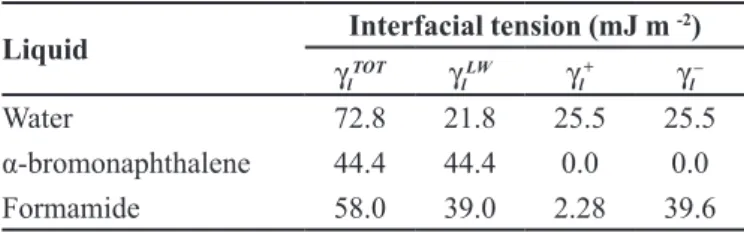
Determination of the total interfacial tension
The total interfacial tension was determined by the sum of the apolar and polar components of the respective surfaces (Eq. 1).
(1)
where γl is the total interfacial tension of the liquid, γlLW is
the interfacial tension of the interactions of the Lifshitz-van der Waals, γl+ is the interfacial tension of the electron
acceptor component of the acid-base component, γl– is the
interfacial tension of the electron donor component of the
acid-base component, θ is the contact angle, and s and l
indicate surface and liquid, respectively.
The three components of the interfacial tension that are necessary to obtain the contact angle formed by
three liquids of diferent polarities were determined. The
amounts of polar liquid and nonpolar components used
are shown in Table I (Van Der Mei, Bos, Busscher, 1998).
The interfacial tension (γsTOT) is the result of the sum
of the two components γsLW and γsAB.
(2)
(3)
(4)
TABLE I - Values for interfacial tension of the components of
the liquid at 25 °C
Liquid Interfacial tension (mJ m
-2)
γl TOT
γl LW
γl
+ γ
l
–
Water 72.8 21.8 25.5 25.5
α-bromonaphthalene 44.4 44.4 0.0 0.0
where γsLW is the interfacial tension of the interactions
of the Lifshitz-van der Waals forces, θB is the contact
angle obtained withα-bromonaphthalene, γsAB is the polar
component of the Lewis acid-base interaction, γs+is the
interfacial tension of the electron acceptor component of the acid-base component, γs– is the interfacial tension of
the electron donor component of the acid-base component, and γsTOT is the total interfacial tension of the surface.
Free energy of the hydrophobic interaction ∆GSASTOT
The total free energy of the interaction among molecules of the surface (s) immersed in water (w) was determined by the sum of the apolar and polar free energies of interaction, ∆GSASLW e ∆GSASAB , respectively (Van Oss, 1995).
(5)
(6)
(7)
Determination of the total free energy of adhesion (∆G adhesion)
Using the values of the components of the interfacial
tension, it is possible to determine the ΔG between two
surfaces (microbial cells (b) and food surfaces (s)):
(8)
(9)
(10)
When free energy is related to the interfacial tension,
ΔGadhesion is then represented by the following:
(11)
(12)
(13)
where γbs is the interfacial tension between the bacterial
surfaces and the adhesion surface, γbl is the interfacial
tension between the bacterial surfaces and the liquid, and
γsl is the interfacial tension between the adhesion surfaces
and the liquid.
T h e f r e e e n e r g y o f a d h e s i o n b e t w e e n t h e
polypropylene surface and isolated and identiied bacteria
from the water were determined to evaluate the inluence
of these factors on the thermodynamics of adhesion. The
value of ΔG of adhesion allows for the assessment of the thermodynamic process: if ΔG adhesion is <0, the process is favorable; if ΔG adhesion is > 0, the process is
unfavorable.
Statistical analysis
The data of physical-chemical analysis and counts of heterotrophic bacteria in purified water at the three distribution points were subjected to analysis of variance, and means were compared by Tukey’s test at 5 % probability using the Statistical Analysis System, version
9.1 (2006), licensed to the Federal University of Viçosa.
RESULTS AND DISCUSSION
Physical-chemical and microbiological quality of drinking water purified by the purification system
The assessment of drinking water that supplies the
puriication system established good physical-chemical
and microbiological quality (Table II). In the period analyzed, the count of heterotrophic bacteria remained below 500 CFU ml-1, a limit set by the Portaria MS n°
2.914 (Brasil, 2011). The average of heterotrophic bacteria
count was 1.24 log CFU ml−1. Analyzing these results, it was noted that the water supply was one of the main factors responsible for good system performance.
ND- Not detected
As a fundamental guideline, potable water is the
starting point for any process of puriication. In addition
to the pre-treatments that are used, routine monitoring of its quality must be implemented to avoid operational problems. In purification system water using reverse osmosis membranes, high levels of hardness and alkalinity provoke fouling of the membranes, reducing their
eiciency and producing a higher frequency of chemical
cleaning of the membranes in the devices.
The determination of pH is important, as it can affect the removal of salts and prevent or promote the deterioration of membranes. The pH of the feed water can
afect the rejection of salt by the membrane because there
is an optimum pH at which maximum rejection occurs. A higher rejection rate of certain ionic constituents, such as
luorides and bicarbonates, is eliminated by the increase
of pH (Castanheira, 2010).
osmosis membranes to assist chemicals in the pre-treatment of the feed water (Ning, Netwig, 2002). In the
water puriication system under study, the manufacturer
recommends that the maximum value ISL should be +0.3. The purified water produced by the system met physical-chemical quality and microbiological quality standards in compliance with the current U.S. and EU Pharmacopoeias. The purified water was analyzed immediately after production by the system without
storage in the tank, presenting 1.78 logCFU 100 mL-1
on average. The physico-chemical analyses of puriied
water samples from the distribution points also met the
requirements and did not show diferences (p≥0.05).
The physico-chemical and microbiological analyses of purified water are of great importance for a quality
product because puriied water is one of the raw materials most often used in the pharmaceutical ield. The quality
of the purified water depends on a number of factors,
such as the type of puriication system and the storage and distribution procedures of the puriied water that is
produced. Moreover, it is necessary to constantly assess
the puriication procedures, including monitoring system,
maintenance and cleaning.
The microbiological analysis of purified water distributed in the three points of use showed approximately the same level of bacterial contamination and did not
present diferences (p≥0.05). The average of the bacterial numbers at points I, II and III were 2.18, 2.19 and 2.20
log CFU mL-1,respectively. In the period analyzed, the
distributed puriied water was kept in recirculation for 12 h, with a mean low velocity in the distribution loop of 0.87 m.s-1.
The results show that during the 40 days of analysis,
for 23 days, the number of bacteria was above the allowed amount (2 log CFU mL-1 or 100 CFU mL-1)
(recommendation of British Pharmacopoeia and USP Pharmacopoeia). The purified water produced by the system presented an acceptable microbiological level, but with storage, there was an increase in the bacterial population of approximately 2 log cycles, which may be
related to the presence of microbial growth and bioilms. However, bioilms in systems for the storage and
distribution of purified water are difficult to detect,
inactivate, and remove. Florjanič and Kristl (2011) showed that the number of bioilm microorganisms inluences the number of planktonic cells in the puriied water. These
authors found a high positive correlation (r between
0.99 and 0.84) between the number of planktonic cells in puriied water (CFU mL-1) and the number of heterotrophic
bacteria in the bioilm (CFU cm-2). These results suggest
that biofilm bacteria are continuously detached to the middle of this liquid
Moreover, Boe-Hansen et al. (2002) reported that the number of planktonic bacteria is due not only to the
detached bacteria of bioilm but also to the multiplication
of bacteria in the liquid medium, which should also be considered
The increase of 2 log cycles distributed in puriied
water mentioned above may be due to the multiplication of bacteria, even if the distribution system promotes water circulation with turbulence and very small concentrations of carbon and ions.
These microorganisms have adapted to strict nutrient conditions and can survive and multiply in these adverse environments. They can also induce adaptive responses
to environmental stresses by expressing speciic genes,
TABLE II - Quality of drinking water that feeds the puriication system
Parameters Mean Standard
deviation
Maximum value
Minimum value
Alkalinity (mg l-1 as CaCO
3) 20.3 ± 3.52 26.4 13.7
Heterotrophic bacteria (log CFU mL−1) 1.24 ± 0.82 2.55 ND Residual Free Chlorine (mg L-1 as Cl
2) 0.84 ± 0.33 1.83 0.62
Conductivity (μS cm-1) 62.77 ± 7.49 76.18 52.24
Hardness (mg L-1 as CaCO
3) 20.00 ± 8.97 51.1 10.1
ISL (Langelier Saturation Index) - 3.76 ± 0.16 -4.07 -3.60
pH 6.17 ± 0.34 7.3 5.7
Silica (mg L-1 as SiO
2) 15.23 ± 2.34 23 11
Total dissolved solids (mg L-1) 49.8 ± 30.0 105.5 17.5
Temperature (°C) 25 ± 2.05 21.3 27.2
resulting in changes in physiology, including metabolic
and structural changes (Yousef, Juneja, 2003).
The storage and distribution system should be
conigured to avoid recontamination of the water after
treatment and should be subject to a combination of
monitoring techniques (WHO, 2005; Clinical and
Laboratory Standards Institute, 2006).
Cleaning of the water purification system
The counts of heterotrophic bacteria in drinking water over 20 consecutive days of sampling showed
a mean of 1.89 log CFU mL-1; and after cleaning, the
drinking water tank monitored for 20 consecutive days
had a mean of 0.58 log CFU mL-1, leading to a reduction
in the average count of 1.32 log CFU mL-1.
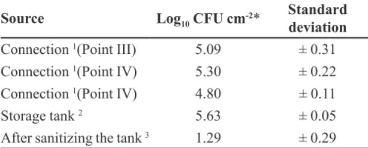
Prior to the sanitization of the storage tank, the swab test was performed to check the level of bacterial contamination on the surfaces in contact with purified water (Table III). To avoid possible contamination and interruption of the supply of purified water after sanitization, the swab was performed only in the area of the tank.
There were differences (p<0.05) in the count of
heterotrophic bacteria after the sanitization procedure. H o w e v e r , a f t e r t h e s a n i t i z a t i o n p r o c e d u r e , t h e
microbiological quality of the puriied water was assessed,
and the results remained within acceptable levels for only 4 days over the 20 days of analysis. There was a gradual increase in the counts of heterotrophic bacteria up to a maximum and after reaching a plateau.
The bacteria present in the purified water may
undergo proliferation and bioilm formation on the surface
of the storage tank. This bioilm is diicult to remove,
even with mechanical and chemical sanitizing rinse, becoming a source of contamination of the stored water.
The bioilm bacteria are more resistant to antimicrobials
than planktonic bacteria.
To maintain the purification of the water in the storage tanks, especially in relation to the microbiological parameters, we must consider that the material used in the distribution loop should prevent the adhesion and
minimize the formation of bioilms.
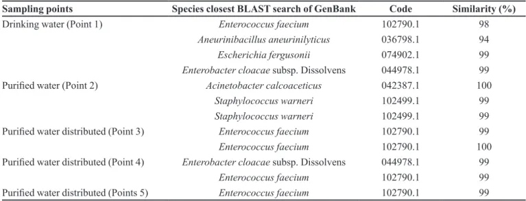
Identification of bacterial biodiversity isolated from the water purification system
Table IV lists the isolated bacterial species of the water collected at various sampling points. Several isolates of Enterococcus faecium, Staphylococcus warneri, Escherichia fergusonii, Enterobacter cloacae, and Acinetobacter ssp. were previously detected in drinking water (Herson et al., 1987; Rice et al., 1991; Adcock,
Saint, 2001; Olofsson, Hermansson, Elwing, 2003;
Bernasconi et al., 2007; Ogier, Serror, 2008). Additionally, the species Staphylococcus warneri has been isolated
from active carbon ilters in water puriication systems
in clinical laboratories (Silva et al., 2006) and bioilms of Acinetobacter spp. were isolated from activated carbon
and sand ilters and water distribution points (Bifulco,
Shirey, Bissonnette et al., 1989; Percival et al., 2004). E. faecium was found in drinking water and distributed purified water, suggesting that this species
passes through all stages in the puriication system.
Studies of the species E. fergusonii showed its ability
to form bioilms, with a larger number of adhered cells at 24 °C compared to 37 °C in minimal glucose medium
relative to the other species of E. coli (Ingle et al., 2011). The levels of nutrients present in drinking water are usually low, with organic carbon concentrations ranging from 0.05 mg L-1 and 12.2 mg L-1 and assimilable organic
carbon fractions of 3 µg L-1 and 500 µg L-1. However,
the concentrations of these organic materials in drinking water support the growth of various heterotrophic bacteria (Camper et al., 1991). Enterobacter cloacae is able to multiply at various concentrations of nutrients and survive chlorination (Herson et al., 1987).
Strains of the species Enterobacter cloacae and A. calcoaceticus produce large amounts of
exopolysaccharides (Bryan, Linhardt, Daniels, 1986;
Rosenberg et al., 1988; Sarafzadeh et al., 2013; Wang,
Yang, Wang, 2013).
According to Rosenberg et al. (1988), strains of A. calcoaceticus adhere to hydrophobic surfaces, such as
TABLE III - Enumeration of heterotrophic bacteria (log CFU
cm-2) in the connections (Point III, IV and V) and storage tank of puriied water
Source Log10 CFU cm
-2* Standard
deviation
Connection 1(Point III) 5.09 ± 0.31
Connection 1(Point IV) 5.30 ± 0.22
Connection 1(Point IV) 4.80 ± 0.11
Storage tank 2 5.63 ± 0.05
After sanitizing the tank 3 1.29 ± 0.29 1Connection specification in polypropylene. 2Storage tank
material (polyethylene). 3Storage tank sanitization with sodium
hypochlorite solution of 500 mg L-1 expressed CRL, pH = 8 *
hydrocarbons, polystyrene and human epithelial cells. The death rates for disinfection of Acinetobacter spp. are similar to those of other heterotrophic bacteria when exposed to chlorine compounds. However, some studies have indicated that Acinetobacter can develop greater resistance to chlorine, chloramines and chlorine dioxide when grown under conditions that favor the development
of bioilms (Percival et al., 2004).
The above statement was confirmed in the study by Simões, Simões and Vieira (2010) They studied the
impact of microbial diversity in bioilms with six species
of bacteria isolated from drinking water as it relates to their resistance to disinfection with sodium hypochlorite. Among these species, the A. calcoaceticus bioilm was
susceptible to disinfection and achieved total inactivation; however, its presence in multi-species bioilms increased
its resistance to disinfection.
A. calcoaceticus species also showed the ability to aggregate strongly compared to other bacteria studied, and their presence in a multi-species community represents an advantage in colonization. This adhesion
mechanism is highly speciic and transmits advantages to
microorganisms, including transfer of chemical signals, exchange of genetic information, protection against adverse environmental conditions, metabolic cooperation
between diferent species and cell diferentiation in some population (Simões, Simões, Vieira, 2008).
Prediction of the adhesion of bacterial isolates to polypropylene
Cells of different species in drinking water and
puriied water showed a value of ΔGTOT>0, considered to
be hydrophilic according to Van Oss (1995). Additionally,
the polypropylene surface is considered to be hydrophilic
due to the value of ΔGTOT>0 (Table V).
The strain of A. calcoaceticus was also considered to be hydrophilic according to research performed by Van der
Mei, Bos and Busscher (1998). However, the numerical
values of hydrophobic interactions of free energy should not be compared to bacteria within the same species
because there are diferences in measurements between
contact angles in water (Simões et al., 2007).
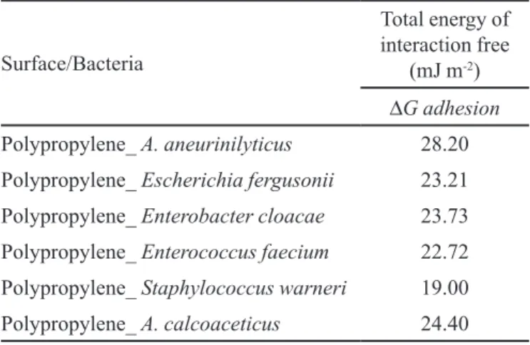
To predict the ability of microorganisms to adhere to the surface, the total free energy between the adhesion of microorganisms and the material of the distribution loop when immersed in water was
calculated (Table VI). The values of ΔG adhesion allow
TABLE IV - Identiication of bacterial species based on the analysis of 16S RNA gene
Sampling points Species closest BLAST search of GenBank Code Similarity (%)
Drinking water (Point 1) Enterococcus faecium 102790.1 98
Aneurinibacillus aneurinilyticus 036798.1 94
Escherichia fergusonii 074902.1 99
Enterobacter cloacae subsp. Dissolvens 044978.1 99
Puriied water (Point 2) Acinetobacter calcoaceticus 042387.1 100
Staphylococcus warneri 102499.1 99
Staphylococcus warneri 102499.1 99
Puriied water distributed (Point 3) Enterococcus faecium 102790.1 99
Enterococcus faecium 102790.1 100
Puriied water distributed (Point 4) Enterobacter cloacae subsp. Dissolvens 044978.1 99
Enterococcus faecium 102790.1 99
Puriied water distributed (Points 5) Enterococcus faecium 102790.1 99
TABLE V - Values of the apolar(∆GSAS
LW) and polar (
∆GSAS AB ) components and of the total free energy of interaction (∆GSAS
TOT ) the isolated bacteria and the surface
Surfaces
Free energy of interaction
(mJ m-2)
∆GSAS LW ∆G
SAS AB ∆G
SAS TOT
Polypropylene -3.61 16.76 13.11
A. aneurinilyticus -0.32 36.87 36.51
Escherichia fergusonii 0.00 21.22 21.25
Enterobacter cloacae -1.19 29.77 28.57
Enterococcus faecium -1.27 28.45 27.18
Staphylococcus warneri -2.98 7.03 4.05
us to evaluate the thermodynamics of adhesion, which
are thermodynamically favorable when ΔG adhesion <
0, suggesting that adhesion occurs spontaneously, with
unfavorable adhesion when ΔG adhesion > 0.
It was observed that the adhesion between the polypropylene and the isolated bacteria was thermodynamically unfavorable. According to Van Oss
(1995), it is well known that an aqueous environment
favors adhesion to hydrophobic surfaces due to the expulsion of water. However, adhesion between hydrophilic surfaces can occur.
The prediction of adhesion potential based on physicochemical properties provides useful information about the possible microbial behavior under actual conditions (Simões et al., 2007). In this work, only one thermodynamic factor was studied, which indicated that
the adhesion is thermodynamically unfavorable; the
adhesion and the formation of biofilms of the species identified on the surface cannot be predicted because the thermodynamic theory does not consider the microbiological aspects of the adhesion.
However, the results of prediction thermodynamics were consistent with the counts of heterotrophic bacteria in polypropylene surfaces (distribution loop), which reached values of approximately 5 log CFU·cm-2. If
the thermodynamics were favorable to the adhesion of bacteria to the surface, this count would be much higher and could reach between 6 log CFU cm-2 to 7 log CFUcm -2. Although these results are consistent, it is important to
note that these values for a surface that comes into contact
with the puriied water can inluence the microbiological
quality.
Several factors can influence the adhesion of
bacteria to surfaces, such as the hydrophobicity of the cells and cellular appendages that contribute to increased adherence (Pang et al., 2005; Herzberg, Elimelech, 2007). Many of the identified bacterial species have cellular
appendages, which can mediate the adhesion process; for
example, the genus Acinetobacter presents polysaccharide
capsules and fimbriae (Towner, 2002; Percival et al., 2004). Extracellular structures, such as pili, fimbriae,
lagella and exopolysaccharides, can liaise between the
cell and the adhesion substrate, neutralizing electrostatic repulsion (Simões, Simões, Vieira, 2010).
Predictions based solely on the physical-chemical properties of the surface and on thermodynamic approaches only provide information on the understanding of the adhesion process and the formation of biofilms. Furthermore, multi-species interactions prevail in the environment, and the strongly adherent bacteria may play a key role in the primary colonization of surfaces with other microorganisms.
CONCLUSION
The water puriication system monitored in this case
study, which has been in operation for two years, produces water that meets the physico-chemical and microbiological quality requirements within the criteria established by pharmacopeia. However, with storage, the distributed
puriied water exhibits microbiological levels above the
permitted standards because they exceeded the maximum of 2 log CFU·mL-1, which may compromise the analytical
results of some research laboratories.
The surfaces of the bacteria isolated in the samples
from the puriication system and the surface of the water
distribution loop are hydrophilic and thermodynamically unfavorable for adhesion.
The results of the thermodynamic predictions were consistent with the counts of heterotrophic bacteria of the polypropylene surface (distribution loop), which reached approximate values of 5 log10 CFU·cm-2.
AKNOWLEDGEMENTS
The authors would like to thanks the inancial support
provided by the Conselho Nacional de Desenvolvimento Cientíico e Tecnológico (CNPq/Brazil) and Fundação de Amparo à Pesquisa do Estado de Minas Gerais (FAPEMIG/MG).
TABLE VI - Values of free energy of adhesion (∆G adhesion)
between the isolated bacterial cells (b) and the surface of the loop distribution (polypropylene) (s) in an aqueous medium (l)
Surface/Bacteria
Total energy of interaction free
(mJ m-2)
∆G adhesion
Polypropylene_ A. aneurinilyticus 28.20
Polypropylene_ Escherichia fergusonii 23.21
Polypropylene_ Enterobacter cloacae 23.73
Polypropylene_ Enterococcus faecium 22.72
Polypropylene_ Staphylococcus warneri 19.00
REFERENCES
ABSOLOM, D.R.; LAMBERTI, F.V; POLICOVA, Z.; ZINGG, W.; VAN OSS, C.J.; NEUMANN, A.W. Surface
thermodynamics of bacterial adhesion. Appl. Environ. Microbiol., v.46, n.1, p.90-97, 1983.
ADCOCK, P.W.; SAINT, C.P. Development of glucosidase agar for the conirmation of water-borne Enterococcus. Water Res., v.35, n.17, p.4243-4246, 2001.
AMERICAN PUBLIC HEALTH ASSOCIATION. Standard methods for the examination of water and wastewater. 21.ed. Washington, DC: American Public Health Association,
2005. 1368 p.
AMERICAN SOCIETY FOR TESTING AND MATERIAL.
Standard Specification for Reagent Water, Document
D1193-91. West Conshohocken, PA: American Society for Testing and Materials (ASTM), 1991. 6 p.
BAKER, G.C.; SMITH, J.J.; COWAN, D.A. Review and
re-analysis of domain-specific 16S primers. J. Microbiol. Meth., v.55, n.3, p.541-555, 2003.
BERESCHENKO, L.A.; STAMS, A.J.M.; EUVERINK, G.J.W.;
VAN LOOSDRECHT, M.C.M. Biofilm formation on reverse osmosis membranes is initiated and dominated by Sphingomonas spp. Appl. Environ. Microbiol., v.76, n.8, p.2623-2632, 2010.
BERNASCONI, C.; VOLPONI, G.; PICOZZI, C.; FOSCHINO,
R. Use of the tna operon as a new molecular target for Escherichia coli detection. Appl. Environ. Microbiol., v.73,
n.19, p.6321-6325, 2007.
BIFULCO, J.M.; SHIREY, J.J.; BISSONNETTE, G.K.
Detection of Acinetobacter spp. in rural drinking water supplies. Appl. Environ. Microbiol., v.55, n.9, p.2214-2219,
1989.
BOE-HANSEN, R.; ALBRECHTSEN, H.-J.; ARVIN, E.; JØRGENSEN, C. Bulk water phase and bioilm growth in
drinking water at low nutrient conditions. Water Res., v.36,
n.18, p.4477-4486, 2002.
BRASIL. Ministério da Saúde. Portaria no 2.914 de 12 de
dezembro de 2011. Dispõe sobre os procedimentos de controle e de vigilância da qualidade da água para consumo e seu padrão de potabilidade. Diário Oficial da União, Brasília, Seção 1, 14 dez. 2011.
BRITISH PHARMACOPOEIA. Purified Water. In: British
Pharmacopoeia 2009. [s.l: s.n.], 2009. p.418-420.
BRYAN, B.A.; LINHARDT, R.J.; DANIELS, L. Variation in
composition and yield of exopolysaccharides produced by
Klebsiella sp. strain K32 and Acinetobacter calcoaceticus BD4. Appl. Environ. Microbiol., v.51, n.6, p.1304-1308,
1986.
BUSSCHER, H.; WEERKAMP, A.; VANDERMEI, H.; VANPELT, A.; DEJONG, H.; ARENDS, J. Measurement
of the surface free-energy of bacterial-cell surfaces and its relevance for adhesion. Appl. Environ. Microbiol., v.48,
n.5, p.980-983, 1984.
CAMPER, A.K; MCFETERS, G.A; CHRACKLINS, W.G.; JONES, W.L. Growth kinetics of coliform bacteria under
conditions relevant drinking water distribution systems to. Appl. Environ.l Microbiol., v.57, n.8, p.2233-2239, 1991.
C A S TA N H E I R A , A . A . Aplicación de membrana de
nanoiltración para eliminar disruptores endocrinos en la potabilización del agua. Catalunya: Universitat Politecnica de Catalunya, 2010.
CHAKRAVORTY, S.; HELB, D.; BURDAY, M.; CONNELL, N.; ALLAND, D.A detailed analysis of 16S ribosomal RNA
gene segments for the diagnosis of pathogenic bacteria. J. Microbiol. Meth., v.69, n.2, p.330-9, 2007.
CHRISTIE-OLEZA, J.A.; PINA-VILLALONGA, J.M.; BOSCH, R.; NOGALES, B.; ARMENGAUD, J.
Comparative proteogenomics of twelve roseobacter
exoproteomes reveals diferent adaptive strategies among
these marine bacteria. Mol. Cell. Proteomics, v.11, n.2, p.M111.013110-M111.013110, 2012.
CLINICAL AND LABORATORY STANDARDS INSTITUTE.
Preparation and testing of reagent water in the clinical laboratory. Wayne, PA: Clinical and Laboratory Standards Institute (CLSI). Document C3-A4, 2006.
FOOD AND DRUG ADMINISTRATION. FDA. Inspection
Guide - Water for Pharmacuetical Use. Silver Spring, MD:
FDA, 1986.
FLORJANIČ, M.; KRISTL, J. The control of bioilm formation by hydrodynamics of puriied water in industrial distribution
HERSON, D.S.; MCGONIGLE, B.; PAYER, M.A.; BAKER, K.H. Attachment as a factor in the protection of Enterobacter cloacae from chlorination. Appl. Environ. Microbiol., v.53,
n.5, p.1178-1180, 1987.
HERZBERG, M.; ELIMELECH, M. Biofouling of reverse osmosis membranes: Role of bioilm-enhanced osmotic
pressure. J. Memb. Sci., v.295, n.1-2, p.11-20, 2007.
INGLE, D.J.; CLERMONT, O.; SKURNIK, D.; DENAMUR, E.; WALK, S.T.; GORDON, D.M. Bioilm formation by and
thermal niche and virulence characteristics of Escherichia spp. Appl. Environ. Microbiol., v.77, n.8, p.2695-2700, 2011.
KIM, M.; MORRISON, M.; YU, Z. Evaluation of different
partial 16S rRNA gene sequence regions for phylogenetic analysis of microbiomes. J. Microbiol. Meth., v.84, n.1,
p.81-87, 2011.
MCFETERS, G.A.; BROADAWAY, S.C.; PYLE, B.H.; EGOZY, Y. Distribution of bacteria within operating laboratory water puriication systems. Appl. Environ. Microbiol., v.59, n.5,
p.1410-1415, 1993.
MEINDERS, J.M.; VAN DER MEI, H.C.; BUSSCHER, H.J. Deposition Eiciency and Reversibility of Bacterial
Adhesion under Flow. J. Colloid Interface Sci., v.176, n.2,
p.329-341, 1995.
NING, R.Y.; NETWIG, J.P. Complete elimination of acid
injection in reverse osmosis plants. Desalination, v.143,
n.1, p.29-34, 2002.
OGIER, J.-C.; SERROR, P. Safety assessment of dairy
microorganisms: the Enterococcus genus. Int. J. Food Microbiol., v.126, n.3, p.291-301, 2008.
OLOFSSON, A.; HERMANSSON, M.; ELWING, H. N-Acetyl-L-Cysteine afects growth , extracellular polysaccharide
production , and bacterial biofilm formation on solid surfaces. Appl. Environ. Microbiol., v.69, n.8, p.4814-4822, 2003.
PANG, C.M.; HONG, P.; GUO, H.; LIU, W.T. Bioilm formation
characteristics of bacterial isolates retrieved from a reverse osmosis membrane. Environ. Sci. Technol., v.39, n.19, p.7541-7550, 2005.
PENNA, V.T.C.; MARTINS, S.A.M.; MAZZOLA, P.G.
Identification of bacteria in drinking and purified water
during the monitoring of a typical water puriication system. BMC Public Health, v.2, p.13, 2002.
PERCIVAL, S.; CHALMERS, R.; EMBREY, M.; HUNTER, P.; SELLWOOD, J.; WYN-JONES, P. Microbiology of waterborne diseases. [s.l.]: Elsevier, 2004. 480p.
PUBLIC HEALTH PROTECTION. Public health engeneering guideline: disinfection of water storage facilites. [s.l.]:
PHP, 2009.
RICE, E.W.; ALLEN, M.J.; BRENNER, D.J.; EDBERG, S.C. Assay for β-glucuronidase in species of the genus
Escherichia and its applications for drinking-water analysis. Appl. Environ. Microbiol., v.57, n.2, p.592-593, 1991.
RIDGWAY, H.F.; RIGBY, M.G.; ARGO, D.G. Adhesion of a
Mycobacterium sp. to cellulose diacetate membranes used in reverse osmosis. Appl. Environ. Microbiol., v.47, n.1,
p.61-67, 1984.
ROSENBERG, E.; RUBINOVITZ, C.; GOTTLIEB, A; ROSENHAK, S.; RON, E.Z. Production of biodispersan by
Acinetobacter calcoaceticus A2. Appl. Environ. Microbiol.,
v.54, n.2, p.317-322, 1988.
SARAFZADEH, P.; HEZAVE, A.Z.; RAVANBAKHSH, M.; NIAZI, A.; AYATOLLAHI, S. Enterobacter cloacae as biosurfactant producing bacterium: diferentiating its efects
on interfacial tension and wettability alteration Mechanisms for oil recovery during MEOR process. Colloids Surf. B: Biointerf., v.105, p.223-9, 2013.
SILVA, C.H.P.M; LINS, A.P.; CRUZ, C.S.O; GREENBERG, W.; STEWART, T. Caracterização dos bioilmes formados em iltros de carvão ativado de sistemas de puriicação de
água em laboratórios clínicos. RBAC, v.38, n.4, p.243-253, 2006.
SIMÕES, L.C.; SIM̃OES, M.; OLIVEIRA, R.; VIEIRA, M.J.
Potential of the adhesion of bacteria isolated from drinking water to materials. J. Basic Microbiol., v.47, n.2, p.174-183, 2007.
SIMÕES, L.C.; SIMÕES, M.; VIEIRA, M.J. Intergeneric
coaggregation among drinking water bacteria: Evidence of a role for Acinetobacter calcoaceticus as a bridging bacterium. Appl. Environ. Microbiol., v.74, n.4,
SIMÕES, L.C.; SIMÕES, M.; VIEIRA, M.J. Influence of
the diversity of bacterial isolates from drinking water
on resistance of bioilms to disinfection. Appl. Environ. Microbiol., v.76, n.19, p.6673-6679, 2010.
SOHRABI, M.R.; MADAENI, S.S.; KHOSRAVI, M.;
GHAEDI, A.M. Chemical cleaning of reverse osmosis
and nanoiltration membranes fouled by licorice aqueous
solutions. Desalination, v.267, n.1, p.93-100, 2011.
TOWNER, J.K. Molecular medical microbiology. [s.l.]: Elsevier, 2002. v.2.
VAN DER MEI, H.C.; BOS, R.; BUSSCHER, H.J. A reference
guide to microbial cell surface hydrophobicity based on contact angles. Coll. Surf. B: Biointerf., v.11, n.4,
p.213-221, 1998.
VAN LOOSDRECHT, M.C.M.; NORDE, W.; LYKLEMA, J.; ZEHNDER, A.J.B. Hydrophobic and electrostatic
parameters in bacterial adhesion - Dedicated to Werner Stumm for his 65th birthday. Aquat. Sci., v.52, n.1,
p.103-114, 1990.
VAN OSS, C.J. Energetics of cell-cell and cell -biopolymer
interactions. Cell Biophys., v.14, p.1-16, 1989.
VAN OSS, C.J. Interfacial forces in aqueous media. Powder
Technol., v.82, n.2, p.209-210, 1995.
WANG, F.; YANG, H.; WANG, Y. Structure characterization
of a fucose-containing exopolysaccharide produced by Enterobacter cloacae Z0206. Carbohydr. Polym., v.92,
n.1, p.503-9, 2013.
WORLD HEALTH ORGANIZATION. WHO good
manufacturing practices : water for pharmaceutical use,
Annex 3 WHO Technical Report Series. Geneva: WHO, 2005.
YOUSEF, A.E.; JUNEJA, V.K. Microbial stress adaptation and food safety. Danvers, MA: CRC Press, 2003.
ZHANG, H.; HUANG, T.; LIU, T. Sediment enzyme activities
and microbial community diversity in an oligotrophic drinking water reservoir, Eastern China. PLoS ONE, v.8,
n.10, e78571, 2013.
Received for publication on 15th April 2016