Plant Pathology (2005) 54, 325–330 Doi: 10.1111/j.1365-3059.2005.01161.x
Blackwell Publishing, Ltd.
Binucleate
Rhizoctonia
sp. AG G causing root rot in yacon
(
Smallanthus sonchifolius
) in Brazil
R. C. Fenille
a, M. B. Ciampi
b, N. L. Souza
b, A. K. Nakatani
band E. E. Kuramae
c*†
a
Laboratorio de Apoio Vegetal, DFA-GO, MAPA, C.P. 149, 74003–010, Goiania, GO; b
Departamento de Producão Vegetal – Defesa Fitossanitaria, Faculdade Ciencias Agronomicas, UNESP, C.P. 237, 18603 –970, Botucatu, SP, Brazil; and c
Centraalbureau voor Schimmelcultures – CBS, Uppsalalaan 8, 3584 CT, Utrecht, Netherlands
A new rot caused by a binucleate Rhizoctonia sp. affecting the tuberous root cortex of the domesticated yacon ( Small-anthus sonchifolius) has been observed in Brazil. Isolates of a binucleate Rhizoctonia sp. were collected from roots with rot symptoms and characterized by the number of nuclei per cell, hyphal anastomosis, RAPD molecular markers, ITS-5·8S rDNA sequence and pathogenicity tests. All isolates had a mean of 1·9–2·2 nuclei per cell and anastomosed with the binucleate Rhizoctonia sp. AG G-tester strain. RAPD analysis was carried out between 11 isolates recovered from yacon and 11 AG (A, Ba, Bb, Bo, C, D, F, G, O, P, Q) standard testers of binucleate Rhizoctonia sp. Genetic similarities of 94·8– 100% were observed among isolates of the binucleate Rhizoctonia sp. from yacon and all isolates were genetically more closely related to the AG G tester than other strains according to upgma analysis using RAPD markers. Homologies of
complete ITS nucleotide sequences were 100% between binucleate isolates of Rhizoctonia sp. from yacon and the AG G tester. According to pathogenicity tests, the isolates caused typical rot symptoms of yacon tubers 90 days after inoculation
Keywords: anastomosis grouping, ITS, RAPD, rDNA
Introduction
Yacon (Smallanthus spp.; syn. Polymnia spp.) is a member of the Asteraceae (Grau & Rea, 1997). Originating in the Andes, where it occurs from Columbia and northern Argentina to Peru and Ecuador, yacon has only recently been introduced to other parts of the world as a novelty root crop and as an experimental source of natural sugars. It is rapidly becoming popular in eastern New Zealand, Asia (Floridata, 2000), Europe, the United States and Japan (National Research Council, 1989). Yacon is grown for edible roots, which are sweet, low in calories and eaten raw. The plant contains sweet-tasting oligofructans, car-bohydrates the human body does not metabolize. Fructans thus pass through the digestive tract unmetabolized.
Several bacteria and fungi have been reported affecting the tubers and stems of yacon. A Fusarium sp. in Peru (Lizárraga et al., 1997) and Erwinia chrysanthemi in Japan (Mizuno et al., 1993) have been identified as causal agents of wilting, while Sclerotinia species cause soft rot of the tubers in Peru (Lizárraga et al., 1997). An Alter-naria species has been found to produce marginal necrosis
of the leaves in Ayacucho, Peru (Barrantes del Aguila, 1988).
Yacon is becoming an important crop in Brazil, mainly in the state of São Paulo. Crown rot and a new rot of the cortex of tubers have been observed in Brazilian yacon fields since 1998, with up to 50% of diseased roots becoming unmarketable and inedible. Binucleate isolates of Rhizoctonia sp. have been obtained from diseased yacon roots (E. E. Kuramae, unpublished data). Binucle-ate Rhizoctonia sp. represent a diverse group of organisms that have been isolated from soil and plants throughout the world (Cubeta et al., 1991). In Brazil, there are reports of R. solani causing disease in bean (Phaseolus vulgaris) (Ceresini & Souza, 1997), peanut (Ceresini et al., 1996), soybean (Fenille et al., 2002) and several different veget-able crops (Bolkan & Ribeiro, 1985; Kuramae et al., 2003). According to the number of nuclei per cell, isolates of Rhizoctonia can be divided into two groups: the binu-cleate Rhizoctonia and the multinucleate R. solani. These species are genetically diverse and the ability to form anas-tomosis groups (AGs) has been used for identification and classification purposes (Parmeter et al., 1969; Ogoshi, 1987; Sneh et al., 1991). Currently, 13 AGs of R. solani, AG-1 to AG-13, have been identified (Carling et al., 2002) and binucleate Rhizoctonia spp. are grouped in seven AGs (CAG1 to CAG7), as described by Burpee et al. (1980) in the USA, and 19 AGs (AG A to AG S), as described *To whom correspondence should be addressed.
326 R. C. Fenille et al.
by Ogoshi (1987) in Japan. The pathogenicity of the binucleate Rhizoctonia group is correlated with anasto-mosis grouping.
The objective of this study was to identify the Rhizoc-tonia isolates collected from diseased roots of yacon by nuclear number, hyphal AGs, random amplified polymor-phic DNA (RAPD) markers and complete ITS-5·8S rDNA (CITS-rDNA) sequence, and to confirm their association with rot of tubers by pathogenicity tests.
Materials and methods
Collection, isolation and identification of Rhizoctonia
One hundred diseased tubers were collected from a 3-ha commercial yacon-growing area in Pardinho, São Paulo, Brazil (899·50 m altitude, 23°04′35″S and 48°22′13″W). Eighty-seven Rhizoctonia isolates were obtained from diseased roots in February 1998, 1 month before harvest. Small sections (0·5 – 0·7 cm long) were cut from the edge of an advancing lesion on each diseased root sample. These samples were surface-sterilized by immersion in 70% ethanol for 30 s and 2% sodium hypochlorite for 30 s, then washed in sterile distilled water and placed directly on Ko and Hora medium (Ko & Hora, 1971), supplemented with 5 mg prochloraz mL−1 (Castro et al., 1988) and 0·240 mg metalaxyl mL−1 (Ceresini et al., 1996). After 24 – 48 h at 26°C, single hyphal tips from the margin of each developing colony were placed on potato dextrose agar (PDA) medium. The purified isolates were stored on PDA medium at room temperature (~20°C) and on rice grains at −20°C. The sources of all isolates, including the tester strains, are shown in Table 1.
The number of nuclei per hyphal cell was determined in 35 isolates by staining vegetative cells with 1 µg mL−1 DAPI (4′,6′-diamidino-2-phenyl-indole) (Kulik & Dery, 1995). Twenty randomly selected cells of each of the 35 isolates were examined by fluorescent microscopy at ×300 magnification to count the number of nuclei.
RAPD analysis
Eleven isolates from yacon (YA18, YA25, YA29, YA45, YA52, YA53, YA61, YA65, YA76, YA77, YA82) were
analysed by RAPD analysis. The binucleate Rhizoctonia isolates from yacon and the standard testers of the binu-cleate Rhizoctonia group (AG A, AG Ba, AG Bb, AG Bo, AG C, AG D, AG F, AG G, AG O, AG P and AG Q) were each grown in 200 mL potato-dextrose broth for 7 days at 26°C in the dark. DNA was isolated as described by Kuramae-Izioka (1997). The RAPD reactions were car-ried out using four primers, OPA-01, OPA-20, OPP-14 and OPP-18 (Operon Technologies Inc.), and amplifica-tion condiamplifica-tions were according to the method of Williams et al. (1990). Negative controls, in which DNA template solution was replaced with sterile water, were included in all experiments to test for contamination. After amplifica-tion, samples were separated by electrophoresis on 1·5% (w/v) agarose gel, stained with ethidium bromide (Sam-brook et al., 1987) and photographed under UV light. Only strongly stained bands were considered for analysis. Comparison of the profiles for each primer was carried out based on the presence (1) or absence (0) of amplified products of the same size. Bands of the same size were scored as identical. Analyses were based on the simple matching coefficient. A dendrogram was derived from the distance matrix by the unweighted pair-group method arithmetic average (upgma) using the NTSYS-pc 1·8
(Numerical Taxonomy and Multivariate Analysis System) computer program (Rohlf, 1992).
Polymerase chain reaction, DNA sequencing and data analysis
PCR amplifications of three selected isolates, YA18, Y29 and YA65, with diverse RAPD profiles, and the 15 AG testers (AG A, AG Ba, AG Bb, AG Bo, AG C, AG D, AG E, AG F, AG G, AG H, AG I, AG K, AG O, AG P, AG Q) were performed using the primer set ITS4 / ITS5 for nuclear ITS and 5·8S rDNA (White et al., 1990). Ampli-fications (50 µL) were performed using 100 ng of genomic DNA, 1·5 mm MgCl2, 2 U Taq polymerase (Gibco Life
Technologies), 0·2 mm each of dNTP, 50 mm KCl, 10 mm
Tris-HCl and 0·2 µm of each primer. MilliQ water was
used instead of target DNA as a negative control. A ther-mocycler was used with the following programme: 2 min at 94°C (initial denaturation cycle); 1 min at 94°C, 1 min at 55°C and 2 min at 72°C (35 cycles); and 5 min at 72°C
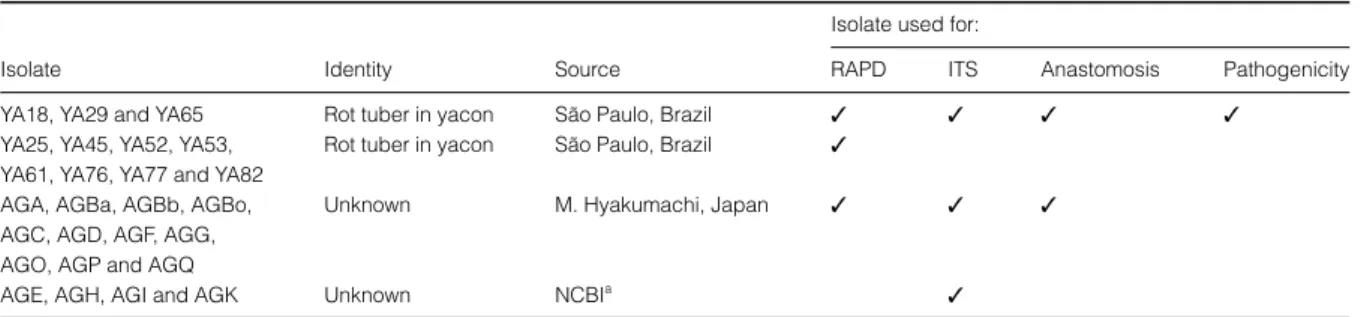
Table 1 Origin of Rhizoctonia isolates from diseased yacon roots and other substrates used for RAPD, ITS, anastomosis and pathogenicity tests
Isolate Identity Source
Isolate used for:
RAPD ITS Anastomosis Pathogenicity
YA18, YA29 and YA65 Rot tuber in yacon São Paulo, Brazil ✓ ✓ ✓ ✓
YA25, YA45, YA52, YA53, YA61, YA76, YA77 and YA82
Rot tuber in yacon São Paulo, Brazil ✓
AGA, AGBa, AGBb, AGBo, AGC, AGD, AGF, AGG, AGO, AGP and AGQ
Unknown M. Hyakumachi, Japan ✓ ✓ ✓
AGE, AGH, AGI and AGK Unknown NCBIa ✓
Rhizoctonia AG G causing yacon root rot 327
(one cycle). Aliquots of each PCR product from these amplifications were separated by electrophoresis on 1·0% (w/v) agarose gel in TBE buffer (Sambrook et al., 1987), stained with ethidium bromide and visualized under UV light.
Each PCR product was purified using MicroSpin S-400 HR columns (Amersham Pharmacia) according to the manufacturer’s instructions. Isolates were sequenced using a double-stranded DNA template of each PCR product (75 ng) and 1 µm of each ITS2, ITS3, ITS4 or
ITS5 primer following the protocol supplied with Amer-sham Premix Dye Terminator (AmerAmer-sham Pharmacia). Sequencing was conducted using a PE Applied Biosystems Model 377 DNA sequencer as recommended by the manufacturer. The four sequence fragments generated by the four primers of each isolate were assembled using
phred/phrap (Ewing et al., 1998) and consed (Gordon
et al., 1998) and all consensus bases of each isolate were of high quality with a phred value greater than 20. The
consensus sequence of each isolate was trimmed in so that only the CITS sequences were analysed. The CITS sequence data of all isolates were aligned by Clustal
W (Thompson et al., 1997). The tree showing the phylo-genetic relatedness between isolates and AG testers was constructed from distance matrix values by the neighbour-joining method and 1000 bootstrap values. The compu-tational analysis to generate the phylogenetic relationship tree between representative AGs and the isolates was performed using paup* (Phylogenetic Analysis Using
Parsimony, version 4·0b5a) (Swofford, 2001) with heuris-tic search, 50 replicates and tree-bisection-reconnection (TBR) as the branch-swapping algorithm.
Anastomosis grouping
For AG tests, three isolates of yacon, YA18, YA29 and YA65, were selected. For each pairing, a 0·3-cm-diameter disc from 2- to 4-day-old colonies of the yacon isolate and the tester strain were positioned 3 cm apart on a sterilized microscope slide (7·5 × 2·5 cm) covered with a thin water-agar layer, pH 8·5. Slides with discs were kept in a satu-rated atmosphere in a Petri plate in the dark at 26°C, and observed for anastomosis after 24 – 48 h (Ceresini et al., 1996). The three binucleate isolates were paired against the AG testers (AG A, AG Ba, AG Bb, AG Bo, AG C, AG D, AG F, AG G, AG O, AG P and AG Q). Anastomosis was regarded as positive when hyphae between the yacon binucleate Rhizoctonia sp. and AG tester made contact with each other and their walls fused, with subsequent plasmolysis of adjacent cells. Hyphal anastomosis was examined at ×400 magnification using light microscopy after staining the vegetative cells with a 0·03% safranin-O aqueous solution and a 3% Ksafranin-OH aqueous solution (Yamamoto & Uchida, 1982).
Pathogenicity test
Three isolates, YA18, YA29 and YA65, used for the anas-tomosis test, RAPD and CITS-rDNA nucleotide sequence
Figure 1Dendrogram showing the levels of genetic relatedness among 11 isolates collected from tubers of yacon (YA) and AG (A, Ba, Bb, Bo, C, D, F, G, O, P, Q) testers of binucleate Rhizoctonia based on RAPD analysis. The tree was constructed from the similarity coefficient using UPGMA.
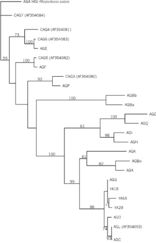
Figure 2Single-most parsimonious tree generated from a branch-swapping algorithm in PAUP obtained with 25 aligned CITS-rDNA
328 R. C. Fenille et al.
analysis were also used for pathogenicity tests. In order to have healthy plants for pathogenicity tests, shoots of 15– 20 cm long were planted into 1500 mL pots containing sterilized soil. After rooting and production of leaves, the new plants were transferred into new 5000 mL pots containing sterilized soil and kept in the glasshouse at 25 ± 2°C. Treatments were replicated three times using a randomized complete block design. Pots filled with the same sterile substrate without the inoculum served as con-trol. The inoculum was prepared by the method of Fenille & Souza (1999) and a 2% aliquot (w/v) from a mixture of the three isolates, YA18, YA65 and YA29, was used. Evaluation was carried out 90 days after inoculation. Plants were carefully removed from pots, the tubers were washed and the incidence of tuber rot recorded.
Results and discussion
Eighty-seven isolates were obtained from diseased tubers. All isolates had dark brown mycelium on PDA medium. Thirty-five of the 87 isolates had 1·9–2·2 mean nuclei per cell, with a few cells of some isolates having three nuclei. Seventy-four per cent of isolates had two nuclei per cell in all cells evaluated; of the remaining 26%, 80% of their cells had two nuclei and 20% had three nuclei per cell.
RAPD analysis showed genetic variability among 11 binucleate Rhizoctonia isolates and 11 testers of the binu-cleate Rhizoctonia group. A total of 63 high-quality poly-morphic amplified DNA fragments were generated by four 10-mer primers. A total of eight (12·7%) polymor-phic RAPD amplified products were generated when the isolates of yacon were compared with one another; this represented 5·2% diversity among binucleate Rhizoctonia isolates from yacon collected from the same field. The
relationship between isolates and standard AG testers of binucleate Rhizoctonia is shown (Fig. 1), and revealed two main clusters: cluster 1 included all isolates from yacon, together with the AG G tester, and cluster 2 the AG Ba, AG Bb, AG F, AG P, AG D, AG Q, AG A, AG Bo, AG O and AG C testers (Fig. 1). The similarity between clusters 1 and 2 was 68·11% by simple matching coefficient analysis. The phylogenetic relationship among the yacon isolates YA18, YA29, YA65 and AG testers, based on CITS-rDNA sequences, is shown in Fig. 2. Isolates YA18, YA29 and YA65 were more similar (> 98·2%) to the AG G tester than to the other testers. The similarity among the three isolates ranged from 97·8 to 99·2%.
Three divergent (according to RAPD analyses) isolates, YA18, YA29 and YA65, showed a C2-type anastomosis reaction (Carling et al., 1988) with the AG G standard tester strain. Hyphae involved in anastomosis were smaller in diameter at the anastomosis point than normal hyphae and dead anastomosing and adjacent cells.
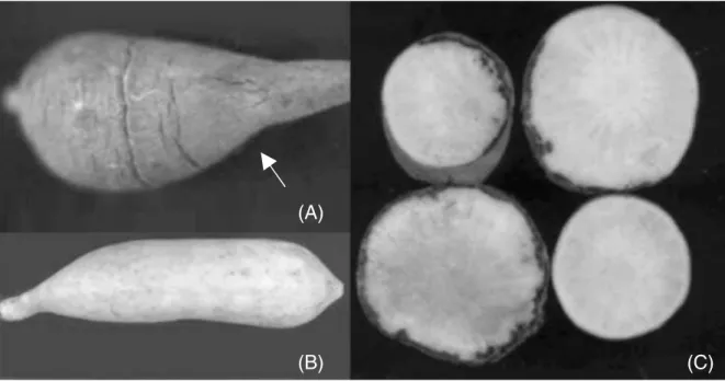
Ninety days after inoculation, yacon tubers showed typical rot symptoms (Fig. 3), including discoloration of the cortex (Fig. 3C). Once the tubers show initial disease symptoms, they became completely rotted after 3 – 4 days in the field. As not only the tubers, but other parts of the plant, can be infected by the pathogen, especially offsets (‘seed’) and the rootstock (‘crown’), the use of healthy yacon offsets and rootstocks for planting is important to reduce incidence of the disease.
The binucleate Rhizoctonia isolates recovered from dis-eased yacon tubers were identified as belonging to anasto-mosis group AG G by nuclei number and CITS-rDNA sequence analysis (data not shown). This is the first report of a binucleate Rhizoctonia sp. AG G causing root rot in yacon in Brazil. Binucleate Rhizoctonia sp. AG G has been
Rhizoctonia AG G causing yacon root rot 329
reported to cause disease of strawberry roots (Martin, 1988): strawberry crops preceded yacon in the field where the binucleate Rhizoctonia sp. AG G isolates were obtained. Thus, the possibility that isolates pathogenic to strawberry contaminated the field cannot be ignored. Also in Brazil, binucleate Rhizoctonia spp. have been isolated together with R. solani in crops such as bean and peanuts with symptoms of pod rot and hypocotyl and root rots, respectively (Ceresini & Souza, 1997), or associated with Pythium aphanidermatum, Sclerotium rolfsii and R. solani in soybean, causing damping-off, in Indonesia (Naito et al., 1993). Future crop management strategies for yacon production, such as crop rotation, should be carried out with a view to decreasing the population of binucleate Rhizoctonia spp. in the soil.
Acknowledgements
The authors wish to thank M. Hyakumachi (Faculty of Agriculture, Gifu University, Gifu, Japan) who supplied tester strains of Rhizoctonia spp.
References
Barrantes del Aguila F, 1988. Enfermedades de cultivos andinos en Ayacucho, Peru. In. Proceedings of the 4th
congreso Int de Cultivos Andinos, 1988, Quito, Peru.
Bolkan HA, Ribeiro WRC, 1985. Anastomosis groups and pathogenicity of Rhizoctonia solani isolates from Brazil. Plant Disease69, 599–601.
Burpee LL, Sanders PL, Cole H Jr, Sherwood RT, 1980. Anastomosis groups among isolates of Ceratobasidium cornigerum (Bourd.) Rogers and related fungi. Mycologia72, 689–701.
Carling DE, Koninaga S, Brainard KA, 2002. Hyphal anastomosis reactions, rDNA-internal transcribed spacer sequences, and virulence levels among subsets of Rhizoctonia solani anastomosis group-2 (AG-2) and AG-BI.
Phytopathology92, 43–50.
Carling DE, Kuninaga S, Leiner RH, 1988. Relatedness within and among intraspecific groups of Rhizoctonia solani. A comparison of grouping by anastomosis and DNA hybridization. Phytoparasitica16, 209–10.
Castro C, Davis JR, Wiese MV, 1988. Quantitative estimation of Rhizoctonia solani AG-3 in soil. Phytopathology78, 1287–92.
Ceresini PC, Fenille RC, Souza NL, 1996. Associação de Rhizoctonia spp. binucleadas e de R. solani Kühn GA 4 HGI à vagens de amendoinzeiro (Arachis hypogaea) no estado de São Paulo. Summa Phytopathologica22, 145–55.
Ceresini PC, Souza NL, 1997. Associação de Rhizoctonia spp. binucleadas e de R. solani Kühn GA 4 HGI e GA 2–2 IIIB ao feijoeiro (Phaseolus vulgaris L.) no estado de São Paulo. Summa Phytopathologica23, 14–24.
Cubeta MA, Echandi E, Gumpertz ML, 1991. Survival of binucleate Rhizoctonia species, biological control agents, in soil and plant debris under field conditions. Biological Control1, 218–26.
Ewing B, Hillier L, Wendl M, Green P, 1998. Basecalling of automated sequencer traces using phred. I. Accuracy assessment. Genome Research8, 175–85.
Fenille RC, Souza NL, 1999. Efeitos de materiais orgânicos e da umidade do solo na patogenicidade de Rhizoctonia solani Kühn GA-4 HGI ao feijoeiro. Pesquisa Agropecuária Brasileira34, 1959–67.
Fenille RC, Souza NL, Kuramae EE, 2002. Characterization of Rhizoctonia solani associated with soybean in Brazil. European Journal of Plant Pathology108, 783–92. Floridata, 2000. Polymnia sonchifolia. [http://
www.floridata.com].
Gordon D, Abajian C, Green P, 1998. Consed: a graphical tool for sequence finishing. Genome Research8, 195–202. Grau A, Rea J, 1997. Yacon. Smallanthus sonchifolius (Poepp.
& Endl.) H. Robinson. In: Hermann M, Heller J, eds. Andean Roots and Tubers: Ahipa, Arracacha, Maca and Yacon. Rome, Italy: International Plant Genetic Resources Institute, 199–242.
Ko W, Hora KF, 1971. A selective medium for the quantitative determination of Rhizoctonia solani in soil. Phytopathology 61, 707–10.
Kulik MM, Dery PD, 1995. Use of DAPI for anastomosis group typing of strains of the fungus Rhizoctonia solani. Biotechnique and Histochemistry70, 95–8.
Kuramae EE, Buzeto AL, Ciampi MB, Souza NL, 2003. Identification of Rhizoctonia solani AG 1-IB in lettuce, AG 4 HG-I in tomato and melon, and AG 4 HG-III in broccoli and spinach, in Brazil. European Journal of Plant Pathology109, 391–5.
Kuramae-Izioka EE, 1997. A rapid, easy and high yield protocol for total genomic DNA isolation from Colletotrichum gloeosporioides and Fusarium oxysporum for RAPD. Revista Unimar19, 683–9.
Lizárraga L, Ortega R, Vargas W, Vidal A, 1997. Cultivo del yacón (Polymnia sonchifolia). In: Proceedings of the Curso Pre Congreso – 9th Congreso Internacional de Cultivos Andinos 1997, Cusco, Peru, 65–7.
Martin SB, 1988. Identification, isolation frequency, and pathogenicity of anastomosis groups of binucleate
Rhizoctonia spp. from strawberry roots. Phytopathology78, 379–84.
Mizuno A, Nakanishi T, Nishiyama K, 1993. Bacterial wilt of yacon strawberry caused by Erwinia chrysanthemi. Annals of the Phytopathological Society of Japan59, 702–8. Naito S, Mohamad D, Nasution A, Purwanti H, 1993.
Soil-borne diseases and ecology of pathogens on soybean roots in Indonesia. JARQ (Japan Agricultural Research Quarterly) 26, 247–53.
National Research Council, 1989. Lost Crops of the Incas: Little-known Plants of the Andes with Promise for Worldwide Cultivation. Washington, DC, USA: National Academy Press.
Ogoshi A, 1987. Ecology and pathogenicity of anastomosis and intraspecific groups of Rhizoctonia solani Kühn. Annual Review of Phytopathology25, 125–43.
Parmeter JR Jr, Sherwood RT, Platt WD, 1969. Anastomosis grouping among isolates of Thanatephorus cucumeris. Phytopathology59, 1270–8.
Rohlf FJ, 1992. NTSYS-PC, Version 1.7. Numerical Taxonomy and Multivariate Analysis System. Setauket, NY, USA: Exeter Software Publications.
330 R. C. Fenille et al.
Sneh B, Burpee L, Ogoshi A, 1991. Identification of Rhizoctonia Species. St Paul, MN, USA: APS Press.
Swofford DL, 2001. PAUP*. Phylogenetic Analysis Using Parsimony (*and Other Methods), Version 4·0b5. Sunderland, UK: Sinauer Associates.
Thompson JD, Gibson TJ, Plewniak F, Jeanmougin F, Higgins DG, 1997. The Clustal X windows interface: flexible strategies for multiple sequence alignment aided by quality analysis tools. Nucleic Acids Research25, 4876–82.
White TJ, Bruns T, Lee S, Taylor JW, 1990. Amplification and direct sequencing of fungal ribosomal RNA genes for
phylogenetics. In: Innis MA, Gelfand DH, Sninsky JJ, White TJ, eds. PCR Protocols: A Guide to Methods and Applications. San Diego, CA, USA: Academic Press, 315–22.
Williams JGK, Kubelik AR, Rafalski JA, Tingey SV, 1990. DNA polymorphisms amplified by arbitrary primers are useful as genetic markers. Nucleic Acids Research18, 6531–5.