The Differences in Ultrasound and
Clinicopathological Features between
Basal-Like and Normal-Basal-Like Subtypes of Triple
Negative Breast Cancer
Ziyao Li1, Min Ren1*, Jiawei Tian1*, Shuangquan Jiang1‡, Yujie Liu1‡, Lei Zhang1‡, Zhenzhen Wang1‡, Qianqian Song2‡, Chong Liu1‡, Tong Wu1‡
1Department of Ultrasound, Second Affiliated Hospital of Harbin Medical University, Harbin, Heilongjiang Province, China,2Department of Biostatistics, College of Public Health of Harbin Medical University, Harbin, Heilongjiang Province, China
‡These authors contributed equally to this work.
*[email protected](JWT);[email protected](MR)
Abstract
Purpose
The aim of this study was to identify the ultrasound features and clinicopathological charac-teristics of basal-like subtype of triple negative breast cancers (TNBCs).
Materials and Methods
This study was approved by the ethical board of the Second Affiliated Hospital of Harbin Medical University. The patients’clinicopathological information was available. The ultra-sound features of 62 tumors from 62 TNBC patients were interpreted. The immunohisto-chemical results of cytokertain5/6 (CK5/6) and Epidermal Growth Factor Receptor (EGFR) were used to classify the tumor into basal-like and normal-like groups. The association of the ultrasound features interpreted by experienced ultrasound doctors with the immunohis-tochemical classification was studied.
Results
Of the 62 TNBC cases, 42 (67.7%) exhibited the basal-like phenotype and 20 (32.3%) ex-hibited the normal-like phenotype based on the immunohistochemical CK5/6 and EGFR markers. Of all the tumors, 90.3% were invasive carcinomas. The basal-like tumors were significantly associated with a maximum diameter on ultrasound of more than 20 mm (36, 85.7%) (P = 0.0014). The normal-like tumors usually exhibited lateral shadows (15, 75%) (P = 0.0115) as well as microlobulated margins (12, 60%) (P = 0.0204) compared to the basal-like subtype. Other ultrasound features showed no significant differences between the two groups.
OPEN ACCESS
Citation:Li Z, Ren M, Tian J, Jiang S, Liu Y, Zhang L, et al. (2015) The Differences in Ultrasound and Clinicopathological Features between Basal-Like and Normal-Like Subtypes of Triple Negative Breast Cancer. PLoS ONE 10(3): e0114820. doi:10.1371/ journal.pone.0114820
Academic Editor:William B. Coleman, University of North Carolina School of Medicine, UNITED STATES
Received:July 28, 2014
Accepted:October 17, 2014
Published:March 3, 2015
Copyright:© 2015 Li et al. This is an open access article distributed under the terms of theCreative Commons Attribution License, which permits unrestricted use, distribution, and reproduction in any medium, provided the original author and source are credited.
Data Availability Statement:All relevant data are within this paper.
Funding:The project was supported by the National Natural Science Foundations of China (Grant NO. 81271647, NO. 81071216 and NO. 81101103). Jiawei Tian received the previous two and Ying Wang received the last one.
Conclusions
Although ultrasound cannot yet be used to differentiate between the basal-like subtype and normal-like subtype of TNBC, ultrasound can be used to provide some useful information to the clinicians.
Introduction
Basal-like tumors comprise an important heterogeneous group of breast cancers associated with aggressive behavior and poor prognosis. They account for 15% of all breast cancers and often present as interval cancers that mainly affect younger patients [1–4]. Basal-like breast cancer usually lacks the expression of estrogen receptor (ER), progesterone receptor (PR) and Human Epidermal Growth Factor Receptor 2 (HER-2), which is generally defined as triple negative breast cancer (TNBC). It has been reported that basal-like breast cancers account for 56–85% of TNBCs, and the remainder of TNBCs, which lack the gene expression profile of the basal-like tumors and are similar to normal mammary stromal cells, are normal-like breast cancers [1,2]. Compared to the basal-like subtype, normal-like breast cancer has a very similar or better prognosis to that of hormone receptor positive breast cancers[1,4,5]. Therefore, the sub-classification of basal-like and normal-like TNBCs is essential in determining treatment and estimating the prognosis.
Gene expression profiling (GEP) analysis is the gold standard for diagnosing basal-like breast cancers. However, GEP is still inaccessible in most global clinical settings because of its high cost and high technological and clinical expertise requirements. With this clinical situa-tion, some researchers have proposed a panel of immunohistochemical markers including ER, PR, HER-2, cytokertain5/6 (CK5/6) and Epidermal Growth Factor Receptor (EGFR) to define the basal-like subtype [6–8]; this panel is cost-effective and widely accessible. With the develop-ment of high frequency ultrasound, breast ultrasound has been demonstrated to be a useful tool for the differential diagnosis of malignant and benign breast lesions, for guiding breast bi-opsy and for the guiding therapeutic treatment and prognosis estimation [9]. However, few studies have been conducted to evaluate its clinical value in the differentiation of basal-like and normal-like TNBC subtypes.
In this study, we aimed to identify the clinicopathological and ultrasound features that are correlated with basal-like tumors in 62 TNBC patients with immunohistochemical examina-tion results as proof of the diagnosis.
Patients and Methods
Patients
criteria were as follows: 1) single and unilateral breast lesions; 2) the ultrasound examination conducted by the particular sonographer with 3 years of experience, as well as the particular ul-trasound machine; 3) standard B-mode and Doppler ulul-trasound examination available; and 4) immunohistochemical markers (ER, PR, HER-2, CK5/6 and EGFR) accessible. The character-istics of the study subjects are illustrated inTable 1.
Breast ultrasound examination
Breast ultrasound examinations were performed by the particular sonographer with 3 years of experience and were interpreted by two sonographers, each with 5 years of experience in the di-agnosis of breast cancer. A HITACHI Vision 900 system (Hitachi Medical System, Tokyo, Japan) with high frequency ultrasound and color Doppler features equipped with a 12–5 MHz linear-array transducer was used to perform the breast ultrasound examination. During the real-time examination, longitudinal and transverse static images were obtained from more than two different planes per lesion. Additionally, cine clips in standardized views were also ob-tained through the masses. In the color Doppler examination, we obob-tained images and cine clips displaying the most abundant flow signals. Every patient’s static images and cine clips were stored as a separate folder in our work station and then added to our prospectively main-tained breast tumor database for subsequent application.
Histological examination and Immunohistochemistry
The histological examination was conducted by the pathology department at our hospital. All patients were examined to determine the histological tumor type, tumor grade for invasive can-cer and lymph node metastasis after the surgery. Invasive cancan-cer was graded as grade 1 (well differentiated), grade 2 (moderately differentiated) or grade 3 (poorly differentiated) according to the Scarff-Bloom-Richardson System [10]. Specimens from the surgically resected lesions
Table 1. Characteristics of the study subjects.
Variables Number of patients Count (%)
Age (years)
ge 45 72.6
>50 17 27.4
Pathological type
Invasive cancer 56 90.3
*DCIS 6 9.70
Tumor grade for invasive cancer
Grade 1 3 5.40
Grade 2 22 39.3
Grade 3 31 55.3
Immunohistochemical markers
EGFR positive 10 23.8
CK5/6 positive 8 19.0
EGFR positive and CK5/6 positive 24 57.2
Lymphatic node metastasis
Yes 26 41.9
No 36 58.1
*DCIS = ductal carcinoma in situ.
were fixed with formalin and embedded in paraffin blocks. Three slices from each patient were selected for immunohistochemistry analyses of membrane and cytoplasm. The immunohisto-chemical classification was performed by two pathologists each with more than 3 years of experience. In cases of inconsistencies, consensus of the two pathologists was used as the final result.
The appropriate antibodies were chosen for ER, PR, HER2, EGFR and CK5/6 immunohisto-chemical staining. The cutoff point for ER and PR negative was less than 10% of the cells with nuclear staining [11,12]. HER2 status was graded as 0, 1+, 2+ or 3+, and 0 and 1 + were deemed to be negative HER2 status [13]. Membrane staining was assessed for EGFR according to DAKO criteria. Any intensity of EGFR in more than 1% of cells was considered to be a positive basal marker, and the detection of CK5/6 cytoplasmic expression in either tumor or surround-ing tissues was considered to be CK5/6 positive [1]. EGFR positive, CK5/6 positive or EGFR and CK5/6 positive TNBCs were classified as basal-like cancers, and TNBCs that were not posi-tive for those markers were classified as normal-like cancers [1,4,5].
Interpretation of ultrasound findings
The interpretation of the ultrasound images was performed by two radiologists (each with more than 5 years of experience in the diagnosis of breast cancer) who were blinded to the immunohistochemistry results. In cases of inconsistencies, consensus between the two radiolo-gists was used as the final result.
Features of tumor size (maximum diameter measured by ultrasound), shape, growth orien-tation, boundary, margin, posterior acoustic echo, lateral acoustic shadow, microcalcifications, and echogenicity were evaluated from the B-mode ultrasound images and categorized using the criteria as illustrated inTable 2. From the color Doppler examination, the blood flow prop-erties of each lesion were scored according to the Alder grading method [14].
Statistical analyses
Chi-squared tests or Fisher exact tests were performed on each single clinicopathological or qualitative ultrasound feature of the two TNBC subtypes to evaluate the significance of differ-ence. For correlations, we performed multivariate regression analysis and expressed the odds ratio (OR) with 95% confidence intervals (CI). The P value was two sided, and a P value0.05 indicated a significant difference.
SPSS software (version 21.0; Chicago, IL, USA) was used for the statistical analysis.
Results
Clinicopathological and immunohistochemistry
In total, 62 patients were analyzed in this study including 48 invasive ductal carcinomas, 6 duc-tal carcinomas in situ, 3 medullary carcinomas, 3 metaplastic carcinomas, 1 clear cell carcino-ma, and 1 apocrine carcinoma. All patients were female and the ages ranged from 32 to 71 years (mean age, 48.8 years). The age range of the 42 patients with basal-like subtype was 32–
Among the 62 TNBCs, 42 were classified as basal-like tumors and 20 were classified as nor-mal-like tumors. In the 42 basal-like tumors, 23.8% (10/42) were EGFR positive only, 19.0% (8/42) were CK5/6 positive only and 57.2% (24/42) were both EGFR and CK5/6 positive.
Ultrasound findings
The results of the chi-squared test and multivariate regression analysis for the ultrasound find-ings are shown in Tables4and5. Forward selection was used in the multivariate regression models comparing the ultrasound findings of the two subtypes to screen the variables, and the standard was P value0.05. The selected parameters include tumor margin, posterior feature, lateral shadow and tumor size. The result of multivariate regression analysis is shown in
Table 5.
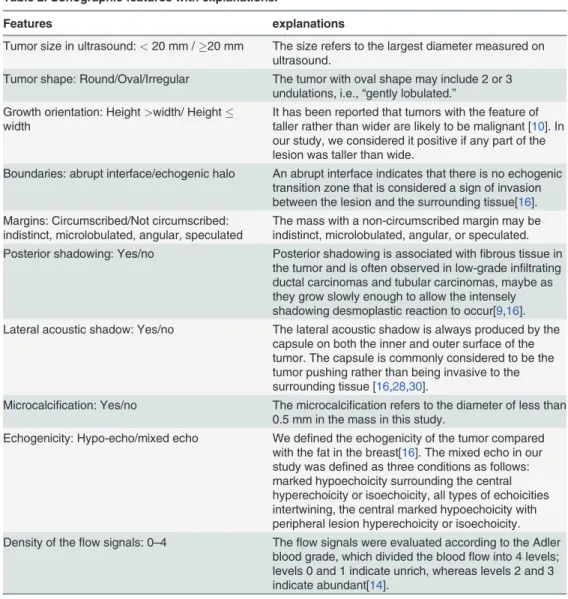
Regarding the ultrasound findings of the two groups, significant differences were observed in the tumor maximum diameter measured on the ultrasound, the tumor margin and lateral acoustic shadow. Tumor size20 mm was significantly more frequent in the basal-like group than in the normal-like group (85.7% vs 35%) (P = 0.0014) (seeFig. 1). According to the results
Table 2. Sonographic features with explanations.
Features explanations
Tumor size in ultrasound:<20 mm /20 mm The size refers to the largest diameter measured on ultrasound.
Tumor shape: Round/Oval/Irregular The tumor with oval shape may include 2 or 3 undulations, i.e.,“gently lobulated.”
Growth orientation: Height>width/ Height width
It has been reported that tumors with the feature of taller rather than wider are likely to be malignant [10]. In our study, we considered it positive if any part of the lesion was taller than wide.
Boundaries: abrupt interface/echogenic halo An abrupt interface indicates that there is no echogenic transition zone that is considered a sign of invasion between the lesion and the surrounding tissue[16]. Margins: Circumscribed/Not circumscribed:
indistinct, microlobulated, angular, speculated
The mass with a non-circumscribed margin may be indistinct, microlobulated, angular, or speculated. Posterior shadowing: Yes/no Posterior shadowing is associated withfibrous tissue in
the tumor and is often observed in low-grade infiltrating ductal carcinomas and tubular carcinomas, maybe as they grow slowly enough to allow the intensely shadowing desmoplastic reaction to occur[9,16]. Lateral acoustic shadow: Yes/no The lateral acoustic shadow is always produced by the
capsule on both the inner and outer surface of the tumor. The capsule is commonly considered to be the tumor pushing rather than being invasive to the surrounding tissue [16,28,30].
Microcalcification: Yes/no The microcalcification refers to the diameter of less than 0.5 mm in the mass in this study.
Echogenicity: Hypo-echo/mixed echo We defined the echogenicity of the tumor compared with the fat in the breast[16]. The mixed echo in our study was defined as three conditions as follows: marked hypoechoicity surrounding the central hyperechoicity or isoechoicity, all types of echoicities intertwining, the central marked hypoechoicity with peripheral lesion hyperechoicity or isoechoicity. Density of theflow signals: 0–4 Theflow signals were evaluated according to the Adler
blood grade, which divided the bloodflow into 4 levels; levels 0 and 1 indicate unrich, whereas levels 2 and 3 indicate abundant[14].
of the analyzed images, we noticed that there were no tumors with circumscribed or indistinct margins in our study. An angular/speculated margin was more common in basal-like tumors
Table 3. The patients’clinical pathological characteristics in the two groups.
Age (Y) The histological type The tumor grade in invasive cancer
Lymphatic metastasis
ym >50 Invasive carcinoma DCIS Grade 1–2 Grade 3 Yes No
Basal-like 28 (67%) 14(33%) 39(93%) 3(7%) 16(41%) 23(59%) 19 (45%) 23 (55%)
Normal-like 17 (85%) 3(15%) 17(85%) 3(15%) 9(53%) 8(47%) 7 (35%) 13 (65%)
P value 0.223 0.377 0.410 0.445
doi:10.1371/journal.pone.0114820.t003
Table 4. The result of chi-squared test of the ultrasound features by groups.
Group Basal-like Normal-like P
Tumor size
<20mm 6 13 0.000**
20mm 36 7
Growth orientation
Aspect ratio>1 8 10 0.012**
Aspect ratio<1 34 10
Lateral acoustic shadow
Yes 19 15 0.028**
No 23 5
Tumor shape
Round/oval 11 8 0.270
Irregular 31 12
Echo pattern
Hypoechoic 9 4 1.000
Mixed/marked hypoechoic 33 16
Tumor margin
Microlobulated 15 12 0.071
Angular/speculated 27 8
Posterior acoustic feature
No change 20 12 0.057
Enhancement 18 3
Attenuation 4 5
Microcalcification
Yes 29 11 0.280
No 13 9
Boundary
Echogenic halo 0 2 0.100
Abrupt interface 42 18
Bloodflow signals
Alder 0–1 16 4 0.154
Alder 2–3 26 16
Note:**indicates significant difference in the two groups.
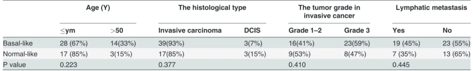
(64.3% of the basal-like subtype versus 40% of the normal-like subtype) (P = 0.0204) (see
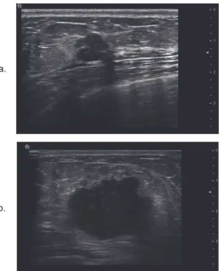
Fig. 2). There were 19 basal-like subtype cases with lateral shadow and 15 normal-like subtype cases with lateral shadow (45.2% vs 75%) (P = 0.0115) (seeFig. 1andFig. 3).
The growth orientation of the tumor in the two subtypes had significant difference based on the chi-squared test (P = 0.012), but it was not involved in the multivariate regression model when we screened variations. So there was no significant difference in multivariate regression analysis for the tumors’growth orientation.
Table 5. The result of the multivariate logistical regression analysis.
variables SE χ2 P-value OR OR (95%CI)
margin 0.9707 5.3757 0.0204 9.494 (1.416,63.641)
Posterior feature 1.5397 3.3286 0.0681 0.060 (0.003,1.232)
Lateral shadow 1.0045 6.3904 0.0115 12.670 (1.769,90.732)
size 0.9857 10.1599 0.0014 23.147 (3.353,159.785)
doi:10.1371/journal.pone.0114820.t005
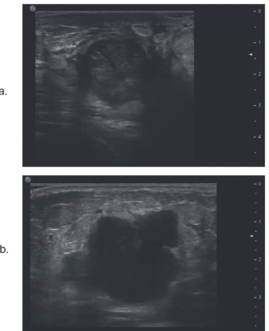
Fig 1. The maximum diameter of the tumor and lateral acoustic shadow feature on ultrasound.a. The maximum diameter of the normal-like breast cancer is 14 mm, and the lateral acoustic shadow is shown. b. The maximum diameter of the basal-like breast cancer is 30 mm. There is no lateral acoustic shadow.
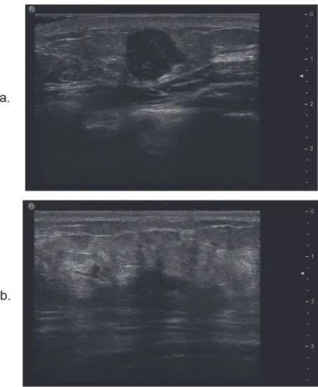
It has been reported that the TNBCs had a tendency to show marked hypoechoicity [15]. In our study, based on the image analysis, we defined the lesion mixed echo as the three condi-tions explained inTable 2. The echo pattern of the tumors in our research all showed hypoe-choicity or mixed ehypoe-choicity, and none showed hyperehypoe-choicity or isoehypoe-choicity. A total of 49 (79%) tumors showed mixed echoicity, 33 (78%) were basal-like cancer and 16 (80%) were nor-mal-like cancer. Based on the echo pattern, the TNBC always presents as predominant hypoe-cho with other mixed ehypoe-cho patterns (seeFig. 4). Other ultrasound features, including the tumor shape, posterior acoustic feature, microcalcifications, boundary, and blood flow signal, had no significant differences between the two groups.
Discussion
Ultrasound and mammography are two vital imaging tools for breast screening. Ultrasound is particularly important as the mammography has some limitations. It can be used to distinguish the malignant from benign lesions, to guide biopsies, to assist in the selection of the appropriate therapeutic method and to aid further management [9,16,17]. Currently, TNBC has become a top global issue. A series of studies have shown that TNBC is a subtype that has a more
Fig 2. The tumor margin on ultrasound.a. It shows that there is a slight lobulated margin of normal-like breast tumor. b. The ultrasound image of a basal-like breast cancer shows that the tumor margin is angular and speculated.
aggressive clinical course compared with other forms of breast cancer and that is associated with relatively high rates of recurrence, distant metastases and low overall survival rates [15,18–20]. The TNBC and basal-like subtype are usually thought to be identical based on clin-ical work-up, but this is not actually true [21,22]. Gene expression profiling (GEP) analysis is the gold standard for diagnosing basal-like breast cancer, but currently, the use of GEP is still limited due to financial concerns and because this technology is often inaccessible in most global clinical settings. Given these circumstances, some researchers have proposed a panel of immunohistochemical markers defining the basal-like subtype, including ER, PR, HER-2, CK5/6 and EGFR, with a sensitivity of 55–76% and a specificity of 100% [6–8]. With this meth-od, the TNBCs are divided into the basal-like subtype and the normal-like subtype. Previous
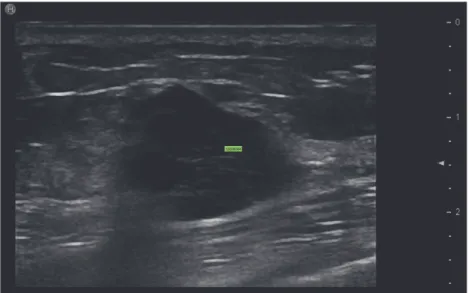
Fig 3. The relationship between lateral acoustic shadow feature on ultrasound and molecular subtypes of breast cancer.a. The ultrasound image of a tumor with lateral acoustic shadow is classified as the normal-like subtype according to the immunohistochemistry results. b. The ultrasound image of a tumor without lateral acoustic shadow is classified as the basal-like subtype according to the immunohistochemistry results.
studies have proved that basal-like breast cancers are particularly more aggressive than the nor-mal-like tumors [1,4,5,7]. We sought to demonstrate whether there is a difference in the ultra-sound and clinicopathological features between the basal-like subtype and the normal-like subtype of TNBC. Although we cannot differentiate the molecular subtypes of breast cancers based on ultrasound imaging, the radiologists should be aware of different subtypes with dis-tinctive ultrasound features that provide more information to clinicians.
Many studies reported that patients with TNBC were younger than those without TNBC [5,18,19,23–25]. In all 62 patients enrolled in our study, the number of patient50 years old ac-counted for 72.6%, and so there was a clear predominance of younger patients. However, this dis-crepancy in the age of onset did not exist in the basal-like and normal-like cancers in our study.
As one of the main prognostic indicators as well as a determining factor for surgical treat-ment planning, the breast cancer size is vital. It is reported that the tumor size measured on ul-trasound has a comparatively good correlation with the tumor size on pathology [26].
Therefore, accurate prediction of tumor size based on ultrasound is essential. Regarding the tu-mor’s maximum diameter measured by ultrasound, the basal-like tumors were usually larger than the normal-like subtype. This may be relative to the highly malignant and invasive nature of basal-like breast cancers. Due to these biological behaviors, the tumors in this subtype always grow fast and may rapidly lead to an invasive stage and present with a larger volume.
Other ultrasound features that were found to be significantly different in the two groups in our study were the tumor margin and lateral acoustic shadow. The angular/speculated margin and no lateral acoustic shadow were more common in the basal-like breast cancers. An angu-lar/speculated margin always indicates that the tumor grows fast and that the growth speed in every direction is not consistent. The lateral acoustic shadow is thought to originate from the capsule, which is on both the tumor’s inner and outer surfaces and is usually a pseudocapsule of compressed adjacent normal breast tissue. Visualization of such a capsule always indicates that the leading edge of the nodule is pushing rather than infiltrating [16]. Therefore, the result of our study was consistent with the property of basal-like and normal-like breast tumors that the basal-like subtype is usually more malignant than the normal-like subtype and it always in-vasive rather than just encroaching.
Fig 4. The echo pattern of the breast tumor with TNBC subtype.We observed the marked hypoechoicity in the tumor, and all three markers including ER, PR, and HER2 are negative.
The growth orientation was found to be different in the two groups based on the chi-squared test result. However, the difference on the multivariate logistical regression was not significant. It has been reported that the malignant nodules are more likely to present with masses that are taller than wider [16,27–29]. This finding is alarming because it suggests growth across normal tissue planes, which are horizontally oriented in patients scanned in the supine position [16,27]. The result of our study demonstrated that an aspect ratio>1 may
re-duce the possibility of a tumor being a basal-like cancer, but it is not an independent influenc-ing factor. Other ultrasound features had no significant differences between the two groups.
There are certainly some limitations to our research, including the retrospective nature of the study. Another important limitation is that because TNBCs account for only 10–15% of all breast cancers and the basal-like subtype or normal-like subtype accounts for a lower propor-tion, the limited number of patients in our cohort is unavoidable. Therefore, we are still in the process of acquiring cases to further support these conclusions. In addition, one of the pur-poses of this study is to attract people’s attention to these two subtypes of breast cancer, so that more people can develop more comprehensive and in-depth exploration in the future. Also, we didn’t test the predictive ability of these distinguishing ultrasound features between the two subtypes and this would be the emphasis of the next step of our study.
Conclusions
Although ultrasound cannot yet be used to differentiate between basal-like subtype and nor-mal-like subtype of TNBC, we have demonstrated some sonographic features with a significant difference. Further study is needed to confirm our findings and to explain the biological rea-sons for these findings.
Acknowledgments
We are grateful to our department for supporting this work, and we thank the pathologists who participated in this study.
Author Contributions
Conceived and designed the experiments: ZYL JWT. Performed the experiments: SQJ YJL LZ. Analyzed the data: QQS. Contributed reagents/materials/analysis tools: ZZW CL. Wrote the paper: ZYL MR. Obtained the patients' follow up information: TW.
References
1. Choccalingam C, Rao L, Rao S (2012) Clinico-Pathological Characteristics of Triple Negative and Non Triple Negative High Grade Breast Carcinomas with and Without Basal Marker (CK5/6 and EGFR) Ex-pression at a Rural Tertiary Hospital in India. Breast Cancer (Auckl) 6: 21–29. doi:10.4137/BCBCR. S8611PMID:22346359
2. Rakha EA, Ellis IO (2009) Triple-negative/basal-like breast cancer: review. Pathology 41: 40–47. doi:
10.1080/00313020802563510PMID:19089739
3. Banerjee S, Reis-Filho JS, Ashley S, Steele D, Ashworth A, et al. (2006) Basal-like breast carcinomas: clinical outcome and response to chemotherapy. J Clin Pathol 59: 729–735. PMID:16556664
4. Dogan BE, Turnbull LW (2012) Imaging of triple-negative breast cancer. Ann Oncol 23 Suppl 6: vi23– 29. PMID:23012298
5. Rao C, Shetty J, Prasad KH (2013) Immunohistochemical profile and morphology in triple—negative breast cancers. J Clin Diagn Res 7: 1361–1365. doi:10.7860/JCDR/2013/5823.3129PMID:23998066
7. Nielsen TO, Hsu FD, Jensen K, Cheang M, Karaca G, et al. (2004) Immunohistochemical and clinical characterization of the basal-like subtype of invasive breast carcinoma. Clin Cancer Res 10: 5367– 5374. PMID:15328174
8. Cakir A, Gonul II, Uluoglu O (2012) A comprehensive morphological study for basal-like breast carcino-mas with comparison to nonbasal-like carcinocarcino-mas. Diagn Pathol 7: 145. doi: 10.1186/1746-1596-7-145PMID:23082819
9. Au-Yong IT, Evans AJ, Taneja S, Rakha EA, Green AR, et al. (2009) Sonographic correlations with the new molecular classification of invasive breast cancer. Eur Radiol 19: 2342–2348. doi:10.1007/ s00330-009-1418-2PMID:19440719
10. Bloom HJ, Richardson WW (1957) Histological grading and prognosis in breast cancer; a study of 1409 cases of which 359 have been followed for 15 years. Br J Cancer 11: 359–377. PMID:13499785
11. Reiner A, Neumeister B, Spona J, Reiner G, Schemper M, et al. (1990) Immunocytochemical localiza-tion of estrogen and progesterone receptor and prognosis in human primary breast cancer. Cancer Res 50: 7057–7061. PMID:2208173
12. Guerra I, Algorta J, Diaz de Otazu R, Pelayo A, Farina J (2003) Immunohistochemical prognostic index for breast cancer in young women. Mol Pathol 56: 323–327. PMID:14645694
13. Taucher S, Rudas M, Mader RM, Gnant M, Dubsky P, et al. (2003) Do we need HER-2/neu testing for all patients with primary breast carcinoma? Cancer 98: 2547–2553. PMID:14669272
14. Adler DD, Carson PL, Rubin JM, Quinn-Reid D (1990) Doppler ultrasound color flow imaging in the study of breast cancer: preliminary findings. Ultrasound Med Biol 16: 553–559. PMID:2238263
15. Ko ES, Lee BH, Kim HA, Noh WC, Kim MS, et al. (2010) Triple-negative breast cancer: correlation be-tween imaging and pathological findings. Eur Radiol 20: 1111–1117. doi:10.1007/s00330-009-1656-3
PMID:19898850
16. Stavros AT, Thickman D, Rapp CL, Dennis MA, Parker SH, et al. (1995) Solid breast nodules: use of sonography to distinguish between benign and malignant lesions. Radiology 196: 123–134. PMID:
7784555
17. Rakha EA, Ellis IO (2007) An overview of assessment of prognostic and predictive factors in breast cancer needle core biopsy specimens. J Clin Pathol 60: 1300–1306. PMID:17630399
18. Krizmanich-Conniff KM, Paramagul C, Patterson SK, Helvie MA, Roubidoux MA, et al. (2012) Triple re-ceptor-negative breast cancer: imaging and clinical characteristics. AJR Am J Roentgenol 199: 458– 464. doi:10.2214/AJR.10.6096PMID:22826413
19. Dent R, Trudeau M, Pritchard KI, Hanna WM, Kahn HK, et al. (2007) Triple-negative breast cancer: clin-ical features and patterns of recurrence. Clin Cancer Res 13: 4429–4434. PMID:17671126
20. Boisserie-Lacroix M, Macgrogan G, Debled M, Ferron S, Asad-Syed M, et al. (2013) Triple-negative breast cancers: associations between imaging and pathological findings for triple-negative tumors com-pared with hormone receptor-positive/human epidermal growth factor receptor-2-negative breast can-cers. Oncologist 18: 802–811. doi:10.1634/theoncologist.2013-0380PMID:23821326
21. Boisserie-Lacroix M, Hurtevent-Labrot G, Ferron S, Lippa N, Bonnefoi H, et al. (2013) Correlation be-tween imaging and molecular classification of breast cancers. Diagn Interv Imaging 94: 1069–1080. doi:10.1016/j.diii.2013.04.010PMID:23867597
22. Billar JA, Dueck AC, Stucky CC, Gray RJ, Wasif N, et al. (2010) Triple-negative breast cancers: unique clinical presentations and outcomes. Ann Surg Oncol 17 Suppl 3: 384–390. doi: 10.1245/s10434-010-1260-4PMID:20853062
23. Rakha EA, El-Sayed ME, Green AR, Lee AH, Robertson JF, et al. (2007) Prognostic markers in triple-negative breast cancer. Cancer 109: 25–32. PMID:17146782
24. Reis-Filho JS, Tutt AN (2008) Triple negative tumours: a critical review. Histopathology 52: 108–118. doi:10.1111/j.1365-2559.2007.02889.xPMID:18171422
25. Thike AA, Cheok PY, Jara-Lazaro AR, Tan B, Tan P, et al. (2010) Triple-negative breast cancer: clinico-pathological characteristics and relationship with basal-like breast cancer. Mod Pathol 23: 123–133. doi:10.1038/modpathol.2009.145PMID:19855377
26. Luparia A, Mariscotti G, Durando M, Ciatto S, Bosco D, et al. (2013) Accuracy of tumour size assess-ment in the preoperative staging of breast cancer: comparison of digital mammography, tomosynthesis, ultrasound and MRI. Radiol Med 118: 1119–1136. doi:10.1007/s11547-013-0941-zPMID:23801389
27. Fornage BD, Sneige N, Faroux MJ, Andry E (1990) Sonographic appearance and ultrasound-guided fine-needle aspiration biopsy of breast carcinomas smaller than 1 cm3. J Ultrasound Med 9: 559–568. PMID:2231854
29. Ueno E, Tohno E, Soeda S, Asaoka Y, Itoh K, et al. (1988) Dynamic tests in real-time breast echogra-phy. Ultrasound Med Biol 14 Suppl 1: 53–57. PMID:3055603
30. Cole-Beuglet C, Soriano RZ, Kurtz AB, Goldberg BB (1983) Fibroadenoma of the breast: sonomammo-graphy correlated with pathology in 122 patients. AJR Am J Roentgenol 140: 369–375. PMID: