CLINICAL SCIENCE
Triple-negative breast carcinomas are a
heterogeneous entity that differs between young
and old patients
Filomena M. Carvalho,ILivia M. Bacchi,IPriscila P. C. Santos,ICarlos E. BacchiII
IDepartment of Pathology, Faculdade de Medicina, Universidade de Sa˜o Paulo, Sa˜o Paulo, SP, Brazil.IIConsultoria em Patologia, Botucatu, SP, Brazil.
OBJECTIVE:To compare the frequency and immunohistochemical profiles of triple-negative breast carcinomas in younger and older women.
METHODS AND RESULTS: We selected patients diagnosed with triple-negative breast carcinomas. The groups examined were women who were 35 years old or younger between 1997 and 2007 (n = 74) and, for comparison, women who were 60 years old or older (n = 19, consecutive cases). All formalin-fixed and paraffin-embedded tumor samples were reviewed and immunohistochemically stained for ER, PR, HER2, Ki-67 antigen, epidermal growth factor receptor, cytokeratin 5/6, p53, vimentin, CD117, and p63 using tissue microarrays blocks. Triple-negative breast carcinomas corresponded to 34.6% (74/213) of the carcinomas from the younger patients and 16.2% (19/117) of the carcinomas from the older patients (p = 0.002). No significant differences in the frequency of the basal phenotype were observed in the two patient groups based on CK5/6 and/or epidermal growth factor receptor expression (74.3%vs.68.4%). However, triple-negative breast carcinomas in the older patients presented a higher frequency of CK5/6 expression compared to those of younger patients (42.1%vs.9.6%; p = 0.005), whereas triple-negative breast carcinomas of younger patients had a higher expression level of epidermal growth factor receptor (71.6%vs.47.3%).
CONCLUSIONS:These results show that there were significant molecular differences between the triple-negative basal-like breast carcinomas that were diagnosed in younger women and those that were diagnosed in older women. These findings may provide a basis for describing the more aggressive phenotype of the triple-negative breast carcinomas observed in younger women.
KEYWORDS: Breast cancer; Triple-negative carcinomas; Basal-like Carcinomas; Young women; Immunohisto-chemistry.
Carvalho FM, Bacchi LM, Santos PPC, Bacchi CE. Triple-negative breast carcinomas are a heterogeneous entity that differs between young and old patients. Clinics. 2010;65(10):1033-1036.
Received for publication onSeptember 15, 2010;First review completed onSeptember 20, 2010;Accepted for publication onSeptember 20, 2010 E-mail: [email protected]
Tel.: 55 11 3061-7254
INTRODUCTION
Breast cancer detected in younger patients has been associated with a more aggressive phenotype.1-3To improve treatment outcomes and to reduce mortality from this disease, a greater understanding of this aggressive pheno-type is needed. Gene-expression profiling using DNA microarrays has identified five subtypes of breast cancer (i.e., luminal A, luminal B, normal breast-like, HER2-over-expression, and basal-like). Each of these subtypes is associated with a distinct prognosis, but the different sub-types share similarities. For example, immunohistochemistry has been used to evaluate the expression of estrogen recep-tors (ERs), progesterone receprecep-tors (PRs), HER2, and Ki-67 to
characterize the different subtypes.4 The basal-like and HER2+ subtypes both have a shorter relapse-free survival
and overall survival than luminal tumors.5,6Triple-negative (TN) breast carcinomas (i.e., ER-negative, PR-negative, and HER2-negative tumors) have been shown to be related to a basal-like phenotype and, accordingly, exhibit more aggres-sive clinical and pathologic features.7Triple-negative
carci-nomas are also more prevalent among specific subgroups of women, particularly younger patients, and have been found to be associated with BRCA1 germline mutations.8-11
Although significant overlap in criteria of classification of TN, basal-like carcinomas, and BRCA1-related tumors has
been shown, these three tumor types are not synonymous. Therefore, no consensus exists regarding the criteria that identify basal-like subgroups of TN carcinomas. Currently, the most useful criteria have been the expression of basal-cytokeratin (CK5/6) and/or epidermal growth factor recep-tor (EGFR);12however, recent studies have indicated that TN carcinomas are not as homogeneous as they were first thought to be.7,13,14 In this study, the frequencies and Copyrightß2010CLINICS– This is an Open Access article distributed under
the terms of the Creative Commons Attribution Non-Commercial License (http:// creativecommons.org/licenses/by-nc/3.0/) which permits unrestricted non-commercial use, distribution, and reproduction in any medium, provided the original work is properly cited.
CLINICS 2010;65(10):1033-1036 DOI:10.1590/S1807-59322010001000019
phenotypic characteristics of TN and basal-like carcinomas in patients who were 35 years old or younger were analyzed and compared with those of TN carcinomas diagnosed in patients who were 60 years old and older.
METHODS AND MATERIALS
This project was approved by the Scientific Committee of the Department of Pathology of the Faculdade de Medicina da Universidade de Sao Paulo and by the Ethical Committee for Research Projects of the Hospital das Clinicas da Faculdade de Medicina da Universidade de Sao Paulo (CAPPesq) (protocol 563/07). Between 1997 and 2007, 213 tumor specimens from patients who were 35 years old or younger were registered at the Consultoria em Patologia, a large reference laboratory located in Botucatu, Sao Paulo, Brazil. An additional 117 tumor samples from patients who were 60 years old or older were also analyzed by the same laboratory in 2006. All tumor samples from the two age groups were reviewed and classified according to histolo-gical type, histolohistolo-gical grade, nuclear grade, the presence of tumor necrosis, the presence of an in situ component, and
any vascular involvement.
For each tumor, representative areas were selected to construct tissue microarrays (TMAs). Briefly, three cylin-ders, each 1.5 mm in diameter, were removed from selected areas of donor blocks and mounted into paraffin blocks at 1-mm intervals using a precision microarray instrument (Beecher Instruments, Silver Spring, MD). A grid system was established, and each core had a coordinate reference (i.e., x-axis, y-axis) for sample identification. Blocks were sealed at 60
˚
C for 10 min, and 5-mm sections of the resultingTMA blocks were prepared using standard techniques and mounted on StarfrostHslides.
Histological sections from the TMA blocks were immu-nostained for ER, PR, HER2, Ki-67 antigen, EGFR, CK5/6, p53, vimentin, CD117, and p63. The sources and dilutions of the antibodies and the epitope retrieval methods that were used are listed in Table 1. Bound antibodies were detected using Novolink system (Leica, USA).
Positive immunohistochemistry staining was used to identify cytoplasmic localization of CK5/6, vimentin, and CD117; nuclear localization of ER, PR, Ki-67, p63, and p53;
and membranous-pattern staining of EGFR and HER2. In this study, only cases that had no ER- or PR-positive cells, were negative for HER2, and were scored as 0 or 1+according to
the guidelines of the American Society of Clinical Oncology (ASCO) and College of American Pathologists (CAP)15were included. Ki-67 expression was scored as either,25% or.
25%. For the other markers, at least 1% of the positive cells with a moderate to strong intensity were considered positive. Statistical analysis was performed using a two-way contingency table and the chi-square test.
RESULTS
Triple-negative breast carcinomas were diagnosed in 74/ 213 (34.7%) patients who were 35 years old or younger and in 19/117 (16.2%) patients who were 60 years old or older (p = 0.002). The median ages of the younger and older patients who were included in the study were 32 years (mean¡SD: 30.9¡3.94; range: 21-35 years) and 73 years (mean¡SD: 73.7¡7.53; range: 61-91 years), respectively. In the younger group, 67/74 (90.5%) tumors were of a ductal histological type; other histological types identified included metaplas-tic (2 cases), pleomorphic lobular (2 cases), secretory (1 case), mucinous (1 case), and medullary (1 case). Regarding the older patients, 15/19 (78.9%) tumors were of a ductal histological type, and 4 (21.1%) were of a pleomorphic lobular type. The pathological features of the tumors that were identified in each patient group are summarized in Table 2. The histological and nuclear grades and the angiovascular involvement were not different between the two patient groups. Tumoral necrosis was more frequently observed in tumors that were derived from younger patients than in those from older patients (52.7% vs. 31.6%, respectively). However, the difference was not statistically significant. The basal phenotype, which was determined by the expression of EGFR and/or CK5/6 according to the criteria of Nielsen et al.,12 was similar for
both groups (74.3% for younger patientsvs.68.4% for older
patients). However, the two markers showed differences in
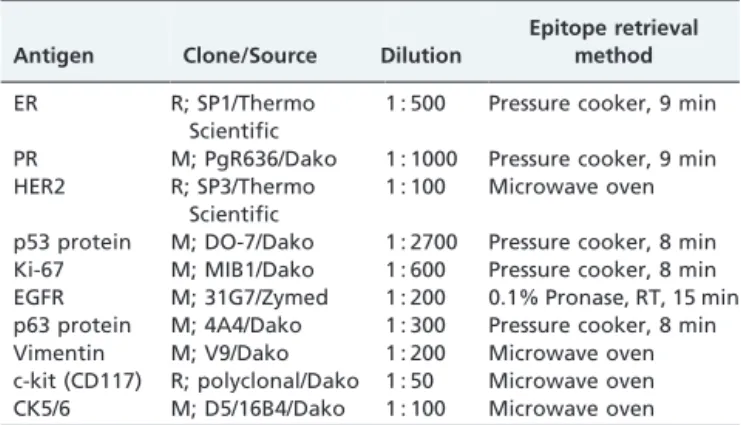
Table 1 -Reagents and methods used for immunohistochemical analysis.
Antigen Clone/Source Dilution
Epitope retrieval method
ER R; SP1/Thermo
Scientific
1 : 500 Pressure cooker, 9 min
PR M; PgR636/Dako 1 : 1000 Pressure cooker, 9 min
HER2 R; SP3/Thermo
Scientific
1 : 100 Microwave oven
p53 protein M; DO-7/Dako 1 : 2700 Pressure cooker, 8 min Ki-67 M; MIB1/Dako 1 : 600 Pressure cooker, 8 min EGFR M; 31G7/Zymed 1 : 200 0.1% Pronase, RT, 15 min p63 protein M; 4A4/Dako 1 : 300 Pressure cooker, 8 min
Vimentin M; V9/Dako 1 : 200 Microwave oven
c-kit (CD117) R; polyclonal/Dako 1 : 50 Microwave oven CK5/6 M; D5/16B4/Dako 1 : 100 Microwave oven
ER: Estrogen receptor; PR: Progesterone receptor; EGFR: Epidermal growth factor receptor; CK: cytokeratin
Pressure cooker: citrate buffer (pH 6) (Tender Cooker, Nordic Wave, USA) Microwave: citrate buffer (pH 6), 15 min (Eletrolux, 900 W)
Table 2 -Pathological and immunohistochemical features of TN Breast carcinomas in younger (#35 years) vs. older ($60 years) patients.
Variable
#35 years (n = 213) N (%)
$60 years (n = 117)
N (%) p-value1
TN carcinomas 74 (34.7) 19 (16.2) 0.0004
Histological grade 3 56 (75.7) 14 (73.7) NS2
Nuclear grade 3 55 (74.3) 14 (73.7) NS
LVI3 12 (16.2) 2 (10.5) NS
Tumoral necrosis 39 (52.7) 6 (31.6) NS
Basal-like molecular profile4 55 (74.3) 13 (68.4) NS
CK5/6 7 (9.4) 8 (42.1) 0.0005
EGFR 53 (71.6) 9 (47.3) 0.0045
Ki-67 (.25%) 51 (68.9) 12 (63.1) NS
p63 1 (1.3) 0 NS
p53 26 (35.1) 7 (36.8) NS
Vimentin 31/71 (43.7)5 5/18 (27.8)5 NS
c-kit 15/71 (21.2)5 7 (36.8) NS
1Pearson’s chi-square test
2Non-significant
3Lymphovascular invasion
4EGFR and/or CK5/6 positive
5Two cases in the younger group and one case in the older group could
not be assessed
Triple-negative breast cancer in young patients
Carvalho FM et al. CLINICS 2010;65(10):1033-1036
the frequency between the two groups (Figures 1 and 2). Tumors that were derived from the younger patients were more frequently EGFR positive (71.6% vs. 47.3%,
respec-tively) and exhibited a lower frequency of CK5/6 expression (9.4% vs. 42.1%, respectively). Regarding the basal-like
tumors of younger patients, 53/55 (96.4%) were associated with EGFR expression, whereas in the older patient group, 9/ 13 (69.2%) expressed EGFR (p = 0.0018). c-KIT expression was also observed more frequently in tumors that occurred in older patients (36.8%) than in younger patients (21.2%), although the difference was not significant. The analysis of other markers, including Ki-67, p63, p53, and vimentin, did not show any significant differences between the two groups.
DISCUSSION
Breast cancer that is detected in younger women is typically associated with aggressive behavior and results in a poor prognosis. Unfortunately, the specific mechanisms responsible for this tumor phenotype remain unclear. This poor understanding is further complicated by the controver-sial results of many studies. Currently, breast cancers that are diagnosed in younger patients are characterized by reduced hormone sensitivity and high HER2/EGFR expression,2,16
although ER-positive carcinomas are still a prevalent
subgroup of tumors in this age group.1,16 Furthermore, luminal carcinomas in younger women are mostly subtype B, which is determined by their frequent co-expression of HER2 and/or their high proliferative activity.16 Triple-negative carcinomas tend to occur less frequently than luminal car-cinomas; however, triple-negative carcinomas are more prevalent in patients who are younger than 35 years old.17
TN carcinomas are considered to be a group of biolo-gically distinct neoplasias that mostly exhibit a basal phenotype and an aggressive biology. Unlike the other subtypes, targeted agents that are specifically aimed at triple-negative breast tumors are not yet available; thus, this deficiency intensifies the need and interest in advancing novel therapeutic strategies beyond chemotherapy for this subset of high-risk patients. Triple-negative is a term based on clinical assays for ER, PR, and HER2, whereas basal-like is a molecular phenotype that was initially defined using cDNA microarrays. However, low-grade tumors, such as adenoid cystic and medullary carcinomas, are also con-sidered TN carcinomas.13,18 Therefore, the best criteria to
characterize the phenotype of basal carcinomas have remained somewhat controversial. Nielsen et al. proposed that the best immunohistochemistry predictors of basal-like gene expression are the positive expression of CK 5/6 and EGFR and the negative expression of HER2 and ER; these criteria were associated with a sensitivity of 76% and a specificity 100%.12In this study, we analyzed these markers; however, it should be noted that other studies have considered the expression patterns of CK5, CK14, CK 17, p-cadherin, p63, c-kit, and vimentin to characterize a basal-like phenotype.19-22Differences in the molecular profile of basal-like carcinomas have also been associated with protein expression patterns of physiological stem/progenitor cells during breast development.23 In this histogenetic model,
adult CK5-positive stem cells are hypothesized to differ-entiate into neoplastic stem cells as a result of EGFR amplification, a mutation of p53, or BRCA inactivation, and this differentiation leads to the generation of basal-like carcinomas. In general, these types of tumors express high levels of p-cadherin, EGFR, and CK5 and an absence or low levels of luminal markers, such as CK 8/18.23 Given the number of markers involved, the possibility that different expression profiles could define distinct subsets of basal-like carcinomas should be considered. For example, Rakha et al. compared tumors with a basal phenotype that were characterized by the expression of basal cytokeratins (CK5/6 or CK14) with cells with a myoepithelial phenotype (p63 or smooth muscle actin).24 The basal phenotype was
associated with a worse outcome, which suggested that although basal and myoepithelial phenotypes share many features, including a similar genetic profile, they possible are distinct tumors.24
Very few studies exist that have examined molecular differences in breast tumors according to age.1-3,9,16,25In the present study, a higher prevalence of TN carcinoma was observed in younger patients (34.3% for younger patientsvs.
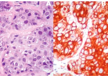
16.3% for older patients), and a basal-like phenotype associated with positive expression of CK5/6 and/or EGFR was present in 74.3% vs. 68.4%, respectively. More importantly, the molecular profiles of the TN carcinomas from younger patients were different from the profiles of the tumors from older patients. Triple negative carcinomas in the younger age group were more frequently EGFR positive (71.6%vs.47.3%, respectively) and less frequently Figure 1 - A high-grade invasive ductal carcinoma (A) with
intense, diffuse CK5/6 immunoexpression (B).
Figure 2 -A high-grade invasive ductal carcinoma with numerous mitotic figures (A) and EGFR immunoexpression (B).
CLINICS 2010;65(10):1033-1036 Triple-negative breast cancer in young patients Carvalho FM et al.
CK5/6 positive (9.4% vs. 42.1%, respectively). The role of
EGFR expression in breast cancer has been investigated by many authors.11,14,26,27 Viale et al. reported a worse
prog-nosis for patients with TN carcinomas that had EGFR-immunoreactivity present in more than 50% of neoplastic cells, which suggested a possible prognostic role for quantification.14Similarly, Rimawi et al. used
radioligand-binding assays to quantify EGFR expression in frozen sections of breast tissue carcinomas and found that higher levels of EGFR were associated with younger and black women, a more aggressive outcome, and lower levels of hormone receptors.27The relationship between EGFR and BRCA1 has been investigated by different groups, and
BRCA1-germline mutations have been associated with a
basal-like phenotype and younger age.8-11 In a study by Collins et al. that analyzed BRCA1 in 144 TN carcinomas, independent of the patient’s age,26 they found a higher prevalence of basal CK and EGFR among TN breast cancers; however, the frequency of expression of these markers was similar in women with and withoutBRCA1mutations.26In
contrast, Arnes et al. found that EGFR was a predictor of
BRCA1status, followed by patient age and ER status.11In
the same study, a worse prognosis was associated with a mutation ofBRCA1and positive expression of EGFR.11
As a receptor tyrosine kinase that plays essential roles in both normal physiological conditions and cancerous condi-tions, EGFR can affect many important characteristics of a cancer’s phenotype, including evasion of apoptosis, prolif-eration, invasion, and metastasis. In a previous study, the frequency of EGFR-positive tumors among young patients was found to be higher than among older women (5.9% vs. 3.3%, respectively), although the difference was not statis-tically significant.16 Based on the results of that study and the data presented in this report, EGFR expression appears to play an important role in the early onset of breast cancer, and we hypothesize that this role is related to intrinsic genetic differences that result in an adverse outcome that is associated with the aggressive breast carcinomas. Further-more, we propose that EGFR immunostaining and/or amplification should be further investigated as predictor of a patient’s response to targeted therapy.
ACKNOWLEDGEMENTS
This work was supported by FAPESP (Fundac¸a˜o de Amparo a Pesquisa do Estado de Sao Paulo): grant#2007-03139-9 (FMC) and scientific initiation scholarship#07/51613-1 (LMB).
REFERENCES
1. El Saghir NS, Seoud M, Khalil MK, Charafeddine M, Salem ZK, Geara FB, et al. Effects of young age at presentation on survival in breast cancer, BMC Cancer. 2006;6:194, doi: 10.1186/1471-2407-6-194.
2. Anders CK, Hsu DS, Broadwater G, Acharya CR, Foekens JA, Zhang Y, et al. Young age at diagnosis correlates with worse prognosis and defines a subset of breast cancers with shared patterns of gene expression, J Clin Oncol. 2008;26:3324-30, doi: 10.1200/JCO.2007.14.2471.
3. Fredholm H, Eaker S, Frisell J, Holmberg L, Fredriksson I, Lindman H. Breast cancer in young women: poor survival despite intensive treatment, PLoS ONE. 2009;4:e7695, doi: 10.1371/journal.pone.0007695. 4. Bhargava R, Striebel J, Beriwal S, Flickinger JC, Onisko A, Ahrendt G,
et al. Prevalence, morphologic features and proliferation indices of breast carcinoma molecular classes using immunohistochemical surrogate markers, Int J Clin Exp Pathol. 2009;2:444-55.
5. Sorlie T, Wang Y, Xiao C, Johnsen H, Naume B, Samaha RR, et al. Distinct molecular mechanisms underlying clinically relevant subtypes of breast cancer: gene expression analyses across three different platforms, BMC Genomics. 200;7:127, doi: 10.1186/1471-2164-7-127.
6. Perou CM, Sorlie T, Eisen MB, van de Rijn M, Jeffrey SS, Rees CA, et al. Molecular portraits of human breast tumours, Nature. 2000;406:747-52, doi: 10.1038/35021093.
7. Rakha EA, El-Sayed ME, Green AR, Lee AH, Robertson JF, Ellis IO. Prognostic markers in triple-negative breast cancer. Cancer. 2007;109:25-32, doi: 10.1002/cncr.22381.
8. Lund MJ, Trivers KF, Porter PL, Coates RJ, Leyland-Jones B, Brawley OW, et al. Race and triple negative threats to breast cancer survival: a population-based study in Atlanta, GA, Breast Cancer Res Treat. 2009;113:357-70, doi: 10.1007/s10549-008-9926-3.
9. Parise CA, Bauer KR, Caggiano V. Variation in breast cancer subtypes with age and race/ethnicity.Crit Rev Oncol Hematol. 2009.
10. Bauer KR, Brown M, Cress RD, Parise CA, Caggiano V. Descriptive analysis of estrogen receptor (ER)-negative, progesterone receptor (PR)-negative, and HER2-negative invasive breast cancer, the so-called triple-negative phenotype: a population-based study from the California cancer Registry. Cancer. 2007;109:1721-8, doi: 10.1002/cncr.22618. 11. Arnes JB, Begin LR, Stefansson I, Brunet JS, Nielsen TO, Foulkes WD,
et al. Expression of epidermal growth factor receptor in relation to BRCA1 status, basal-like markers and prognosis in breast cancer, J Clin Pathol. 2009;62:139-46, doi: 10.1136/jcp.2008.056291.
12. Nielsen TO, Hsu FD, Jensen K, Cheang M, Karaca G, Hu Z, et al. Immunohistochemical and clinical characterization of the basal-like subtype of invasive breast carcinoma. Clin Cancer Res. 2004;10:5367-74, doi: 10.1158/1078-0432.CCR-04-0220
13. Moinfar F. Is ‘basal-like’ carcinoma of the breast a distinct clinicopatho-logical entity? A critical review with cautionary notes, Pathobiology. 2008;75:119-31, doi: 10.1159/000123850.
14. Viale G, Rotmensz N, Maisonneuve P, Bottiglieri L, Montagna E, Luini A, et al. Invasive ductal carcinoma of the breast with the ‘‘triple-negative’’ phenotype: prognostic implications of EGFR immunoreactiv-ity, Breast Cancer Res Treat. 2009;116:317-28, doi: 10.1007/s10549-008-0206-z.
15. Wolff AC, Hammond ME, Schwartz JN, Hagerty KL, Allred DC, Cote RJ, et al. American Society of Clinical Oncology/College of American Pathologists guideline recommendations for human epidermal growth factor receptor 2 testing in breast cancer, J Clin Oncol. 2007;25:118-45, doi: 10.1200/JCO.2006.09.2775.
16. Bacchi LM, Corpa M, Santos PP, Bacchi CE, Carvalho FM. Estrogen receptor-positive breast carcinomas in younger women are different from those of older women: A pathological and immunohistochemical study. Breast. 2010;19:137-41, doi: 10.1016/j.breast.2010.01.002. 17. Rhee J, Han SW, Oh DY, Kim JH, Im SA, et al. The clinicopathologic
characteristics and prognostic significance of triple-negativity in node-negative breast cancer. BMC Cancer. 2008;8:307, doi: 10.1186/1471-2407-8-307.
18. Diaz LK, Cryns VL, Symmans WF, Sneige N. Triple negative breast carcinoma and the basal phenotype: from expression profiling to clinical practice, Adv Anat Pathol. 2007;14:419-30, doi: 10.1097/PAP. 0b013e3181594733.
19. Fulford LG, Easton DF, Reis-Filho JS, Sofronis A, Gillett CE, Lakhani SR, et al. Specific morphological features predictive for the basal phenotype in grade 3 invasive ductal carcinoma of breast, Histopathology. 2006;49:22-34, doi: 10.1111/j.1365-2559.2006.02453.x.
20. Matos I, Dufloth R, Alvarenga M, Zeferino LC, Schmitt F. p63, cyto-keratin 5, and P-cadherin: three molecular markers to distinguish basal phenotype in breast carcinomas. Virchows Arch. 2005;447:688-94, doi: 10. 1007/s00428-005-0010-7.
21. Livasy CA, Karaca G, Nanda R, Tretiakova MS, Olopade OI, Moore DT, et al. Phenotypic evaluation of the basal-like subtype of invasive breast carcinoma. Mod Pathol. 2006;19:264-71, doi: 10.1038/modpathol.3800528. 22. Fadare O, Tavassoli FA. The phenotypic spectrum of basal-like breast cancers: a critical appraisal. Adv Anat Pathol. 2007;14:358-73, doi: 10. 1097/PAP.0b013e31814b26fe.
23. Korsching E, Jeffrey SS, Meinerz W, Decker T, Boecker W, Buerger H. Basal carcinoma of the breast revisited: an old entity with new interpre-tations. J Clin Pathol. 2008;61:553-60, doi: 10.1136/jcp.2008.055475. 24. Rakha EA, Putti TC, Abd El-Rehim DM, Paish C, Green AR, Powe DG,
et al. Morphological and immunophenotypic analysis of breast carcino-mas with basal and myoepithelial differentiation. J Pathol 2006;208:495-506, doi: 10.1002/path.1916.
25. McAree B, O’Donnell ME, Spence A, Lioe TF, McManus DT, Spence RA. Breast cancer in women under 40years of age: A series of 57 cases from Northern Ireland. Breast. 2010;19:97-104, doi: 10.1016/j.breast.2009.12. 002.
26. Collins LC, Martyniak A, Kandel MJ, Stadler ZK, Masciari S, Miron A, et al. Basal cytokeratin and epidermal growth factor receptor expression are not predictive of BRCA1 mutation status in women with triple-negative breast cancers. Am J Surg Pathol. 2009;33:1093-7, doi: 10.1097/ PAS.0b013e3181b7cb7a
27. Rimawi MF, Shetty PB, Weiss HL, Schiff R, Osborne CK, Chamness GC, et al. Epidermal growth factor receptor expression in breast cancer association with biologic phenotype and clinical outcomes. Cancer. 2010;116:1234-42, doi: 10.1002/cncr.24816.
Triple-negative breast cancer in young patients
Carvalho FM et al. CLINICS 2010;65(10):1033-1036