1113
IMMUNOCHEMICAL AND PCR ANALYSIS OF STAPHYLOCOCCUS AUREUS ENTEROTOXIN
B (SEB) IN MILK AND FRUIT JUICES COLLECTED IN LAHORE, PAKISTAN
NADIA DAR1, MEHWISH JAVED2 and ZAHOOR QADIR SAMRA2
1 Department of Biology, Jinnah Degree College for Women, Mozang Road, Lahore, Pakistan
2 Institute of Biochemistry and Biotechnology, Quaid-i-Azam Campus, University of the Punjab, Lahore 54590, Pakistan
Abstract - Enterotoxins secreted by S. aureus are known as a food-poisoning agent that is associated with various gastro-intestinal pathological conditions. In this study, a one-step immunodetection method was devised for routine checking of SEB in milk and fruit juices available locally. Antibodies against recombinant SEB were raised, purified, and cross reactivity was checked against clinically important bacteria (Shigella flexneri, Streptococci, Salmonella typhi, Klebsiella and Bacillus subtilis). Purified anti-SEB antibodies were conjugated with gold nanoparticles (Ab-GNPs) for direct detection of SEB in samples. SEB (33%, 4.76% and 15%) was found in non-sterilized milk (118), sterilized milk (42) and juices (60), respec-tively. Coagulase, MSA tests and PCR amplification of 725 bp of the SEB gene confirmed the presence of S. aureus in the collected samples positive for SEB. Immunoassay is easy, reliable and less time consuming and will be helpful to detect the SEB in food samples at local level.
Key words: SEB, gold nanoparticles, antibodies, food, Pakistan
INTRODUCTION
Staphylococcus aureus (S. aureus) is considered as a pathogen and food-poisoning illness-causing agent. The food poisoning is associated with the ingestion of food contaminated with the S. aureus enterotox-ins. Enterotoxins are stable and retain their biologi-cal activity during heat treatment and normal cook-ing. Generally, staphylococcal enterotoxins (SEs) are resistant to proteases and retain toxic proper-ties in dairy products. Indeed, enterotoxins in food have caused outbreaks where the incriminated food had already undergone heat treatment (Asao et al., 2003).
Gastrointestinal disturbance is the major symp-tom of the food poisoning caused by SE. Even a low dose of non-degraded toxin is sufficient to cause
food poisoning and disturb normal physiological conditions (Asao et al., 2003). Among SE, SEB is a low molecular weight protein of nearly 28 KDa and is important due to its rapid production in dairy products. SEB can be associated with atopic eczema (Breuer et al., 2000; Bunikowski et al., 1999; Mem-pel et al., 2003), rheumatoid arthritis (Howell et al., 1999; Uematsu et al., 1991), and toxic shock syn-drome (Herz et al., 1999). SEB has also been reported as potential bioweapon (Henghold, 2004; Ler et al., 2006; Rosenbloom et al., 2002; Wiener, 1996).
1114 NADIA DAR ET AL.
Omoe et al., 2003; Lina et al., 2004; Thomas et al., 2006). As superantigens, SEB can initiate the activa-tion and proliferaactiva-tion of T-cells through interacactiva-tion with receptors on T-cells, and can cause toxic shock syndrome (Marrack and Kappler, 1990). This condi-tion can cause pyrogenicity, enhance lethal endotox-in shock, and endotox-induce the release of endotox-inflammatory cy-tokines such as tumor necrosis factor and interleukin 1 (IL-1) (Bohach et al., 1990).
The polymerase chain reaction (PCR) has been developed to detect SEB producing S. aureus (Hohn-son et al., 1991; Sharma et al., 2000). However, this assay is labor-intensive, time-consuming and re-quires post-PCR electrophoresis to detect the am-plified products. The real-time PCR assay is used for the quantitative detection of enterotoxigenic S. aureus but this method needs expensive equipment (Sharma et al., 2000). Loop-mediated isothermal amplification (LAMP) of DNA is time consuming for the routine diagnostic analysis of S. aureus (Klotz et al., 2003; Notomi et al., 2000). The sensitivity of the LAMP assay is generally higher than the conven-tional PCR assay and can detect enterotoxigenic S. aureus strains within 60 min. Immuno-PCR is used to detect S. aureus and is also a cumbersome pro-cedure (Goto et al., 2007). The magnetic bead im-munoassay for SEB detection is difficult for routine analysis by technicians (Alefantis et al., 2004).
PCR and RT-PCR assay are used to detect the SE gene, however, they require skill and equipment that are not easily available in under-developed countries. Another approach is using gold nanoparticles in the detection system, which is easy and no specific skill is required. The gold nanoparticles can be synthesized easily and used for antibody or protein immobiliza-tion due to their physical adsorpimmobiliza-tion mechanism.
In the Lahore metropolitan area, the available milk, milk products and juices are prepared in un-hygienic conditions. It has been observed that a large number of humans, especially children, suffer from gastrointestinal problems from April to September. The objective of this study was to devise an easy and cheap method for routine detection of SEB in
food. Prior detection of SEB in food will be helpful to monitor food poisoning as well as to reduce gas-trointestinal pathological conditions.
MATERIALS AND METHODS
Sample collection and processing
Raw milk samples (n = 118, cow and buffalo) were collected under sterile conditions from different distribution shops in the Lahore metropolitan area, Pakistan. Boiled and then cooled milk samples (n = 42) were also collected from shops near bus stops and railway station. Sealed juices (n = 60) of differ-ent fruits, sold at bus stops and the railway station, were also collected. 100 ml of each milk sample was kept in an ice bath and transported to the laborato-ry for storage at 4°C. The samples were centrifuged at 7000 rpm for 15 min at 4°C. The cream layer was removed and the remaining part was stored at -20°C until further processing. The collected juice samples were also stored at -20°C until further process. The juice and milk samples were diluted (1:5 dilution) with sterilized normal saline (0.85 % NaCl) for immunochemical assays. 0.5 ml aliquots of samples was used to inoculate a 5.0 ml mannitol salt medium and kept for 16 h at 37oC. The next day,
the growing culture was centrifuged at 7000 rpm at 4oC. Bacterial pellets were separated and used for
chromosomal DNA extraction and PCR analysis of SEB gene.
Maintenance and culturing of bacterial strains
IMMUNOCHEMICAL AND PCR ANALYSIS OF STAPHYLOCOCCUS AUREUS ENTEROTOXIN B (SEB) IN MILK AND FRUIT JUICES 1115
sterilized milk for 16 h at 37°C separately. The cell-free supernatants were separated by centrifugation of the culture at 8000 rpm for 20 min at 4°C and used in immunoassays to test the specificity of antibodies.
Preparation of SEB
An E. coli strain (BL21-codon plus) carrying a pET28a-SEB hybrid vector expressing recombinant SEB was obtained from Dr. Zahoor Q. Samra (Mo-lecular biotechnology lab, IBB, University of the Punjab). The recombinant SEB was purified on nickel-agarose affinity resin as described (Sambrook and Russel, 2001). The fractions collected from the nickel resin were loaded on a Sephadex G-75 col-umn (1.5 x 30 cm) equilibrated with 20 mM Tris-Cl, pH 7.4 buffer, and 1.0 ml fractions were collected. The collected fractions were checked on 10% SDS-PAGE (Laemmli, 1970). The 28 KDa protein bands were pooled and the protein concentration was
de-Fig. 1. Western blot analysis for specificity of anti- SEB antibod-ies.
Purified SEB, S. aureus (IBB-2011) producing SEB and culture supernatant of contaminated milk with S. aureus were separated on a 10% SDS-polyacrylamide gel, followed by Western blot analysis. Lane 1 - molecular weight protein marker (Fermen-tas, SM0671); lane 2 - recombinant SEB; lane 3 - supernatant of growing culture of S. aureus (IBB2011); lane 4 - culture superna-tant of contaminated milk with S. aureus (IBB 2011). A 28 KDa protein band indicated the specificity of antibody in pure and contaminated samples. A light band of the blue formazan reac-tion product was observed below the 28 KDa protein in lanes 3 and 4. This band may be the degraded product of SEB protein.
Fig.2. Immunoslot blot analysis of SEB.
Detection of SEB in milk and fruit samples by gold conjugated anti-SEB antibodies. Slot 1 - negative control (milk sample con-taminated with non-pathogenic E.coli (DH5α); slot 2 - purified SEB; slot 3, growing culture of S. aureus (IBB-2011); slots 4 to 9 - non-sterilized milk; slots 10 to 12 - boiled then cooled milk; slots 13-24 - juice samples. The reactivity indicated the direct detec-tion of SEB in the contaminated samples. Ab-GNPs did not show reactivity with culture supernatant of Shigella flexneri, Strepto-cocci, Salmonella typhi, Klebsiella and Bacillus subtilis (data not shown).
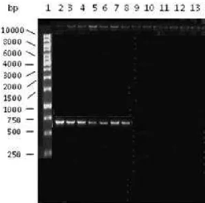
Fig. 3. Agarose gel electrophoresis analysis of PCR amplification of the SEB gene. 5 µl of each amplified sample was loaded. Lane 1 - DNA marker (Fermentas); lane 2 - S. aureus IBB 2011; lanes 3-5 - S. aureus isolated from milk samples; lanes 6–8 - S. aureus
1116 NADIA DAR ET AL.
termined by the Bradford reagent assay using bovine serum albumin as standard (Bradford, 1976). The SEB protein concentration was adjusted to 1.0 mg/ ml and stored at -20°C until further use.
Production and affinity purification of antibodies
Polyclonal antibodies against the SEB antigen were developed in mice (Balb/C, 5 to 6 weeks old) by adopting the guidelines for laboratory animals. 5.0 ml of purified recombinant SEB was mixed with 2.0% buffered-formalin (v/v, in 10 mM Tris-Cl, pH 7.4) to diminish the toxicity to animals. Formalin-treated SEB protein was precipitated with 80% am-monium sulphate saturation. The precipitated SEB protein was collected at 13000 rpm for 20 min at 4oC. The pellet was resuspended in 10 mM Tris-Cl,
pH 7.4, and dialyzed against the same buffer. The formalin-treated SEB protein was mixed with Fre-und’s complete adjuvant in 1:1 ratio and used as an immunogen. Each mouse received 50 to 60 µg pro-tein/per administration, and a total six injections were subcutaneously applied at two-week intervals. Blood (0.1 ml) was drawn from the mouse tail and checked for the anti-SEB antibody titer by enzyme-linked immunosorbent assay (ELISA). After check-ing the antibody titer, blood was isolated from the heart and serum was separated (Harlow and Lane, 1988). The purified recombinant SEB antigen was conjugated with CNBr-activated sepharose 4B ac-cording to the manufacturer’s instruction (Pharma-cia) and used to affinity purify the SEB anti-bodies (Harlow and Lane, 1988). The concentration of purified antibodies was checked by Bradford rea-gent assay and stored at -20°C until further proc-ess.
Characterization of antibodies Enzyme-linked immunosorbent assay
Ten μl of purified recombinant SEB (0.1 mg/ml) and culture supernatant of bacterial strains (S. aureus IBB 2011, Shigella flexneri, Streptococci, Salmonella typhi, Klebsiella and Bacillus subtilis) was mixed separately with 0.09 ml of 0.05 M carbonate buffer, pH 9.0, and was absorbed onto microtiter ELISA plates for one hour at 37°C. The wells were treated with 3% bovine serum albumin in Tris-buffered saline-Tween 20, (TBST) for 45 min at 37°C. The plates were washed with TBST and then affinity-purified mouse anti-SEB antibody (1:2000 dilutions) was added and kept at 37°C for 50 min. After washing with TBST, rabbit anti-mouse IgG antibody – alkaline phosphatase con-jugated (1:5000 dilution), was added and incubated at 37°C for 40 min. After the washings with TBST, a substrate of alkaline phosphatase (para-nitrophenyl phosphate in 0.01 M ethanolamine buffer pH 9.00, 1.0 mM MgCl2) was added to each well (Harlow and
Lane, 1988). The pre-immune serum collected before the immunization was used as control.
Western blot analysis
The monospecificity of anti-SEB antibodies was also analyzed by Western blot analysis. Purified recom-binant SEB and the culture supernatants of growing a native strain of S. aureus (IBB-2011) in minimal salt medium and LB medium, were run on 10% SDS-poly acrylamide gel (Laemmli, 1970) and then transferred onto a nitrocellulose (NC) membrane (Towbin et al., 1979). Non-specific sites on the NC-membrane were blocked with 3% BSA in TBST for 45 min at 37°C. The NC membrane was further incubated with
puri-Table 1. Tests of SEB in milk and juice samples.
No. Samples size positive (n = )
Immunoslot blot analysis & %
MSA test positive & %
Coagulase test positive & %
PCR Assays & %
1 Milk* (118) 39 (33.05%) 63 (53.38%) 39 (33.05%) 39 (33.05%)
2 Milk** (42) 3 (7.14%) 11 (26.19%) 3 (7.1466%) 3 (7.14%)
3 Juices (60) 9 (15%) 17 (28.33) 9 (15%) 9 (15%)
fied anti-SEB antibodies (1:2000 dilution) and then with rabbit anti-mouse IgG antibody – alkaline phos-phatase conjugated (1:5000 dilution). After extensive washing, the blots were exposed to nitroblue tetrazo-lium and 5-bromo 4-choloro 3-indolyl phosphate as substrates and the results were noted.
Preparation of colloidal gold nanoparticles and conjugation
Colloidal gold nanoparticles (20-25 nm) were pre-pared by reducing chloroauric acid (HAuCl4)
(Walk-er, 1994). Chloroauric acid (0.1 g) was dissolved in 1 l of double-distilled deionized water and boiled for 30 min; 12.5 ml of 1% trisodium citrate was added under vigorous stirring. The gold solution turned from yellow to dark blue and finally to red. The gold nanoparticle solution was cooled to room tempera-ture and pH 7.5 was adjusted with 0.1 M K2CO3
so-lution. The final color of the prepared gold-particle solution was deep red and stored at 4°C in an air-tight container. Affinity-purified anti-SEB antibody was conjugated with gold nanoparticles. The amount of antibody necessary to stabilize the gold solution was determined by flocculation test. Briefly, two-fold dilutions of anti-SEB antibody were made. Gold so-lution (2.5 ml) was added to each 0.5 ml of diluted antibodies and the color of the solution was noted. The lowest concentration of antibody that stabilizes the red-colored gold solution was noted.
For conjugation, 0.1 ml of affinity-purified anti-body (20 µg) was mixed with 1.0 ml of gold-nano-particle solution and agitated for 30 min at 25°C. The solution was centrifuged at 13000 rpm for 35 min at 4°C. Supernatant was discarded and the red pellet of antibody-gold complex (Ab-GNPs) was resuspended in 0.5 ml of stabilizing buffer (10 mM Tris-Cl, pH 7.5, 0.0125 gm polyethylene glycol, 0.15 M sodium chloride, and 2.0 mg NaN3 dissolved in 25 ml
deion-ized H2O) and stored at 4oC.
Immunodot blot analysis for Ab-GNPs complex
Nitrocellulose membrane was cut (3.0 cm x 5.0 cm) and 5.0 µ of purified recombinant SEB antigen was
applied to the membrane and allowed to dry. The membrane was stained with acidic Ponceau-S stain (0.1% in 1.0% acetic acid solution) for 5 min in or-der to check the presence of antigen. The membrane was destained with several changes of Tris-buffered saline. The membrane was dipped in blocking buffer (3% BSA in TBS or 2% gelatin in TBS) for 30 min at room temperature to block the nonspecific bind-ing sites. After washbind-ing with 1 x TBS, 0.01 ml of Ab-GNPs solution was diluted in 0.5 ml Tris-buffered-saline solution and added to the membrane for 10-15 min. The blots were rinsed with TBS and the color was noted.
Detection limit of SEB
A serial dilution of purified recombinant SEB anti-gen (1, 5, 10, 20, 30, 40, 50 ng/ml in 0.85 % normal saline) was made. Ten µl of each dilution was applied onto the nitrocellulose membrane and used for im-munodot blot reaction as described above. The dep-osition of Ab-GNPs complex on a minimum amount of SEB in immunodot blot was noted.
Immunoslot blot analysis for SEB antigen in samples
Diluted samples (0.2 ml) of milk and juices were ab-sorbed in a slot blot apparatus (Bethesda Research Laboratories, USA) containing the NC membrane according to the manufacturer’s instructions. The NC membranes were rinsed with TBST and stained with Ponceau S to check for the presence of proteins. The blots were rinsed again with TBST and incubated with blocking buffer (3% BSA in TBST) for 40 min at room temperature. The blots were further incubat-ed with Ab-GNPs for 15 min at room temperature and results were noted. The same quantity of diluted samples was also checked for the presence of SEB by commercially available SEB test strips (Standard di-agnostic Inc, Korea).
Confirmation of S. aureus in food samples
1118 NADIA DAR ET AL.
PCR tests to confirm our immunodetection results. All tests were conducted in triplicate.
MSA test
Food samples (milk and juices; 0.2 ml) were taken in a tube containing 5 ml mannitol salt medium (1% mannitol, 7.5% NaCl, 0.1% beef extract, 1% peptone, 0.0025% phenol red, pH 7.4), and incubated at 37°C. The growing culture was further spread on minimal salt-agar medium and incubated for 24-30 h at 37°C. The growing culture was further processed for Gram staining (Benson, 1998).
Blood agar and coagulase test
A single colony from the MSA medium was picked and spread on the blood agar plate. The plate was kept for 24 h at 37°C. The same colony was used to inoculate 1 ml of human serum and kept at 37°C.
PCR analysis
The bacterial pellets isolated from the milk and juice samples were also processed for SEB gene detection by PCR. The chromosomal DNA of bacterial strains (S. aureus IBB 2011, bacterial culture isolated from milk and juice samples and the above-mentioned clinically important bacterial strains) was isolated as described (Qiagen, USA) and used for amplification of the SEB gene. The primers for the SEB gene were designed using the available nucleotide sequence of SEB gene at the NCBI (accession number AY852244). F-primer, (5/-ATGGAGAGTCAACCAGATC-3/) and
R-primer, (5/-TCACTTTTTCTTTGTCGTAAC-3/)
and were custom synthesized from Fermentas. The SEB gene was amplified in iCycler (Biorad) by using 1 µl of template DNA (0.1 mg/ml), 2.5 units of Taq DNA polymerase, PCR buffer 1 x, 0.25 mM dNTPs,, 2.5 mM MgCl2, 0.5 µM of each forward and the
re-verse primer. The conditions for amplification are as follows: initial denaturation at 95°C for 4 min fol-lowed by 30 cycles of denaturation at 95°C for 1 min, annealing at 56°C for 1 min, and elongation at 72°C for 10 min. After amplification, an aliquot of PCR was mixed with commercially available DNA
load-ing dye (Fermentas) and analyzed on a 1% agarose, 0.5 x Tris-Borate-EDTA buffer.
RESULTS AND DISCUSSION
Characterization and immunodetection of SEB
The recombinant SEB was used to develop the poly-clonal antibodies and its monospecificity was also characterized by Western blot analysis (Fig. 1). Af-finity-purified anti-SEB antibodies were conjugated with gold nanoparticles for direct detection of tox-ins in the milk and juice samples. The development of a pink color on the samples in immunodot blot indicated the specific binding of the antibody with SEB. The minimum appropriate detection limit in the positive control was 1-5 ng/ml. A light color of the Ab-GNPs complex was observed on the blot with 1.0 ng/ml of pure SEB. An appropriate detection was observed in 5 ng/ml. Milk and juice samples were collected from different public areas to examine the presence of SEB by Ab-GNPs (Fig 2).
Immunoslot blot analysis indicated that out of 118 raw milk samples, 39 (33.05%) were contaminat-ed with SEB, 3 (7.14%) out of the 42 boilcontaminat-ed-coolcontaminat-ed milk samples, and 9 (15%) out of 60 juice packets were also contaminated with SEB. The overall per-centage contamination of SEB in the milk and juices were 33% and 15%, respectively (Table 1).The culture supernatants of other microbes (described above) were also tested to cross-check reactivity by Ab-GNPs. It was observed that no other microbes tested for immunochemical reaction showed any reaction except S. aureus IBB 2011. Commercially available immunotest strips also detected the SEB in the sam-ples and similar results were observed.
Biochemical characterization of S. aureus in test samples
co-agulation of plasma confirmed the presence of S. aureus in the samples. Sixty three (53.38%) non-ster-ilized milk samples were positive for the MSA test, and 11 (26.19%) of the boiled-cooled milk samples were positive. A total 17 (28.33%) juice samples were also positive for the MSA test. The coagulase test was positive in 39 (33.05%), 3 (7.14%) and 9 (15%) non-sterilized milk samples, boiled-cooled milk samples and juice samples, respectively. The samples posi-tive for the coagulase test were also tested for the SEB gene of S. aureus. The appearance of a 725 bp PCR product in the tested samples (milk and juic-es) further indicated the contamination of S. aureus (Fig. 3). PCR analysis revealed that 39 (33.05%) of the non-sterilized milk samples, 3 (7.14%) boiled-cooled milk samples and 9 (15%) juice samples were contaminated with S. aureus. The contamination rate was found to be high in the milk samples. The other clinically important bacteria did not show a positive PCR test.
The presence of enterotoxin SEB of S. aureus in milk and fruit juices is a major problem of public health and the local food industry. The food poison-ing due to S. aureus toxin occurs in children and adults depending on the sources that help in the sur-vival of S. aureus. At the moment, there is no effec-tive test available to early check the SEB contamina-tion of food. Generally, biochemical tests are used to check S. aureus contamination/presence of SEB. The expertise for other methods for routine checking of SEB or S. aureus (PCR, immune-PCR) is not avail-able locally. There is a need to devise a simple and reliable method to check the SEB in food and clini-cally important samples.
In this study, gold conjugated anti-SEB antibod-ies were prepared to examine the presence of SEB in milk and juice samples. Our studies indicated a 33% and 15% SEB contamination in the milk and juice samples, respectively. Clinically important microbes did not show any cross reactivity with Ab-GNPs rea-gent, which indicates the specificity and reliability of the devised detection system. The biochemical and PCR tests further provided supporting evidences for the immunodetection of SEB in the samples.
The presence of S. aureus was found to be higher in milk compared to juices. The SEB contamination in milk and juices may be due to the handling of the raw materials under unhygienic conditions and the use of non-sterilized contaminated water. The quan-tity of enterotoxin SEB and S. aureus in the samples depends on many factors: (i) a contaminated carrier, (ii) unhygienic conditions in factories as well as in household production, (iii) ignorance about hygiene, (v) uses of contaminated water and (vi) sterilization procedure.
The presence and proliferation of S. aureus in food for human consumption is a major health prob-lem for children here. The contamination level of SEB should confirm whether a disease is due to S. au-reus infection or to other microbial contamination. Other staphylococcus strains, such as S. intermedius and S. hyicus, are also enterotoxigenic. S. interme-dius is generally considered a veterinary pathogen and has been isolated and characterized from but-ter and margarines. Food poisoning due to S. aureus generally occurs in meat and meat products, cream-filled milk products as well as salads. All these items become contaminated at home or by food supplier companies at local level if handled in unhygienic conditions. It is also reported that if the fermenta-tion of milk products is not completed (e.g. failure of lactic acid consumption), the proliferation of S. aureus is rapid.
immuno-1120 NADIA DAR ET AL.
chemical assays are more sensitive, specific and easy to handle. They can detect a very small amount of antigen in samples.
The direct detection of SEB in food samples by this Ab-GNPs complex is not the replacement of other available detection methods for SEB. Detec-tion of a low concentraDetec-tion of SEB in flow immuno-chromatography assay (1.0 ng/ml) has been report-ed by utilizing the combination of two antibodies (Hwa et al., 2010; Schutt et al., 2002). In this immu-noslot blot assay or immunodot blot assay, the SEB was detected by utilizing only the labeled antibody specific for SEB. Ab-GNPs reagent is reliable, easy to handle and less time is required to detect the SEB in the samples.
CONCLUSION
Anti-SEB antibodies were conjugated successfully with gold nanoparticles. The Ab-GNPs were used to detect SEB in milk and fruit juice samples. The per-centage contamination of SEB in food was observed to be high, which reflects unhygienic conditions. The devised method may be used for early detection of SEB in food samples.
Acknowledgments - The authors are thankful to the Institute of Biochemistry and Biotechnology, University of the Punjab, Lahore, for providing the chemicals and instruments for this study. We are grateful to our colleagues for the constructive discussions during this study.
REFERENCES
Alefantis, T., Grewal, P., Ashton, J., Khan, A.S., Valdes, J.J., and
V.G Del Vecchio (2004), A rapid and sensitive magnetic bead-based immunoassay for the detection of staphylo-coccal enterotoxin B for high-through put screening. Mol Cell Probes; 18, 379-382.
Asao, T., Kumeda Y., Kawai, T., Shibata, T., Oda, H., Haruki, K., Nakazawa, H., andSKozaki (2003). An extensive outbreak of staphylococcal food poisoning due to low-fat milk in Ja-pan: estimation of enterotoxin A in the incriminated milk and powdered skim milk. Epidemiol. Infect; 130, 33-40.
Benson, H.J (1998). Microbiological applications, 7th edn,
McGraw Hill companies, Inc, USA.
Bohach, G.A., Fast, D.J., Nelson, R.D., and P.MSchlievert (1990). Staphylococcal and streptococcal pyrogenic toxins in-volved in toxic shock syndrome and related illnesses. Crit. Rev. Microbiol;17, 251-272.
Bradford, M.M (1976). A rapid and sensitive method for the quantification of microgram quantities of protein utiliz-ing the principle of protein-dye-bindutiliz-ing. Anal. Biochem;
72, 248-251.
Breuer, K., Wittmann, M., Bosche, B., Kapp, A., and T. Werfel
(2000). Severe atopic dermatitis is associated with sensiti-zation to staphylococcal enterotoxin B (SEB). Allergy; 55, 551–555.
Bunikowski, R., Mielke, M., Skarabis, H., Herz, U., Bergmann, R. L., Wahn, U., and H. Renz (1999). Prevalence and role of serum IgE antibodies to the Staphylococcus aureus-derived superantigens SEA and SEB in children with atopic der-matitis. J. Allergy Clin. Immunol; 103, 119–124.
Goto, M., Hayashidani, H., Takatori, K., and Y. Hara-Kudo
(2007). Rapid detection of enterotoxigenic Staphylococcus aureus harbouring genes for four classical enterotoxins, SEA, SEB, SEC and SED, by loop-mediated isothermal amplification assay. Letters in Appl. Micro; 266-8254.
Harlow, D and D. Lane (1988). Antibodies: A Lab Manual of Gold Spring Harbor Laboratory. Gold Spring Harbor, NY,
Henghold, W.B. (2004). Other biologic toxin bioweapons: ricin,
staphylococcal enterotoxin B, and trichothecene mycotox-ins. Dermatol. Clin;22, 257-262.
Herz, U, Bunikowski, R, Mielke, M., and H.Renz (1999). Contri-bution of bacterial superantigens to atopic dermatitis. Int. Arch. Allergy Immunol; 118, 240-241.
Howell, M.D, Diveley, J.P, Lundeen, K.A, Esty, A, Winters, S.T, Carlo, D.J, and S.W. Brostoff (1991). Limited T-cell tor beta-chain heterogeneity among interleukin 2 recep-tor-positive synovial T cells suggests a role for superan-tigen in rheumatoid arthritis. Proc. Natl. Acad. Sci;88, 921-10,925.
Hwa, S.R., Shek, T.S., Jiang, C.D., and Wen, H.Y (2010). Gold nanoparticle-based lateral flow assay for detection of staphylococcal enterotoxin B. Anal Meth; 118 (2), 462-466.
Johnson, W.M., Tyler, S.D., Ewan, E.P., Ashton, F.E., Pollard, D.R.,
and K.R, Rozee (1991). Detection of genes for enterotox-ins, exfoliative toxenterotox-ins, and toxic shock syndrome toxin 1 in
Staphylococcus aureus by the polymerase chain reaction. J Clin Microbiol; 29: 426-430.
Laemmli, U.K (1970). Cleavage of structural proteins during the assembly of the head of bacteriophage T4. Nature; 227,680-685.
Lee, J.C, Takeda, S., Livolsi, P.J., and L. C.Paoletti (1993). Effects of in vitro and in vivo growth conditions on expression of type-8 capsular polysaccharide by Staphylococcus aureus.
Infect. Immun;61, 1853-1858.
Ler, S.G, Lee, F.K, and P.Gopalakrishnakone (2006). Trends in detection of warfare agents. Detection methods for ricin, staphylococcal enterotoxin B and T-2 toxin. J. Chromatogr. A; 1133, 1-12.
Lina, G., Bohach, G.A., Nair, S.P., Hiramatsu, K., Jouvin-Marche, E. and R.Mariuzza (2004). International nomenclature committee for Staphylococcal superantigens. Standard nomenclature for the superantigens expressed by Staphy-lococcus. J Infect Dis; 189, 2334-2336.
Marrack, P., and K.Kappler (1990). The staphylococcal entero-toxins and their relatives. Science, 248, 705-711.
Mempel, M., Lina, G., Hojka, M., Schnopp, C., Seid, H.P., Scha-fer, T., Ring, J., Vandenesch, F., and D. Abeck (2003). High prevalence of superantigens associated with the egc locus in Staphylococcus aureus isolates from patients with atopic eczema. Eur. J. Clin. Microbiol. Infect. Dis; 22, 306-309.
Notomi, T., Okayama, H., Masubuchi, H., Yonekawa, T., Wa-tanabe, K., Amino, N., and T. Hase (2000). Loop-mediated isothermal amplification of DNA. Nucleic Acids Res; 28, e63.
Omoe K, Hu DL, Takahashi-Omoe H, Nakane A and K.
Shina-gawa (2003). Identification and characterization of a new staphylococcal enterotoxin-related putative toxin encoded by two kinds of plasmids. Infect Immun, 71, 6088-6094.
Rosenbloom, M., Leikin, J.B., Vogel, S.N., and Z.A. Chaudry
(2002). Biological and chemical agents: a brief synopsis.
Am. J. Ther;9, 5-14.
Sambrook, J and D. Russel (2001). Molecular cloning. Cold spring harbor laboratory press, N.Y. A lab manual.
Schutt, U., Langfeldt, N., Peruski, A.H., and H.Meyer (2002). De-tection of Staphylococcal enterotoxin B (SEB) by enzyme linked immunosorbent assay and by a rapid hand-held as-say. Clin Lab; 48 (7-8), 395-400.
Sharma, N.K., Rees, C.E., and C.E. Dodd (2000). Development
of a single-reaction multiplex PCR toxin typing assay for
Staphylococcus aureus strains. Appl Environ Microbiol; 66, 1347-1353.
Thomas, D.Y., Jarraud, S., Lemercier, B., Cozon, G., Echasserieau, K., Etienne, J., Gougeon, M.L, and G.Lina, (2006). Staph-ylococcal enterotoxin-like toxins U2 and V, two new staphylococcal superantigens arising from recombina-tion within the enterotoxin gene cluster. Infect Immun, 74, 4724-4734.
Towbin, H.K,. Staehelin, T., and Gordon, J (1979). Electrophoretic transfer of proteins from polyacrylamide gels to nitrocel-lulose membrane. Proc Natl. Acad Sci;76, 4350-4359.
Uematsu, Y, Wege, H, Straus, A, Ott, M., Bannwarth, W., Lanch-bury, J., Panayi, G, and M. Steinmetz (1991). The T-cell receptor repertoire in the synovial fluid of a patient with rheumatoid arthritis is polyclonal. Proc. Natl. Acad. Sci;
88, 8534-8538.
Walker, J.M (1994). Basic protein and peptide protocols;
Meth-ods in molecular biology. Humana press Inc. Totowa, NJ.