abstract
Stress distribution on dentin-cement-post
interface varying root canal and glass iber post
diameters. A three-dimensional inite element
analysis based on micro-CT data
Priscilla Cardoso LAZARI1, Rodrigo Caldeira Nunes de OLIVEIRA2, Rodolfo Bruniera ANCHIETA1, Erika Oliveira de ALMEIDA1, Amilcar Chagas FREITAS JUNIOR3, Sidney KINA1, Eduardo Passos ROCHA1
1- Department of Dental Materials and Prosthodontics, Araçatuba Dental School, Univ. Estadual Paulista, Araçatuba, SP, Brazil. 2- São Leopoldo Mandic, Campinas, SP, Brazil.
3- School of Health Sciences, Potiguar University - UnP, Natal, RN, Brazil.
Corresponding address: Eduardo Passos Rocha - Faculdade de Odontologia de Araçatuba - UNESP - Departamento de Materiais Odontológicos e Próteses - Rua Jose Bonifacio, 1193 - 16015-050 - Araçatuba - SP - Brasil -Phone: +55-18-36363290 - e-mail: [email protected]
Submitted: February 27, 2013 - Modiication: June 19, 2013 - Accepted: August 23, 2013
O
bjective: The aim of the present study was to analyze the inluence of root canal and glass iber post diameters on the biomechanical behavior of the dentin/cement/post interface of a root-illed tooth using 3D inite element analysis. Material and Methods: Sixmodels were built using micro-CT imaging data and SolidWorks 2007 software, varying
the root canal (C) and the glass iber post (P) diameters: C1P1-C=1 mm and P=1 mm; C2P1-C=2 mm and P=1 mm; C2P2-C=2 mm and P=2 mm; C3P1-C=3 mm and P=1 mm; C3P2-C=3 mm and P=2 mm; and C3P3-C=3 mm and P=3 mm. The numerical analysis was conducted with ANSYS Workbench 10.0. An oblique force (180 N at 45°) was applied to
the palatal surface of the central incisor. The periodontal ligament surface was constrained
on the three axes (x=y=z=0). Maximum principal stress (σmax) values were evaluated for
the root dentin, cement layer, and glass iber post. Results: The most evident stress was observed in the glass iber post at C3P1 (323 MPa), and the maximum stress in the cement layer occurred at C1P1 (43.2 MPa). The stress on the root dentin was almost constant in all models with a peak in tension at C2P1 (64.5 MPa). Conclusion: The greatest discrepancy
between root canal and post diameters is favorable for stress concentration at the post surface. The dentin remaining after the various root canal preparations did not increase the stress levels on the root.
Key words: Dental pins. Dental cements. Finite element analysis. Tooth root. Post and core technique.
IntroductIon
The reestablishment of function through direct
and/or indirect restorations in endodontically
treated teeth remains a challenge11,19. Several studies have demonstrated reduced resistance to failure in teeth that are endodontically treated8.
Tooth architecture is modiied during endodontic
treatment as a result of caries removal, access to the canal, and instrumentation in the root canal17. All these factors are associated with the higher internal stress induced in these teeth since this force
is directly proportional to the fracture resistance and volume of the remaining dental structure12.
This fact becomes critical when excessive reduction
of the thickness of the root canal walls occurs as a result of severe caries, over-instrumentation, previous restoration with large-diameter posts, or internal root resorptions30. These characteristics may increase the risk of fracture in these teeth relative to teeth with more pulp vitality23.
The biomechanics of the endodontically treated
teeth are also inluenced since the insertion of
posts, modiies the mechanical behavior of the tooth
and the distribution of the masticatory efforts1,23. According to some authors, a high number of vertical fractures are observed in roots with metallic posts with a high elastic modulus that distribute and concentrate stress in the apical third of the root11,20,28. Thus, the use of intra-radicular posts with an elastic modulus similar to dentin, such as
glass iber posts, promotes better stress distribution
in the remaining dental structure and along the cementation interface7,28. In addition, it has been suggested that the use of these posts with a resin cement increases the fracture resistance of the root7,11,17,20.
Another important factor is the reduced root fracture susceptibility when using these posts, since fracture of the post or cementation interface may occur prior to root fracture, allowing of restoration of the tooth29. However, despite all these beneits
of glass iber posts (GFP), their selection and
indication are still not fully understood23. Little
information exists regarding the selection of glass iber post diameter for use in lared root canals8,18.
For example, it is unclear whether a large- or
small-diameter post should be used with large small-diameter canals1,5,7,8.
Recent studies show that bonding of the post to
the root canal occurs mainly by friction retention, and micromechanical interlocking via the hybrid layer occurs only in the coronal third of the root10.
Furthermore, the cement ilm thickness should
be carefully evaluated when deciding the post diameter. Higher stresses can occur during cement
curing, mainly because the C-Factor in the canal can exceed 200, compromising the bond strength25.
The aim of the present study was to evaluate the stress behavior of the adhesive interface in the root
canal of a non-vital maxillary central incisor restored
with a ceramic crown, varying the root canal and the
glass iber post diameters. The following hypotheses were tested: 1 – The discrepancy between the post
and root canal diameters does not increase the
stress level on the post interface; 2 – The stress
on the remaining root is directly proportional to the increase in root canal diameter, regardless of the post used.
MaterIal and Methods
This study was approved by the Human Research Ethics Committee at the School of Dentistry of Araçatuba, Univ. Estadual Paulista – UNESP (process #2008/01845). A right maxillary central incisor from the human tooth bank at the Department of Dental Materials and Prosthodontics was used to
build the models.
Based on a micro-computed tomography image (µCT), a total of 720 slices were obtained after
tooth scanning (CT40, Scanco Medical, Bassersdorf, Switzerland). For reconstruction of the solid model, 82 slices were serially selected for tridimensional
reconstruction using SolidWorks 2007 software
(SolidWorks Corp., Concord, MA, USA). These
included all the structures of the natural tooth (enamel, crown and root dentin, dental pulp, and periodontal ligament)4,21.
Based on the previous methodology, six
geometric models were built. In all models, the
tooth was considered as root-illed with a glass iber post inserted, and the remaining crown was
restored with a core and an all-ceramic crown of feldspathic ceramic.
The canal diameter varied (C1=1 mm; C2=2 mm; C3=3 mm) while the glass iber post diameter remained constant (1 mm) to evaluate the inluence
of root canal diameter (C) on stress distribution in
the dentin-cement-post interface (Figure 1). To evaluate the inluence of the glass iber post diameter (P) on stress distribution in the dentin/ cement/post interface, we varied the post diameter (P1=1 mm; P2=2 mm; P3=3 mm) while the canal diameter remained constant (D3=3 mm) (Figure 1).
The tooth reduction and the periodontal ligament
thickness (0.25 mm) were comparable in all models.
The major differences between the models were
root canal diameter, glass iber post diameter, and
thickness of the cementation interface.
After the elaboration of the models, the iles were transferred to the inite element software in IGES format (ANSYS Inc., Canonsburg, PA, USA) to determine the regions and generate the inite
element mesh.
The mechanical properties [Elastic modulus (E)
and Poisson’s ratio (v)] were obtained from speciic
Models Description
C1P1 Root canal and glass iber post with 1 mm of diameter
C2P1 Root canal with 2 mm of diameter and glass iber post with 1 mm of diameter
C3P1 Root canal with 3 mm of diameter and glass iber post with 1 mm of diameter
C2P2 Root canal with 2 mm of diameter and glass iber post with 2 mm of diameter
C3P2 Root canal with 3 mm of diameter and glass iber post with 2 mm of diameter
C3P3 Root canal with 3 mm of diameter and glass iber post with 3 mm of diameter
Figure 1- Description of the models with different root canal (C) and glass iber post (P) diameters
Stress distribution on dentin-cement-post interface varying root canal and glass iber post diameters. A three-dimensional inite element analysis based on micro-CT
literature (Table 1)5,21,22,considering isotropic, homogeneous, and linearly elastic parameters. All structures in these models were considered perfectly integrated5.
The periodontal ligament surface was ixed on x, y and z axes for all models (x=y=z=0). A distributed load of 180 N was applied to the lingual surface of the tooth, in 45° angulation to the tooth’s long axis for all models (Figure 2)27.
In order to achieve the convergence of analysis
(6%)4,21, the mesh was composed of tetrahedral elements of 0.2 mm in size. The models had up to
267,091 elements and 423,719 nodes (Figure 3). Maximum principal stress (σmax) values were evaluated for the root dentin, cement layer, and
glass iber post. According to, these analysis criteria
are appropriate for predicting failures in non-ductile materials, such as dentin and cement layers1,3,16.
results
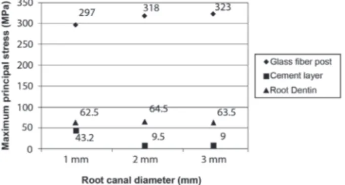
Variation of the root canal diameter (1, 2, and 3 mm) with a constant glass iber post diameter of 1 mm
Different behaviors of the glass iber post and
the cementation interface for a range of root canal
Figure 2- As a boundary condition, the external surface of the periodontal ligament was constrained (left), y=x=z=0. A load of 180 N was applied to a speciic area at the lingual surface of the crown, 45° to the tooth’s long axis (right)
Figure 3- Finite element mesh of the teeth structures. Root dentin (left); Cement line (middle); Glass iber post (right)
Figure 4- Maximum principal stress (MPa) for various
root canal diameters (1 mm, 2 mm, and 3 mm) with a constant glass iber post diameter of 1 mm
Material E (GPa) v
Dentin21 18.6 0.31
Periodontal ligament21 6.9x10⁻⁵ 0.45
Gutta-percha21 1.4x10⁻¹ 0.45
Glass iber post5 40 0.30
Enamel21 80 0,33
Feldspathic ceramic23 69 0.30
Composite resin5 12 0.30
Resin cement5 8 0.30
diameters with constant post diameters.
Increasing stress was observed in the glass iber post with increasing canal diameter. The highest σmax values were observed in model C3P1 (323 MPa), followed by C2P1 and C1P1 (Figure 4) in the incisal region of the post (Figure 5).
However, this behavior was not observed for the cement layer. On the contrary, the thinner the cementation interface (reduced canal diameter), the
higher the σmax level, as observed in models C1P1 (43 MPa), C2P1 (9.5 MPa), and C3P1 (9 MPa). The stress levels differ about 21% between the model
with the highest stress and the model with the
lowest stress (Figure 4).
The maximum σmax was observed in the palatine surface, mainly in the middle third of the cement
layer for C2P1 and C3P1, while in C1P1, the peak
of stress was observed in the coronal area of both
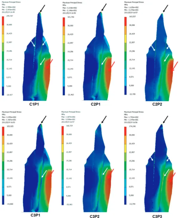
Figure 5- Stress distribution in root-illed models. The stress distributions in the glass iber posts were similar for all models
(palatine surface in the coronal and middle thirds) and the peak of maximum principal stress occurred in the incisal area of the post (black arrow). Note that the stress distributions in the cement layer (white arrows) indicate the stress concentration in the cement line. For C2P1, C3P1, and C3P2, it occurs in the middle third in the palatine surface, while for C1P1, C2P2, and C3P3, the stress concentrates at the middle and near the coronal thirds (white arrows). The stress peak in the dentin was concentrated among the coronal and middle thirds, mainly at the lingual dentin walls (red arrows)
Stress distribution on dentin-cement-post interface varying root canal and glass iber post diameters. A three-dimensional inite element analysis based on micro-CT
the buccal and palatine surfaces (Figure 5).
Variation of the glass iber post diameter (1, 2, and 3 mm) with a constant root canal diameter of 3 mm
The structures of the post/cement interface exhibited different mechanical behaviors when we varied the glass iber post diameter and kept the
root canal diameter constant.
The results showed decreased stress with
increasing glass iber post diameter (Figure 6).
However, although the difference was not as
signiicant, the stress in C3P3 was higher than in C3P2 (Figure 6).
The highest stress level (323 MPa) occurred in C3P1; this was nearly twice the stress observed in C3P2 and C3P3.
Similarly, as the post diameter increased, so did
the stress in the cement layer, which conirms that
the thinner the cement layer, the higher the stress
concentration on it (Figure 6).
The peak of stress occurred in the apical third
of the buccal surface of the cement layer for C3P1 and C3P2, while for C3P3 the peak of stress was
found in the coronal third of the buccal and palatine surfaces.
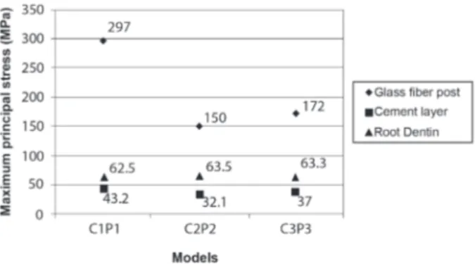
Variation of the root canal and the glass iber post diameters
Considering the use of a fiber post with a
diameter similar to the canal lumen (C1P1, C2P2, and C3P3), the conservative endodontic treatment with the greatest quantity of dental structure (C1P1)
presented the worst mechanical behavior among the models with regard to stress concentration in
the post and cement layer (Figure 7).
The lowest stress concentration was obtained by simulation of intermediary clinical conditions
in C2P2.
Unlike the structures of the glass iber post and
cement layer, stress in the remaining root dentin stayed nearly constant in all models, regardless
of the amount of dentin or the glass iber post diameters (Figures 4, 6, and 7). The highest stress
levels on root dentin were found at the coronal
region of the palatal surface for all models (Figure
7).
dIscussIon
In the present study, regardless of the clinical situation simulated, the highest stress concentration
was observed in the glass iber post. Any discrepancy
between the canal and post diameters was unfavorable, mainly in the coronal portion of the post where higher stress levels were found2,28.
Some authors explain this behavior by the
transference of load from structures with a lower elastic modulus to structures with a higher elastic modulus, which favors stress concentration in the post1, since the elastic modulus of the post (40
GPa) is higher than that of dentin (18.6 GPa) and resin cement (8 GPa). Furthermore, the moment
arm generated a greater concentration of tensile stresses at the lingual surface of the post.
Thus, the irst hypothesis of this study was
rejected, since stress generated in the post is inversely proportional to the diameter for large canals.
On the other hand, when the post and canal
diameters were similar (C1P1, C2P2, C3P3),
the stress values on the post were reduced in Figure 6- Maximum principal stress (MPa) for various
glass iber post diameters (1 mm, 2 mm, and 3 mm) with a constant root canal diameter of 3 mm. Comparing these models, the variation in post diameter did not play a role in stress levels occurring in the dentin. The higher stress levels were found for the 1 mm diameter, showing that a large discrepancy between the root canal and post diameters is less favorable for post integrity. Furthermore, the higher stress level on the cement layer occurred with a 3 mm post
Figure 7- Maximum principal stress (MPa) for various
comparison to the previous situation. This suggests that thinner posts are more susceptible to damage than thicker posts, mainly when there is a difference between the root canal and post diameters.
The higher stress levels on the post may have resulted from the occlusal loading direction17.
Considering that the forces in maxillary central incisors are oblique to the long dental axis during
chewing, theoretically the post may undergo a virtual rotation with the fulcrum in the cervical region of the dentin, increasing the stress
concentration on the post’s external surface17. Some studies have reported proper mechanical
resistance of resin-reinforced GFP submitted to forces directed to the long axis of the reinforcing ibers9. However, under oblique loads, similar to
the maxillary incisors, the resistance of the posts
is reduced and may lead to fracture or debonding
with excessive and repeated forces9.
In the present study, the simulation of a thicker cement layer seems to decrease the stress concentration on it. In a thin resin cement layer, there may be higher friction between the cement and dentin that prevents post displacement under loading and generates higher stress levels12. By contrast, in a thick resin cement layer, the deformation capacity of the cement layer is higher because of its lower elastic modulus, resulting in a lower stress concentration12. The thicker cement may absorb the stress generated in the restoration
after oblique loading6.
On the other hand, Spazzin, et al.23 (2009) found that the thickness of the cement layer did
not signiicantly inluence stress concentration at
the cement interface, although the stress was more evident when a thick cement layer was simulated. These authors reported that the elastic modulus of the cement layer is more important to the stress concentration in the root-filled teeth than the thickness of this layer.
Other authors evaluated a customized glass iber post and non-customized glass iber posts using FEA2 and in vitro13. Under incisal loading, the thickest
cement layer present in the non-customized GFP
models showed a lower stress level. However, they found higher interfacial stresses on the cement layer and coronal dentin when polymerization shrinkage
was simulated for non-customized GFP models,
showing the worst results for thicker cement layers2.
These authors showed that the friction between post
cement and root is higher when a thin cement ilm
is present, improving post retention, and cement thickness plays an important role in the reduction of stress during cement curing. In addition, the other
authors showed that customized GFP improved the
post retention compared to non-customized posts13. A thick cement layer may jeopardize the longevity of the restorations since adhesive failures
are more common in the cementation interface after cyclic loading12,24.The positioning of the maxillary central incisor allows repetitive stress induction in the post, compression in the buccal surface, and tensile stress in the palatal surface26.
Literature reports that the ultimate tensile strength (UTS) of resin cement with an elastic
modulus of 8 GPa is about 12 MPa15.Simulations
of thin cement layers (C1P1, C2P2, and C3P3)
generated stresses almost three times higher than
the maximum rupture load, reaching values of about 43 MPa. However, the stresses did not reach
the limit for cement rupture in the simulations with thicker cement layers.
The increasing canal diameter did not increase the stress surrounding the root dentin, which maintained constant in all models. Similarly, when the post diameter was increased, stress on the
remaining root structure did not increase. Following
these results, the second hypothesis was rejected. However, some authors found that large-diameter posts decrease stress in the remaining root because the larger contact surface with the post improves stress dispersion and distribution18.
Although the stresses achieved the limit of
maximum tensile load for dentin in all models (40 to 60 MPa)14, roots with less dentin do not support loads in the same way as roots with more dentin. The literature clearly shows that the greater the volume of dentin, the greater the fracture resistance of the remaining structure30. Accordingly, the
models with lower amounts of dentin (C3P1, C3P2, and C3P3) are more prone to failure in the root
dentin than the models with greater amounts of
residual dentin (C1P1, C2P1 and C2P2).
Additional information has been presented
concerning the inluence of root canal and glass iber post diameters on stress distribution in a maxillary central incisor; however, direct comparison between
these results and clinical conditions should be made with caution since all structures were considered as bonded, isotropic materials in this study. This
situation may inluence the results, since failures
can initiate many defects in the cementation
interface. Furthermore, although the models were built based on micro-CT data, no experimental in
vitro validation was performed. Further laboratory
and clinical studies are necessary to conirm the
present results.
conclusIon
Within the limitations of the present study, it is possible to conclude that the greatest discrepancy between root canal and post diameters is favorable for stress concentration at the post surface. The amount of dentin remaining after the different root canal preparations did not increase the stress levels
Stress distribution on dentin-cement-post interface varying root canal and glass iber post diameters. A three-dimensional inite element analysis based on micro-CT
on the root.
acKnowledgeMents
This study was supported by the São Paulo Research Foundation (FAPESP, Brazil, grant no. 2008/00209-9) and by the Coordination of Higher Education and Graduate Training (CAPES, Brazil, grant no. BEX 2325-05-5).
references
1- Akkayan B, Gülmez T. Resistance to fracture of endodontically treated teeth restored with different post systems. J Prosthet Dent. 2002;87:431-7.
2- Anchieta RB, Rocha EP, Almeida EO, Freitas AC Jr, Martin M Jr, Martini AP, et al. Inluence of customized composite resin ibreglass
posts on the mechanics of restored treated teeth. Int Endod J.
2012;45:146-55.
3- Anchieta RB, Rocha EP, Ko CC, Sundfeld RH, Martin Junior M, Archangelo CM. Localized mechanics of dentin self etching adhesive system. J Appl Oral Sci. 2007;15:321-6.
4- Archangelo CM, Rocha EP, Anchieta RB, Martin M Jr, Freitas AC Jr, Ko CC, et al. Inluence of buccal cusp reduction when
using porcelain laminate veneers in premolars. A comparative
study using 3-D inite element analysis. J Prosthodont Res. 2011;55:221-7.
5- Asmussen E, Peutzfeldt A, Sahai A. Finite element analysis of stresses in endodontically treated, dowel-restored teeth. J Prosthet Dent. 2005;94:321-9.
6- Ausiello P, Apicella A, Davidson CL. Effect of adhesive layer properties on stress distribution in composite restorations - a 3D inite element analysis. Dent Mater. 2002;18:295-303.
7- Barjau-Escribano A, Sancho-Bru JL, Forner-Navarro L, Rodríguez-Cervantes PJ, Pérez-Gónzález A, Sanchez-Marín FT. Inluence of prefabricated post material on restored teeth: fracture strength and stress distribution. Oper Dent. 2006;31:47-54. 8- Boschian Pest L, Guidotti S, Pietrabissa R, Gagliani M. Stress
distribution in a post-restored tooth using the three dimensional
inite element method. J Oral Rehabil. 2006;33:690-7.
9- Christensen GJ. Post concepts are changing. J Am Dent Assoc. 2004;135:1308-10.
10- Cury AH, Goracci C, Lima Navarro MF, Carvalho RM, Sadek FT, Tay FR, et al. Effect of hygroscopic expansion on the push-out
resistance of glass ionomer-based cements used for the luting of
glass iber posts. J Endod. 2006;32:537-40.
11- Dietschi D, Duc O, Krejci I, Sadan A. Biomechanical
considerations for the restoration of endodontically treated
teeth: a systematic review of the literature, Part II (Evaluation of
fatigue behavior, interfaces, and in vivo studies). Quintessence
Int. 2008;39:117-29.
12- Egilmez F, Ergun G, Cekic-Nagas I, Vallittu PK, Lassila LV. Inluence of cement thickness on the bond strength of
tooth-colored posts to root dentin after thermal cycling. Acta Odontol
Scand. 2013;71:175-82.
13- Faria-e-Silva AL, Pedrosa-Filho CF, Menezes MS, Silveira DM, Martins LR. Effect of relining on iber post retention to root canal. J Appl Oral Sci. 2009;17:600-4.
14- Lertchirakarn V, Palamara JEA, Messer HH. Anisotropy of tensile strength of root dentin. J Dent Res. 2001;80:453-6. 15- Liu HL, Lin CL, Sun MT, Chang YH. Numerical investigation of
macro- and micro-mechanics of a ceramic veneer bonded with various cement thicknesses using the typical and submodeling
inite element approaches. J Dent. 2009;37:141-8.
16- Martini AP, Anchieta RB, Rocha EP, Freitas Junior AC, Almeida EO, Sundfeld RH, et al. Inluence of voids in the hybrid layer based on self-etching adhesive systems: a 3-D FE analysis. J Appl Oral Sci. 2009;17:19-26.
17- Meira JB, Espósito CO, Quitero MFZ, Poiate IA, Pfeifer CS,
Tanaka CB, et al. Elastic modulus of posts and the risk of root
fracture. Dent Traumatol. 2009;25:394-8.
18- Okamoto K, Ino T, Iwase N, Shimizu E, Suzuki M, Satoh G, et al. Three-dimensional inite element analysis of stress distribution in composite resin cores with iber posts of varying diameters. Dent Mater J. 2008;27:49-55.
19- Oliveira FG, Anchieta RB, Rahal V, Alexandre RS, Machado LS, Sundefeld ML, et al. Correlation of the hybrid layer thickness and
resin tags length with the bond strength of a self-etching adhesive
system. Acta Odontol Latinoam. 2009;22:177-81.
20- Pegoretti A, Fambri L, Zappini G, Bianchetti M. Finite element analysis of a glass ibre reinforced composite endodontic post. Biomaterials. 2002;23:2667-82.
21- Rocha EP, Anchieta RB, Freitas AC Jr, Almeida EO, Cattaneo PM, Chang Ko C. Mechanical behavior of ceramic veneer in zirconia-based restorations: a 3- dimensional inite element analysis using microcomputed tomography data. J Prosthet Dent. 2011;105:14-20.
22- Sorrentino R, Aversa R, Ferro V, Auriemma T, Zarone F, Ferrari M, et al. Three-dimensional inite element analysis of strain and stress distributions in endodontically treated maxillary central
incisors restored with different post, core and crown materials.
Dent Mater. 2007;23:983-93.
23- Spazzin AO, Galafassi D, Meira-Júnior AD, Braz R, Garbin CA. Inluence of post and resin cement on stress distribution of maxillary central incisors restored with direct resin composite. Oper Dent. 2009;34:223-9.
24- Tait CM, Ricketts DN, Higgins AJ. Weakened anterior roots - intraradicular rehabilitation. Br Dent J. 2005;198:609-17. 25- Tay FR, Loushine RJ, Lambrechts P, Weller RN, Pashley DH. Geometric factors affecting dentin bonding in root canals: a theoretical modeling approach. J Endod. 2005;31:584-9. 26- Toksavul S, Toman M, Uyulgan B, Schmage P, Nergiz I. Effect of luting agents and reconstruction techniques on the fracture resistance of prefabricated post systems. J Oral Rehabil. 2005;32:433-40.
27- Tortopidis D, Lyons MF, Baxendale RH, Gilmour WH. The
variability of bite force measurement between sessions, in different
positions within the dental arch. J Oral Rehabil. 1998;25:681-6. 28- Watanabe MU, Anchieta RB, Rocha EP, Kina S, Almeida EO, Freitas AC Jr, et al. Inluence of crown ferrule heights and dowel material selection on the mechanical behavior of root-illed teeth: a inite element analysis. J Prosthodont. 2012;21:304-11. 29- Yang HS, Lang LA, Guckes AD, Felton DA. The effect of thermal change on various dowel-and-core restorative materials. J Prosthet Dent. 2001;86:74-80.