Cellular Prion Protein Expression Is Not Regulated by the
Alzheimer’s Amyloid Precursor Protein Intracellular
Domain
Victoria Lewis1,2, Isobel J. Whitehouse1, Herbert Baybutt3, Jean C. Manson3, Steven J. Collins2, Nigel M. Hooper1*
1Institute of Molecular and Cellular Biology, Faculty of Biological Sciences, University of Leeds, Leeds, United Kingdom,2Department of Pathology, University of Melbourne, Parkville, Victoria, Australia,3Neuropathogenesis Unit, Roslin Institute, University of Edinburgh, Midlothian, United Kingdom
Abstract
There is increasing evidence of molecular and cellular links between Alzheimer’s disease (AD) and prion diseases. The cellular prion protein, PrPC, modulates the post-translational processing of the AD amyloid precursor protein (APP), through its inhibition of the b-secretase BACE1, and oligomers of amyloid-b bind to PrPC which may mediate amyloid-b
neurotoxicity. In addition, the APP intracellular domain (AICD), which acts as a transcriptional regulator, has been reported to control the expression of PrPC. Through the use of transgenic mice, cell culture models and manipulation of APP expression and processing, this study aimed to clarify the role of AICD in regulating PrPC. Over-expression of the three major isoforms of human APP (APP695, APP751and APP770) in cultured neuronal and non-neuronal cells had no effect on the level of endogenous PrPC. Furthermore, analysis of brain tissue from transgenic mice over-expressing either wild type or familial AD associated mutant human APP revealed unaltered PrPClevels. Knockdown of endogenous APP expression in cells by siRNA or inhibition of c-secretase activity also had no effect on PrPC levels. Overall, we did not detect any significant difference in the expression of PrPCin any of the cell or animal-based paradigms considered, indicating that the control of cellular PrPClevels by AICD is not as straightforward as previously suggested.
Citation:Lewis V, Whitehouse IJ, Baybutt H, Manson JC, Collins SJ, et al. (2012) Cellular Prion Protein Expression Is Not Regulated by the Alzheimer’s Amyloid Precursor Protein Intracellular Domain. PLoS ONE 7(2): e31754. doi:10.1371/journal.pone.0031754
Editor:Sophie Mouillet-Richard, INSERM, UMR-S747, France
ReceivedDecember 6, 2011;AcceptedJanuary 12, 2012;PublishedFebruary 21, 2012
Copyright:ß2012 Lewis et al. This is an open-access article distributed under the terms of the Creative Commons Attribution License, which permits unrestricted use, distribution, and reproduction in any medium, provided the original author and source are credited.
Funding:The authors gratefully acknowledge the financial support of the Medical Research Council (G0802189 to NMH) and Alzheimer’s Research UK (NMH and JCM). VL is supported by NHMRC (National Health and Medical Research Council) Training Fellowship (#567123). SJC is supported by NHMRC Practitioner Fellowship (#400183). The funders had no role in study design, data collection and analysis, decision to publish, or preparation of the manuscript. Competing Interests:The authors have declared that no competing interests exist.
* E-mail: [email protected]
Introduction
Alzheimer’s disease (AD) and prion diseases fall within the spectrum of neurodegenerative diseases which are causally linked to misfolded and aggregated proteins. Due to similarities in various structural elements and proteolytic processing events involving the major proteins involved in these diseases, potential links and parallels in both disease mechanisms and possible therapeutic avenues have been proposed [1,2,3,4]. Increasingly, recent studies have shown more direct molecular links between AD and prion diseases, and the proteins at the centre of these diseases; namely the amyloid precursor protein (APP) and its proteolytic cleavage product the amyloid-b(Ab) peptide which deposits as plaques in the AD brain, and the normal cellular prion protein (PrPC) and the disease-associated isoform PrPSc, which accumulates in prion diseases. A substantive molecular link was provided when PrPC was shown to modulate production of Ab from wild type APP, through an interaction with the b-secretase BACE1 [5], later demonstrated to be a mechanism for altered trafficking and localisation of BACE1 resulting in reduced Ab production [6]. Additionally, several groups have now presented evidence that PrPCcan bind oligomeric forms of Ab[7,8,9,10], although there is conflicting data regarding the downstream consequences of this
binding. Some results suggest that Aboligomer synaptic toxicity is mediated through its binding to PrPC[7,11,12], whereas others have reported that Ab oligomer neurotoxicity is independent of PrPCexpression [8,9]. Whilst perhaps explained by methodolog-ical differences, these opposing results underscore the complexity in the possible interactions between these two key proteins and diseases.
In addition to Ab, a number of other proteolytic fragments are generated from APP. Cleavage of the full length APP by eithera -secretase or BACE1 produces large soluble N-terminal ectodo-mains, and C-termimal membrane-bound stubs, denoted C83 and C99, respectively. Both C83 and C99 can be cleaved by thec -secretase complex to produce the APP intracellular domain (AICD) [13]. This latter fragment appears to act as a transcrip-tional regulator after forming a complex with Fe65 and Tip60 [14]. In particular AICD has been shown to regulate the expression of the Ab degrading enzyme neprilysin [15,16]. Interestingly, it appears to be only the AICD produced from the combined action of BACE1 and c-secretase on APP that is transcriptionally active [17,18,19].
There are three major isoforms of APP expressed in the brain, APP695, APP751and APP770, which are produced via alternative
major neuronal splice variant. Recently, we reported that only the AICD produced from theb- andc-secretase cleavage of APP695,
and not that produced from the other two isoforms, is transciptionally active as assessed by its ability to upregulate neprilysin expression [19]. This transcriptionally active AICD was only produced in neuronal (SH-SY5Y and N2a) cell lines and was not functional in non-neuronal human embryonic kidney (HEK293) cells [19]. Further, AICD produced from the familial AD associated Swedish mutant form of APP695, known to be
subject to increased BACE1 cleavage compared to wild type APP695[21], was more transcriptionally active relative to wild type
APP695[19].
The molecular and cellular links between APP and PrPCwere extended recently when PrPC expression was reported to be regulated by AICD [22]. Overexpression of APP751in HEK cells
triggered a significant increase in PrPCimmunoreactivity, while a reduction in PrPCwas observed in APP deficient fibroblasts. The c-secretase inhibitor DAPT significantly reduced PrPC levels in primary neurons, implicating a role for AICD in controlling the expression of PrPC [22]. The aim of the present study was to clarify the role of AICD in the regulation of PrPC and to
specifically determine whether, similar to the control of neprilysin expression [19], there was an APP isoform effect.
Results
Over-expression of APP does not alter endogenous PrPC protein expression
Initially we sought to replicate the findings of Vincent et al. [22] by expressing APP751 in HEK cells. In addition, we looked to
advance this research by determining whether the control of PrPC expression by AICD was specific to a particular APP isoform. HEK cells stably over-expressing either APP695, APP751 or
APP770, alongside a vector only control were assessed for total
cell associated PrPC and APP protein levels by western blotting (Fig. 1A and B). Surprisingly, in contrast to previously published results [22], although there was a significant 2–3-fold increase in APP in the cells transfected with any of the three APP isoforms, there was no significant difference in PrPClevel in any of the APP isoform expressing cells when compared to the Hyg vector-only controls.
We have recently shown that transcriptionally active AICD is only produced by the BACE1 and c-secretase cleavage of the
Figure 1. Over-expression of APP isoforms in HEK cells does not alter endogenous PrPC.(A) Representative western blot of APP and PrPC
(antibody 3F4) in HEK cells stably transfected with either the vector alone (Hyg) or one of the APP isoforms (APP695, APP751, APP770), and subsequent
b-actin staining to allow adjustments for equal protein loading. Approximate molecular weights (kDa) are indicated. (B) Quantification of APP and PrPCprotein levels expressed relative to Hyg control cells (dashed line). Data from 4 independent experiments. Statistical analysis by one way ANOVA
with Dunnett’s post test comparison to the Hyg cells, ***p,0.001, **p,0.01, n.s. not significant. doi:10.1371/journal.pone.0031754.g001
APP695 isoform in neuronal cells [19]. In light of this, and the
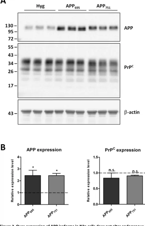
negative result observed in the non-neuronal HEK cells, we utilized mouse neuronal N2a cells over-expressing human APP695
or APP751 to again assess total PrP C
and APP protein levels (Fig. 2A and B). Despite a significant 2.5-fold increase in APP expression in the N2a cells transfected with the cDNAs encoding either APP695or APP751, there was no difference in endogenous
PrPC levels when comparing the APP over-expressing cells with each other or the vector only controls.
To further examine the effect of APP over-expression on PrPC levels, two transgenic mouse models were investigated. PrPCand APP protein levels were evaluated in brain homogenates from I5 mice which over-express wild type human APP, J20 mice which over-express human APP containing the Swedish/Indiana familial AD mutations [23] and non-transgenic matched genetic back-ground control mice (Fig. 3A and B). Despite a significant 2.8 -fold increase in APP in the transgenic I5 mice, as compared to the non-transgenic mice, there was no difference in brain PrPC levels.
Figure 2. Over-expression of APP isoforms in N2a cells does not alter endogenous PrPC.(A) Representative western blot of APP and PrPC
(antibody 6H4) in N2a cells stably transfected with either the vector alone (Hyg) or one of the APP isoforms (APP695, APP751), and subsequentb-actin
staining. Approximate molecular weights (kDa) are indicated. (B) Quantification of APP and PrPCprotein levels expressed relative to Hyg control cells
(dashed line). Data from 3 independent experiments. Statistical analysis by one way ANOVA with Dunnett’s post test comparison to the Hyg cells, *p,0.05, n.s. not significant.
doi:10.1371/journal.pone.0031754.g002
Analysis of the J20 mice, although only involving two animals, reinforced this conclusion. Collectively these over-expression experiments indicate that control of PrPC expression does not appear to involve AICD in either cell-based or transgenic animal paradigms.
Reduction of AICD production through APP gene silencing or c-secretase inhibition does not alter expression of endogenous PrPC
In light of the above results, we considered whether the level of AICD required to regulate PrPCexpression in the cell lines or the transgenic mice were already maximal from the endogenous APP, such that the AICD produced from the over-expressed APP was not having any additional affect on PrPC expression. Thus we sought to investigate a possible role for endogenous AICD in the control of PrPCexpression. First, to reduce endogenous APP levels and thereby remove the substrate for AICD production, N2a cells were treated with siRNA against murine APP. Cells were
harvested, lysed and PrPC and APP levels measured by western blotting (Fig. 4A and B). After directed siRNA treatment there was a significant 70% decrease in total APP levels (endogenous AICD level is below the limits of detection by immunoblot; data not shown). However, the amount of PrPC remained unchanged following siRNA knockdown of endogenous APP.
In order to test further for a possible involvement of endogenous AICD in controlling PrPCexpression, both HEK and N2a cells were treated with DAPT, a cell permeablec-secretase inhibitor. Again, whole cell lysates were assessed for PrPC and APP expression, as well as the levels of C83 and C99, by western blotting (Fig. 5A and B). Although DAPT treatment inhibitedc -secretase activity, as shown by the significantly increased C83 and C99 levels (9.4-fold in the HEK cells and 17.8-fold in the N2a cells), there was no difference in endogenous PrPCprotein levels in the DAPT treated cells as compared to the untreated cells (Fig. 5C and D). Together these results indicate that in both a neuronal and a non-neuronal cell line, endogenous AICD is also not involved in the control of PrPCprotein expression.
Figure 3. Unaltered PrPCprotein levels in transgenic mice over-expressing human wild type or familial AD mutant APP.(A) Western blot of APP and PrPC(antibody 6D11) in I5 (n = 3) and J20 (n = 2) transgenic, and age-matched non-transgenic control, mouse brain homogenates, with membrane re-probing forb-actin. Approximate molecular weights (kDa) are indicated. (B) Quantification of APP and PrPCprotein levels
expressed relative to the control mice (dashed line). Error bars represent6SD. Statistical analysis by unpaired t-test, **p,0.01, n.s. not significant. doi:10.1371/journal.pone.0031754.g003
Discussion
Similarities in the pathogenesis of the protein-misfolding neurodegenerative illnesses, especially AD and prion diseases, and possible connections between these diseases have long been contemplated [1,2,3,4]. Elucidation of any functional links between these diseases is an important research goal, with determination of the most appropriate protein or process to target for development of therapeutics being paramount. Links in the pathologies of AD and prion diseases have been determined, with various reports of AD features in prion disease brains [24,25,26], and PrPClocalised in Abplaques in AD brain [27,28]. In addition, a polymorphism at codon 129 of the prion protein gene, known to influence susceptibility to sporadic and iatrogenic human prion disease [29,30], may also influence susceptibility and the pathophysiology of AD [31,32,33]. Interestingly there is some indication of a more direct interaction between Aband PrPSc, with the finding of an acceleration and exacerbation of both AD and prion disease pathologies in animals engineered to have both of these diseases, and enhanced protein misfolding due to cross-seeding events stimulating oligomerization in vitro [34]. This
propensity for cross-seeding highlights the importance for a more complete understanding of interactions between these key proteins and any resultant downstream consequences.
Recent studies have provided evidence of direct interactions between the proteins central to AD and prion diseases. Various studies have determined that the cellular prion protein can act as a receptor for Ab, with Ab oligomers binding to PrPC with high affinity, although there are conflicting views as to the physiological significance of this binding. Some results suggest that Absynaptic toxicity is mediated through its binding to PrPC[7,11,12], which specifically impacts on spatial learning and memory in vivo[35],
whereas others have reported that Ab oligomer neurotoxicity occurs independently [8,9]. Confounding the relationship between these key proteins, and in apparent contrast to PrPCmediating Ab neurotoxicitiy, PrPChas been shown to decrease production of Ab from wild type APP through its interaction with the b-secretase BACE1 [5]. This interaction, mapped to the BACE1 pro-domain,
leads to slowed BACE1 trafficking following exit from the ER, thereby increasing its localization in the trans-Golgi network and reducing levels at the cell surface and consequently in endosomes where APPb-cleavage occurs [6]. Importantly, these studies also ascertained links in the pathology of AD and prion diseases. It was found that human prion disease-associated mutations in PrPCdid not inhibit BACE1, and scrapie infected mice brains contained dramatically higher Ab levels [5], suggesting a loss of PrPC function perhaps as a result of PrPC-PrPScconversion during prion disease progression.
PrPCmay be a key therapeutic target for sporadic AD, and the recent report that PrPCexpression was controlled by AICD in ac -secretase dependent manner [22] presented a potential avenue for achieving this. Further, a possible feedback model reconciling the control of APP processing and PrPC expression in both normal conditions and in the presence of increased Absuch as that seen in AD was proposed [36]. Therefore our study was carried out to further understand the relationship between AICD production and PrPCexpression. However, utilizing a range of experimental approaches we found no evidence for AICD involvement in PrPC expression. This is despite using a cellular system (N2a cells expressing APP695) in which we have proven that AICD is
transcriptionally active [19]. If AICD is involved in regulating the transcription of PrPC, then the mechanism underlying this is more complex than that involved in regulating the expression of neprilysin and is not readily reproduced in cultured cells or transgenic mice over-expressing human APP. Our findings have implications for the continued investigation and design of possible AD therapeutics.
Materials and Methods
Ethics statement
All experimental procedures performed on mice were approved by the Roslin Institute (University of Edinburgh) Ethical Review Process Committee and carried out under the UK Home Office License 60/3478. Human embryonic kidney (HEK) cells were obtained from the European Collection of Cell Cultures and
Figure 4. Knockdown of APP expression in N2a cells has no effect on PrPCprotein levels.(A) Representative western blots of APP and PrPC(antibody 6H4) in N2a cells treated with APP directed siRNA, non-coding control siRNA, and a no RNA transfection control (H2O control), and
subsequentb-actin staining. Approximate molecular weights (kDa) are indicated. (B) Quantification of APP and PrPCprotein levels expressed relative
to the H2O control cells. Data from 3 independent experiments. Statistical analysis by one way ANOVA with Dunnett’s post test comparison to the
H2O control cells, **p,0.01, n.s. not significant.
doi:10.1371/journal.pone.0031754.g004
murine neuroblastoma (N2a) cells were obtained from Dr Lehmann, Universite´ Montpellier, France [37].
Cell culture
HEK cells and N2a cells stably over-expressing the human APP isoforms (APP695, APP751and APP770), alongside the vector-only
(Hyg), were generated by electroporation as described previously [19]. Cells were maintained in Dulbecco’s modified Eagle’s medium (DMEM; Lonza, Basel, Switzerland) containing 10% (v/v) fetal bovine serum (FBS; Biosera, East Sussex, UK) and 1% penicillin/streptomycin (Lonza), in a humidified incubator at 37uC, 5% CO2.
APP gene silencing
To ablate endogenous APP expression in the N2a cells, cells were grown to 80% confluency in growth medium prior to treatment with 50 nM final concentration of murine APP directed siRNA, non-coding siRNA or siRNA-free controls following the manufacturer’s instructions (Thermo Fisher Scientific, Lafayette, CO, USA). Briefly, sub-confluent cell monolayers were washed gently with OptiMEM (GIBCO, Invitrogen, Glasgow, UK), before further incubation in OptiMEM for approximately 30 min (37uC, 5% CO2) during siRNA preparation. A 10mM siRNA solution in
16siRNA buffer of either murine APP directed siRNA TARGETplus SMARTpool) or non-coding siRNA control
(ON-Figure 5. Inhibition ofc-secretase activity does not alter the expression of PrPC.Representative western blots showing total cell-associated APP, PrPC(antibody 6H4) and C-terminal APP fragments (C83/99) in control (
2) and DAPT treated (+) HEK (A) and N2a (B) cells, with membrane re-probing forb-actin. Approximate molecular weights (kDa) are indicated. Quantification of C83/99, APP and PrPCprotein levels in DAPT treated HEK
(C) and N2a (D) cells, expressed relative to control cells (dashed line). Data from 3 (HEK) or 4 (N2a) independent experiments. Statistical analysis by one way ANOVA with Dunnett’s post test comparison to the control cells, ***p,0.001, **p,0.01, n.s. not significant.
doi:10.1371/journal.pone.0031754.g005
TARGETplus Non-targeting pool) was prepared, and diluted to 1mM in OptiMEM. For the RNA-free control, sterile RNase-free water was diluted 1:10 in OptiMEM. DharmaFECT Transfection Reagent-1 was diluted 1:40 in OptiMEM, mixed gently and incubated for 5 min at room temperature. Equal volumes of the diluted siRNA/control and DharmaFECT solutions were then mixed and incubated for 20 min at room temperature, prior to the addition of 46volumes of OptiMEM containing 10% (v/v) FBS. The OptiMEM was then removed from the cells, and replaced with the OptiMEM/FBS/siRNA complexes (or control) for 72 h.
Inhibition ofc-secretase
To inhibit endogenousc-secretase activity, HEK or N2a cells were grown to 90–95% confluency prior to treatment. The cell monolayer was then washed twice with PBS prior to incubating the cells in serum-free OptiMEM containing a final concentration 10mM N-[N-(3,5-difluorophenacetyl)-L-alanyl]-S-phenylglycine t-butyl ester (DAPT; Sigma-Aldrich, Dorset, UK), or an equal volume of dimethyl sulfoxide (DMSO, as the control) for 24 h.
Cell lysis, SDS-PAGE and immunoblotting
When confluent and/or after appropriate treatments as described above, cells were washed twice in phosphate-buffered saline (PBS with Ca2+
and Mg2+
; Lonza), harvested by scraping into PBS, and pelleted at 5006g for 3 min. Cell pellets were lysed for 30 min on ice in cold lysis buffer (25 mM Tris/HCl, pH7.5, 150 mM NaCl, 5 mM EDTA, 1% (v/v) Triton X-100) containing CompleteTM protease inhibitor cocktail (Roche, West Sussex, UK), prior to centrifugation for 5 min at 10006g. Post-nuclear supernatants were assessed for total protein content using a bicinchoninic acid protein assay (Sigma-Aldrich). Cell lysate containing 50mg total protein was resolved on 7–17% (APP and PrPC) or 16.5% (C83/99) polyacrylamide SDS gel, then electro-transferred to Hybond-P polyvinylidene difluoride membrane (PVDF; Amersham Life Sciences, Buckinghamshire, UK). PVDF membranes were blocked for 1–2 h at room temperature in PBS containing 0.1% Tween-20 (PBST) and 5% (w/v) skimmed milk powder, prior to incubation with primary antibody overnight at 4uC. For detection of APP the membrane was incubated with the monoclonal antibody 22C11 (Millipore, Billerica, MA, USA), and for detection of PrPCthe membrane was incubated with either the monoclonal antibody 3F4 (Covance, Princeton, NJ, USA), 6D11 (Eurogentec Ltd, Hampshire, UK) or 6H4 (Prionics, Zurich, Switzerland), as indicated in the figure legends. For detection of C83/99, a polyclonal anti-C-terminal APP antibody A8717
(Sigma-Aldrich) was used. After washing off non-specifically bound primary antibody with PBST, membranes were incubated in peroxidase-conjugated rabbit-anti-mouse or goat-anti-rabbit secondary antibodies (Sigma-Aldrich), before further washes with PBST and detection using enhanced chemiluminescence (Pierce ECL substrate; Thermo Fischer Scientific). To assess and correct for protein loading, membranes were stripped at low pH (1% (v/v) aqueous HCl) for approximately 30 min, re-blocked and probed with an anti-b-actin antibody (clone AC-15, Sigma-Aldrich) and the secondary antibody described above. All chemiluminescent images were captured by a Fujifilm LAS-3000 Intelligent Dark Box.
Transgenic mice and tissue homogenisation
Animals were obtained from The Jackson Laboratory (Bar Harbor, ME, USA), and all care was carried out in strict accordance with institutional guidelines. Transgenic I5 mice (Line B6.Cg-Tg(PDGFB-APP)5 Lms/J, stock number 004662), which over-express wild type human APP, and J20 mice (Line B6.Cg-Tg(PDGFB-APPSwInd)20 Lms/2J, stock number 006293), which over-express human APP containing the Swedish (K670N/ M671L) and Indiana (V717F) familial AD mutations [23], were crossed with inbred 129P2 mice, and genotyped to confirm the
APPgene sequence. Brain hemispheres from the I5 (8 weeks old),
J20 (5–9 weeks old) and age-matched non-transgenic littermate controls were homogenized in 2% (w/v) SDS solution containing protease inhibitors, and homogenates centrifuged at 100,0006g
for 1 h at 4uC. The resultant supernatant was assayed for total protein and assessed for PrPCand APP protein by SDS-PAGE and western blotting as described above for cell lysates.
Densitometry and statistical analysis
Quantification and densitometric analyses were carried out using Image J v1.42q. Within each experiment, data was normalised to b-actin, and expressed relative to the control samples. Statistical analyses were performed in GraphPad Prism v5.03. All quantitative data are expressed as the mean6 SEM, unless stated otherwise.
Author Contributions
Conceived and designed the experiments: VL JCM SJC NMH. Performed the experiments: VL IJW HB. Analyzed the data: VL IJW HB JCM NMH. Wrote the paper: VL JCM SJC NMH.
References
1. Wisniewski T, Sigurdsson EM (2007) Therapeutic approaches for prion and Alzheimer’s diseases. FEBS J 274: 3784–3798.
2. Checler F, Vincent B (2002) Alzheimer’s and prion diseases: distinct pathologies, common proteolytic denominators. Trends Neurosci 25: 616–620.
3. Taylor DR, Hooper NM (2007) Role of lipid rafts in the processing of the pathogenic prion and Alzheimer’s amyloid-beta proteins. Semin Cell Dev Biol 18: 638–648. 4. Barnham KJ, Cappai R, Beyreuther K, Masters CL, Hill AF (2006) Delineating common
molecular mechanisms in Alzheimer’s and prion diseases. Trends Biochem Sci 31: 465–472. 5. Parkin ET, Watt NT, Hussain I, Eckman EA, Eckman CB, et al. (2007) Cellular prion protein regulates beta-secretase cleavage of the Alzheimer’s amyloid precursor protein. Proc Natl Acad Sci USA 104: 11062–11067.
6. Griffiths HH, Whitehouse IJ, Baybutt H, Brown D, Kellett KA, et al. (2011) Prion protein interacts with BACE1 protein and differentially regulates its activity toward wild type and Swedish mutant amyloid precursor protein. J Biol Chem 286: 33489–33500.
7. Lauren J, Gimbel DA, Nygaard HB, Gilbert JW, Strittmatter SM (2009) Cellular prion protein mediates impairment of synaptic plasticity by amyloid-beta oligomers. Nature 457: 1128–1132.
8. Balducci C, Beeg M, Stravalaci M, Bastone A, Sclip A, et al. (2010) Synthetic amyloid-beta oligomers impair long-term memory independently of cellular prion protein. Proc Natl Acad Sci USA 107: 2295–2300.
9. Calella AM, Farinelli M, Nuvolone M, Mirante O, Moos R, et al. (2010) Prion protein and Abeta-related synaptic toxicity impairment. EMBO Mol Med 2: 306–314.
10. Chen S, Yadav SP, Surewicz WK (2010) Interaction between human prion protein and amyloid-beta (Abeta) oligomers: role of N-terminal residues. J Biol Chem 285: 26377–26383.
11. Barry AE, Klyubin I, Mc Donald JM, Mably AJ, Farrell MA, et al. (2011) Alzheimer’s disease brain-derived amyloid-beta-mediated inhibition of LTP in vivo is prevented by immunotargeting cellular prion protein. J Neurosci 31: 7259–7263.
12. Freir DB, Nicoll AJ, Klyubin I, Panico S, Mc Donald JM, et al. (2011) Interaction between prion protein and toxic amyloid beta assemblies can be therapeutically targeted at multiple sites. Nat Commun 2: 336.
13. Vardy ER, Catto AJ, Hooper NM (2005) Proteolytic mechanisms in amyloid-beta metabolism: therapeutic implications for Alzheimer’s disease. Trends Mol Med 11: 464–472.
14. Cao X, Sudhof TC (2001) A transcriptionally active complex of APP with Fe65 and histone acetyltransferase Tip60. Science 293: 115–120.
15. Belyaev ND, Nalivaeva NN, Makova NZ, Turner AJ (2009) Neprilysin gene expression requires binding of the amyloid precursor protein intracellular domain to its promoter: implications for Alzheimer disease. EMBO Rep 10: 94–100.
16. Pardossi-Piquard R, Petit A, Kawarai T, Sunyach C, Alves da Costa C, et al. (2005) Presenilin-dependent transcriptional control of the Abeta-degrading enzyme neprilysin by intracellular domains of betaAPP and APLP. Neuron 46: 541–554.
17. Goodger ZV, Rajendran L, Trutzel A, Kohli BM, Nitsch RM, et al. (2009) Nuclear signaling by the APP intracellular domain occurs predominantly through the amyloidogenic processing pathway. J Cell Sci 122: 3703–3714. 18. Hoey SE, Williams RJ, Perkinton MS (2009) Synaptic NMDA receptor
activation stimulates alpha-secretase amyloid precursor protein processing and inhibits amyloid-beta production. J Neurosci 29: 4442–4460.
19. Belyaev ND, Kellett KA, Beckett C, Makova NZ, Revett TJ, et al. (2010) The transcriptionally active amyloid precursor protein (APP) intracellular domain is preferentially produced from the 695 isoform of APP in a beta-secretase-dependent pathway. J Biol Chem 285: 41443–41454.
20. Tanaka S, Nakamura S, Ueda K, Kameyama M, Shiojiri S, et al. (1988) Three types of amyloid protein precursor mRNA in human brain: their differential expression in Alzheimer’s disease. Biochem Biophys Res Commun 157: 472–479.
21. Citron M, Oltersdorf T, Haass C, McConlogue L, Hung AY, et al. (1992) Mutation of the beta-amyloid precursor protein in familial Alzheimer’s disease increases beta-protein production. Nature 360: 672–674.
22. Vincent B, Sunyach C, Orzechowski HD, St George-Hyslop P, Checler F (2009) p53-Dependent transcriptional control of cellular prion by presenilins. J Neurosci 29: 6752–6760.
23. Mucke L, Masliah E, Yu GQ, Mallory M, Rockenstein EM, et al. (2000) High-level neuronal expression of abeta 1–42 in wild-type human amyloid protein precursor transgenic mice: synaptotoxicity without plaque formation. J Neurosci 20: 4050–4058.
24. Hainfellner JA, Wanschitz J, Jellinger K, Liberski PP, Gullotta F, et al. (1998) Coexistence of Alzheimer-type neuropathology in Creutzfeldt-Jakob disease. Acta Neuropathol 96: 116–122.
25. Brown P, Gibbs CJ, Jr., Rodgers-Johnson P, Asher DM, Sulima MP, et al. (1994) Human spongiform encephalopathy: the National Institutes of Health series of 300 cases of experimentally transmitted disease. Ann Neurol 35: 513–529.
26. Tsuchiya K, Yagishita S, Ikeda K, Sano M, Taki K, et al. (2004) Coexistence of CJD and Alzheimer’s disease: an autopsy case showing typical clinical features of CJD. Neuropathology 24: 46–55.
27. Takahashi RH, Tobiume M, Sato Y, Sata T, Gouras GK, et al. (2010) Accumulation of cellular prion protein within dystrophic neurites of amyloid plaques in the Alzheimer’s disease brain. Neuropathology 3: 2080214. 28. Voigtlander T, Kloppel S, Birner P, Jarius C, Flicker H, et al. (2001) Marked
increase of neuronal prion protein immunoreactivity in Alzheimer’s disease and human prion diseases. Acta neuropathologica 101: 417–423.
29. Deslys JP, Marce D, Dormont D (1994) Similar genetic susceptibility in iatrogenic and sporadic Creutzfeldt-Jakob disease. J Gen Virol 75: 23–27. 30. Windl O, Dempster M, Estibeiro JP, Lathe R, de Silva R, et al. (1996) Genetic
basis of Creutzfeldt-Jakob disease in the United Kingdom: a systematic analysis of predisposing mutations and allelic variation in the PRNP gene. Hum Genet 98: 259–264.
31. Riemenschneider M, Klopp N, Xiang W, Wagenpfeil S, Vollmert C, et al. (2004) Prion protein codon 129 polymorphism and risk of Alzheimer disease. Neurology 63: 364–366.
32. Del Bo R, Scarlato M, Ghezzi S, Martinelli-Boneschi F, Fenoglio C, et al. (2006) Is M129V of PRNP gene associated with Alzheimer’s disease? A case-control study and a meta-analysis. Neurobiol Aging 27: 770 e1–770 e5.
33. Gacia M, Safranow K, Styczynska M, Jakubowska K, Peplonska B, et al. (2006) Prion protein gene M129 allele is a risk factor for Alzheimer’s disease. J Neural Transm 113: 1747–1751.
34. Morales R, Estrada LD, Diaz-Espinoza R, Morales-Scheihing D, Jara MC, et al. (2010) Molecular cross talk between misfolded proteins in animal models of Alzheimer’s and prion diseases. J Neurosci 30: 4528–4535.
35. Gimbel DA, Nygaard HB, Coffey EE, Gunther EC, Lauren J, et al. (2010) Memory impairment in transgenic Alzheimer mice requires cellular prion protein. J Neurosci 30: 6367–6374.
36. Kellett KA, Hooper NM (2009) Prion protein and Alzheimer disease. Prion 3: 190–194.
37. Nishida N, Harris DA, Vilette D, Laude H, Frobert Y, et al. (2000) Successful transmission of three mouse-adapted scrapie strains to murine neuroblastoma cell lines overexpressing wild-type mouse prion protein. J Virol 74: 320–5.