Discovery of New Mitogenic Growth Factors for Mouse
Hepatocytes: Mitogenic Effects of FGF1/2 and PDGF
William C. Bowen1, Amantha W. Michalopoulos1, Anne Orr1, Michael Q. Ding1, Donna B. Stolz2,
George K. Michalopoulos1*
1Department of Pathology, University of Pittsburgh School of Medicine, Pittsburgh, Pennsylvania, United States of America,2Department of Cell Biology, University of Pittsburgh School of Medicine, Pittsburgh, Pennsylvania, United States of America
Abstract
Chemically defined serum-free media for rat hepatocytes have been useful in identifying EGFR ligands and HGF/MET signaling as direct mitogenic factors for rat hepatocytes. The absence of such media for mouse hepatocytes has prevented screening for discovery of such mitogens for mouse hepatocytes. We present results obtained by designing such a chemically defined medium for mouse hepatocytes and demonstrate that in addition to EGFR ligands and HGF, the growth factors FGF1 and FGF2 are also important mitogenic factors for mouse hepatocytes. Smaller mitogenic response was also noticed for PDGF AB. Mouse hepatocytes are more likely to enter into spontaneous proliferation in primary culture due to activation of cell cycle pathways resulting from collagenase perfusion. These results demonstrate unanticipated fundamental differences in growth biology of hepatocytes between the two rodent species.
Citation:Bowen WC, Michalopoulos AW, Orr A, Ding MQ, Stolz DB, et al. (2014) Development of a Chemically Defined Medium and Discovery of New Mitogenic Growth Factors for Mouse Hepatocytes: Mitogenic Effects of FGF1/2 and PDGF. PLoS ONE 9(4): e95487. doi:10.1371/journal.pone.0095487
Editor:Antonio Facchiano, IDI, Istituto Dermopatico dell’Immacolata, Italy
ReceivedDecember 30, 2013;AcceptedMarch 27, 2014;PublishedApril 17, 2014
Copyright:ß2014 Bowen et al. This is an open-access article distributed under the terms of the Creative Commons Attribution License, which permits unrestricted use, distribution, and reproduction in any medium, provided the original author and source are credited.
Funding:This study was supported by NIH grants DK35373 and CA103958 (GKM). The funders had no role in study design, data collection and analysis, decision to publish, or preparation of the manuscript.
Competing Interests:The authors have declared that no competing interests exist.
* E-mail: [email protected]
Introduction
Primary cultures of rodent and human hepatocytes have been used as tools for studies of metabolism, drug toxicity, bile acid synthesis, and many other aspects of hepatic physiology. In addition, growth of rat hepatocytes in primary culture and in chemically defined (serum-free) media has been a very useful tool for determining growth factors (Hepatocyte Growth Factor (HGF), and Epidermal Growth Factor Receptor (EGFR) ligands) that are direct mitogens for rat hepatocytes [1]. These studies paved the progress made for determining the role of EGFR and MET (the HGF receptor) receptors and their ligands in liver regeneration and hepatocyte growth [2–5]. While the conditions for hepatocyte growth in chemically defined media have been well defined for the rat [1], definition of similar conditions for mouse hepatocytes has been elusive. Response of mouse hepatocytes to established mitogens such as HGF and EGF in most media has been mediocre at best compared to rat hepatocytes.
The work presented in this manuscript describes a chemically defined serum-free medium that allows growth of mouse hepatocytes in primary culture and high response to growth factors comparable to those seen with the rat. The medium was substantially modified from the hepatocyte growth medium (HGM) used for rat hepatocytes [1]. During the course of thee studies, we also determined that whereas EGFR ligands and HGF are the major direct mitogens for rat hepatocytes, Fibroblast Growth Factors 1 and 2 (FGF1, FGF2) are equally strong direct mitogens for mouse hepatocytes. Platelet Derived Growth Factor
(PDGF) also has a weaker direct mitogenic effect. These findings present hitherto undiscovered differences in growth biology of hepatocytes between the two rodent species most commonly used for studies of liver regeneration and carcinogenesis. This work needs to be extended to human hepatocytes. Our work also describes technical difficulties in applying common methodologies used to assess cell proliferation in culture.
Materials and Methods
Ethics Statement
Animals (mice) were housed and treated according to institu-tional guidelines of the University of Pittsburgh IACUC commit-tee (Protocol 1105844). This follows guidelines established by NIH for ethical use of animals in biomedical research. The IACUC committee and the aforementioned protocol specifically approved this study.
Male Fisher 344 rats (2–3 months of age) were purchased from Harlan. Male FVB mice (2–3 months of age) were purchased from Charles River. Animals were allowed food and water ad libitum and handled according to the Ethics Statement above.
Methods for Alleviation of Animal Suffering
Isolation and Culture of Hepatocytes
Hepatocytes were isolated by adaptation of the calcium two-step collagenase perfusion technique as previously described [6]. Hepatocytes were plated on collagen-coated six-well plates (BD Biosciences, San Jose, CA) at 250,000 cells/well. After the initial 2-hour attachment period, the medium was changed to formulations as described in Results. There was no serum added at any time during or after plating of hepatocytes. The various growth factors, for either mouse or rat hepatocytes, were added at 24 hours after plating. Media were changed every 48 hours, except for the first medium change, which was done at 24 hours after plating and was used to introduce the added growth factors. Cells were harvested for assessment of cell proliferation at the times indicated in the relevant figures.
Formulation of the HGM (Hepatocyte Growth Medium) and MHGM (Mouse Hepatocyte Growth Medium)
The formulation of HGM medium has been previously described and was the basis of modifications for the formulation of the MHGM medium.
HGM medium. DMEM (Dulbecco’s minimal essential me-dium), Hepes, glutamine, and antibiotics were purchased from GIBCO/BRL (Gaithersburg, MD). ITS mixture (Insulin, Trans-ferrin, Selenium) was purchased from Boehringer Mannheim. All other additives were cell culture grade (Sigma). Unless otherwise indicated for specific experiments, the basal HGM medium consisted of DMEM supplemented with: purified bovine albumin 2.0 gm/L, glucose 2.25 gr/L, galactose 2.0 gr/L, ornithine 0.1 gm/L, proline 0.030 gm/L, nicotinamide 0.610 gm/L, ZnCI2 0.544 mg/L, ZnSO4:7H2O 0.750 mg/L, CuSO4:5H2O 0.20 mg/L, MnSO4 0.025 mg/L, glutamine 5.0 mM, ITS 1.0 gm/L, (rh-insulin 5.0 mg/L, human transferrin 5.0 mg/L [30% diferric iron saturated], selenium 5.0mg/L), and dexameth-asone 1027 M. Penicillin and streptomycin were added to the
basal HGM at 100 mg/L each. The mixed basal HGM was sterilized by filtration through a 0.22-mm low protein-binding filter system, stored at 4uC, and used within 4 wk. The growth factors, as required, were added to medium fresh at the specified concentrations every time the medium was changed.
MHGM medium. MHGM was derived by using E-MEM (from Sigma) instead of DMEM as the basis for the formulation. Nicotinamide was omitted and dexamethasone was added at a final concentration of 1026 M (ten times more than HGM).
Glucose concentration for MHGM 4.5 mg/ml) was also adjusted to two-fold that of HGM.
Hepatocyte Proliferation Assay
Measurement of DNA synthesis.
1. Thymidine incorporation. H3]Thymidine was added to the medium at 24 hours at a concentration of 2.5mCi/ml. The medium was removed when the cultures were harvested and hepatocytes were fixed with ice-cold 5% trichloroacetic acid. Trichloroacetic acid was removed and the plates were washed in running tap water and air-dried completely. Then 750mL 0.33 M NaOH was added to each well for 30 minutes to solubilize the cells. The solution was transferred to a new tube and 250mL of 40% TCA/1.2 M HCl was added for precipitation. The tubes were centrifuged at 12,000 g for 10 minutes and the pellets were dissolved in 500mL 0.33 M NaOH. A 200-mL aliquot was used to measure G3 cpm in a Beckman LS6000IC scintillation counter (Beckman Coulter, CA) and 100mL was used to determine optical density value of total DNA. Data are plotted as CPM/mg DNA.
BRDU incorporation into DNA was assessed by the use of the Invitrogen BRDU staining kit (from Life Technologies).
2. Bromodeoxyuridine (BRDU) and Ki67 immunohistochemistry. Ki-67 antibodies were obtained from Santa Cruz. BRDU immunohistochemistry was performed using the specific kit from Becton-Dickinson.
Concentrations of mitogenic growth factors. We per-formed dose-response studies (data not shown) and assessed that 40 ng/per ml exceeded the highest dose response for each growth factor. They were all added to the medium at 40 ng/ml to assure maximal response.
Transmission electron microscopy. Hepatocytes grown on tissue culture collagen coated plates were fixed in 2.5% glutaraldehyde in 100 mM PBS (8 gm/l NaCl, 0.2 gm/l KCl, 1.15 gm/l Na2HPO4
.
7H2O, 0.2 gm/l KH2PO4, pH 7.4 ) over-night at 4uC. Hepatocyte monolayers were then washed in PBS three times then post-fixed in aqueous 1% osmium tetroxide, 1% Fe6CN3 for 1 hr. Cells were washed 3 times in PBS then dehydrated through a 30–100% ethanol series then several changes of Polybed 812 embedding resin (Polysciences, Warring-ton, PA). Cultures were embedded in by inverting Polybed 812-filled BEEM capsules on top of the cells. Blocks were cured overnight at 37uC, then cured for two days at 65uC. Monolayers were pulled off the coverslips and re-embedded for cross section. Ultrathin cross sections (60 nm) of the cells were obtained on a Riechart Ultracut E microtome, post-stained in 4% uranyl acetate for 10 min and 1% lead citrate for 7 min. Sections were viewed on a JEOL JEM 1011 transmission electron microscope (JEOL, Peobody MA) at 80 KV. Images were taken using a side-mount AMT 2 k digital camera (Advanced Microscopy Techniques, Danvers, MA).
Results
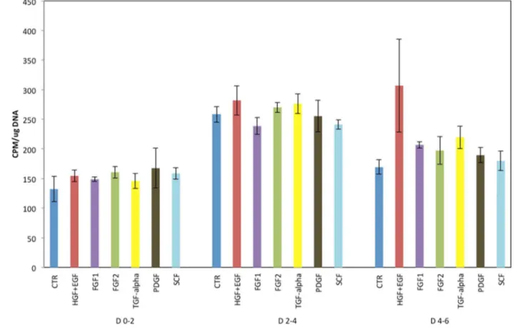
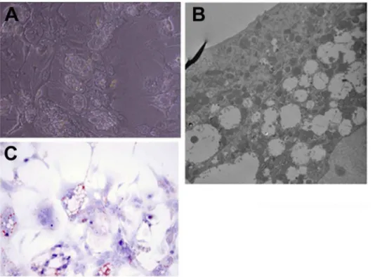
We also observed that even starting from 24 hours in culture, mouse hepatocytes kept in (rat-defined) HGM medium had poor response to growth factors, as shown in Figure 1. A better response was seen when the basic formulation of the mouse medium (to be referred as MHGM) was changed from the HGM basal medium of DMEM to Eagle’s MEM (EMEM). We did observe however that addition of HGF and EGF into that medium, while having minimal effect, was causing substantial vacuolation of the cytoplasm of the mouse hepatocytes (Figures 2 A and B). Vacuoles did not contain triglycerides, as evidence by an Oil Red-O stain (Figure 2 C). The vacuoles were present within the cytoplasm of hepatocytes and did not represent dilated bile canaliculi or autophagic vacuoles. The vacuoles did not appear when nicotin-amide was removed from the medium. It should be noted that no such effect with nicotinamide was observed with rat hepatocytes and, to the contrary, presence of nicotinamide was essential for rat hepatocyte proliferation [1].
Another important factor for the MHGM media formulation was the concentration of the steroid dexamethasone. Increasing dexamethasone concentration from 1027M (in the HGM) to 1026
M in the MHGM medium substantially suppressed the sponta-neous proliferation rate of mouse hepatocytes without affecting the response to growth factors (Figure 3).
The detail final formulation and comparison of the ingredients between the rat hepatocyte-defined HGM medium and the mouse hepatocyte-optimized MHGM medium are described in Materials and Methods.
Growth Factors Stimulating Proliferation of Mouse Hepatocytes in the MHGM Medium
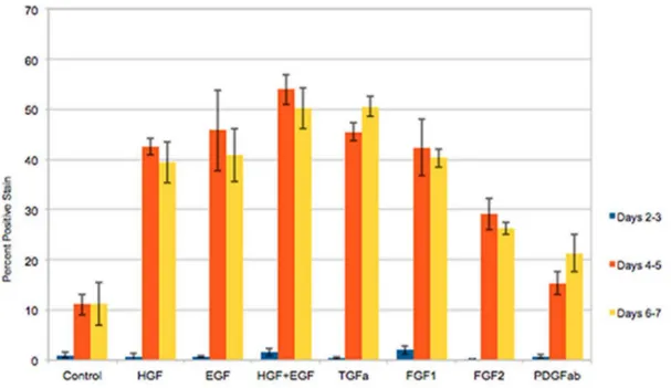
Proliferation of mouse hepatocytes exposed to different growth factors in the MGM medium is shown in Figure 4. Hepatocytes were exposed to 2-day pulses of BRDU at the indicated days and the nuclear labeling index of the cells was assessed. The results show hepatocyte proliferation rates in response to EGF, HGF, (EGF plus HGF), and TGFa, primarily at days 4–7 in culture. Minimal spontaneous proliferation was seen in control cultures without growth factor addition. We also noticed that FGF1 and FGF2 had a strong mitogenic effect, something not seen with rat hepatocytes. Measurable mitogenic effect was also seen with PDGF AB. Immunohistochemical stains for desmin showed that the small effect of PDGF AB on cell proliferation was on desmin-negative cells (hepatocytes) and not on contaminant (desmin positive) stellate cells. There was no further stimulation of hepatocyte proliferation by addition of higher concentrations of PDGF (data not shown).
It should be noted that the results of these studies were obtained by two-day BRDU pulses. When, instead of BRDU, we used immunohistochemistry for the cell cycle specific nuclear protein Ki67, most of the cells appearred positive, whether they were in control or growth factor-exposed cultures. We consider the increased expression of Ki67 as part of the overall change in cell cycle related protein expression induced by the collagenase perfusion and the culture itself. Expression of cell cycle genes (but not DNA synthesis) has been observed in the past with rat
Figure 1. Proliferation of mouse hepatocytes maintained in the rat hepatocyte (HGM) medium.Y-axis indicates incorporation of tritiated thymidine permg DNA. The growth factors added to the medium are listed under the X-axis. CTR: Control cultures, with no growth factors added. Growth factors were added at 24 hours after plating of hepatocytes. D 0–2 (etc.) indicates days in culture after addition of the growth factors. (For growth factor concentration, please see Materials and Methods).
hepatocytes [9,10]. Since BRDU clearly reflects DNA synhesis, we considered that as the method of choice to measure the effect of growth factors on mouse hepatocyte proliferation.
Rat Hepatocytes in the Mouse Medium Perform as Expected and Reflect Species Differences
We were concerned that the mitogenic effects of FGF1/2 and PDGF AB on mouse hepatocytes might have been missed with rat
hepatocytes, due to differences in the cell culture medium. To that end, we assessed proliferation of rat hepatocytes in the MHGM medium, in exactly the same conditions as with the mouse hepatocytes. The results are shown in Figure 5. Though overall proliferation rates were lower than those seen in the HGM medium, rat hepatocytes responded as previously described, with very little proliferation in response to FGF1 and much less to FGF2. No substantial proliferation was induced by PDGF AB. The lower proliferations rates of rat hepatocytes in MHGM may be due to the absence of nicotinamide and the higher concentra-tion of dexamethasone, both of which are likely to negatively affect rat hepatocyte proliferation, as we have previously described [1].
Interactions between FGF2 and PDGF
Previous studies have shown that in several cell types there is interaction between PDGF BB and FGF2, as a result of which FGF2 is often found to inhibit the effects of PDGF BB and vice versa [11–13](see Discussion). In order to investigate such interaction between FGF2 and PDGF, we explored the rate of DNA synthesis (assessed by BRDU incorporation) in cultures of mouse hepatocytes kept in the chemically defined MHGM medium, and exposed to PDGF AA, PDGF BB in the presence or absence of FGF2 (Fig. 6). Our results do not show negative interactions between FGF2 and PDGF AA or BB in terms of stimulation of DNA synthesis.
Discussion
Our findings demonstrate that there are fundamental differ-ences in response to growth factors between mouse and rat hepatocytes in culture, probably as a result of different responses to degradation of extracellular matrix and release of bound growth
Figure 2. Intense vacuolation of hepatocyte cytoplasm in a preliminary formulation of the mouse hepatocyte growth medium (MHGM).Elimination of nicotinamide removed this cytoplasmic change. A: Phase contrast photomicrograph of the hepatocytes in culture showing intense valuation of the cytoplasm (Magnification: 100X). B: Electron micrograph (5,000X) of hepatocytes carrying vacuoles. There is no apparent connection with bile canaliculi or autophagosomes. C: Oil Red-O stain for lipid demonstrates that the vacuoles observed did not contain lipids. doi:10.1371/journal.pone.0095487.g002
Figure 3. Suppression of spontaneous proliferation of mouse hepatocytes in the presence of 1026 M dexamethasone. Tritiated thymidine was added continually in the cultures, from 4 hours after cell plating following perfusion of the liver by collagenase. Cultures were harvested at the indicated time points in the X-axis. Addition of HGF and EGF is indicated as ‘‘GFs’’. (For growth factor concentration, please see Materials and Methods).
factors and/or integrin signaling via collagenase perfusion. Our findings also demonstrate that the family of fibroblast growth
factors (FGF) plays a more important role in mouse hepatocyte growth biology.
Figure 4. Mouse hepatocyte proliferation in the final formulation of the MHGM medium.Y-axis indicates percent of labeled nuclei due to incorporation of bromodeoxyuridine into DNA. The growth factors added to the medium are listed under the X-axis. CTR: Control cultures, with no growth factors added. Growth factors were added at 24 hours after plating of hepatocytes. D 0–2 (etc.) indicates days in culture after addition of the growth factors. (For growth factor concentration, please see Materials and Methods).
doi:10.1371/journal.pone.0095487.g004
Figure 5. Rat hepatocyte proliferation in the MHGM medium. Y-axis indicates percent of labeled nuclei due to incorporation of bromodeoxyuridine into DNA. The scale of Y-axis is kept identical to that of figure 4, for direct comparison of the responses of mouse and rat hepatocytes. The growth factors added to the medium are listed under the X-axis. CTR: Control cultures, with no growth factors added. Growth factors were added at 24 hours after plating of hepatocytes. D 0–2 (etc.) indicates days in culture after addition of the growth factors. (For growth factor concentration, please see Materials and Methods).
Previous studies have demonstrated that FGF1 and FGF2 may contribute to hepatocyte growth. Detail studies have shown that hepatocytes express the fibroblast growth factor receptor FGFR4 but not the receptors FGFR1 and FGFR2 [14]. The effects of FGFR4 are dependent on association with the transmembrane proteinbklotho [15]. This protein is often deleted in hepatocel-lular carcinomas and the results suggest that the interaction of bklotho and FGFR4 suppress proliferation of neoplastic hepato-cytes. FGFR1 and FGFR2 are expressed in other hepatic cell types (endothelial and stellate cells) [16]. FGF1 and FGF2 expression by hepatocytes increases during liver regeneration [17]. The increased expression occurs after hepatocytes have already committed to enter into S phase. Genetic deletion of FGFR4 has been associated with defects in cholesterol and bile acid metabolism but there were no demonstrable effects on hepatocyte proliferation in response to injury [14]. In another study, however, hepatocyte-targeted expression of a dominant negative FGFR2 suppressed and delayed liver regeneration [18]. FGF1 and FGF2 have been associated with induction of the hepatic primordium from the foregut endoderm [19]. However, mice with combined deletion of FGF1 and FGF2 have normal embryonic development, suggesting that the absence of FGF1 and FGF2 is compensated by other members of the 23-member family of FGFs, which use the same receptors as FGF1 and FGF2 [16]. Combined FGF1/FGF2 knockout mice do show increased hepatocyte sensitivity to CCl4 but have a decreased level of stellate cell-derived matrix collagen type 1 synthesis in response to long term CCl4 exposure [16]. Recent studies have also focused on mouse FGF15, the ortholog of human FGF19. Expression of these FGFs is induced by bile acids on enterocytes of the ileum. Migration to the liver has endocrine effects on hepatocyte-mediated cholesterol and bile acid metab-olism. Mice with systemic genetic elimination of FGF15 had delayed liver regeneration [20]. Our findings and the studies cited above suggest that the FGF family members have effects on hepatocyte growth that need to be further studied and understood
in the context of liver regeneration and chronic liver injury. The likelihood of important mitogenic effects of FGF members on hepatocytes is further enhanced by a recent analysis of the signal transduction pathways of receptor tyrosine kinases. Based on the inferred signaling networks, that study demonstrated that EGFR, MET and FGFR1 belong to the same subclass in terms of intracellular signaling [21].
Much less evidence from the literature exists on effects of PDGF on hepatocytes. Transgenic over-expression of PDGF C in hepatocytes was associated with increased activation of stellate cells, hepatic fibrosis and steatosis, and development of hepato-cellular carcinoma. It was not clear whether the increased HCC development was due to direct effects of PDGF or to increased synthesis of HGF by stellate cells [22]. Hepatocyte targeted genetic elimination of PDGFRa is associated with delay in liver regeneration and compensatory increases in EGFR and MET [23]. There is also increased expression of PDGFRain mice with hepatocyte-targeted genetic elimination of beta catenin [24]. Given the smaller response to PDGF compared to the other mitogenic growth factors, it is conceivable that there is a specific sub-class of hepatocytes, perhaps related to specific lobular localization, that have the capacity to respond to PDGF.
Several previous studies have indicated that PDGF BB and FGF2 exert negative effects on each other’s activities in several cell types. PDGF BB inhibits angiogenic effects of FGF2 both in vitro and in vivo, and this action is likely mediated by PDGFRa, since neutralization of the latter eliminates the inhibitory effects of PDGF BB [13]. The interaction is complex and involves both the ligands and their receptors. FGF2 and PDGF BB bind directly with each other [11]. In addition, there is strong evidence that FGFR1 and PDGFRaform heterodimers, through which PDGF BB and FGF2 can have very subtle effects on angiogenesis [12]. The absence of effects of PDGF BB on FGF2 stimulated hepatocyte DNA synthesis probably reflects different patterns of receptor interaction. Since FGF2 and PDGF BB bind directly, this
Figure 6. Interactions between FGF2 and PDGF AA and BB in stimulation of DNA synthesis in mouse hepatocyte cultures.Y-axis indicates percent of labeled nuclei due to incorporation of bromodeoxyuridine into DNA. The growth factors added to the medium are listed under the X-axis.
cannot be the reason for the observed lack of interaction in our study. Localization of receptors on plasma membrane in hepatocyte cultures, however, is much different than in hepato-cytes in intact liver [25]. The change of the hepatocyte shape from cuboidal (in vivo) to a flattened epithelial cell in culture has been found to have complex effects on intermolecular protein associ-ations and other structural changes, including absence of bile canaliculi, etc. Addition of complex matrix material restores such structures bu it also inhibits hepatocyte DNA synthesis, thus not allowing a direct study of the PDGF BB and FGF2 interactions in hepatocytes maintaing proper cell polarity.
In summary, our current study provides information about the conditions required in cell culture for study of mouse hepatocyte growth biology and documents that members of the FGF family and, to a lesser extent, PDGF, should be considered further by in vivo studies for playing a role in liver regenerative responses.
Author Contributions
Conceived and designed the experiments: GKM WCB. Performed the experiments: AWM AO MQD DBS. Analyzed the data: GKM. Contributed reagents/materials/analysis tools: DBS. Wrote the paper: GKM DBS.
References
1. Block GD, Locker J, Bowen WC, Petersen BE, Katyal S, et al. (1996) Population expansion, clonal growth, and specific differentiation patterns in primary cultures of hepatocytes induced by HGF/SF, EGF and TGF alpha in a chemically defined (HGM) medium. J Cell Biol 132: 1133–1149.
2. Michalopoulos GK (2013) Principles of liver regeneration and growth homeostasis. Compr Physiol 3: 485–513.
3. Michalopoulos GK (2007) Liver regeneration. J Cell Physiol.
4. Michalopoulos GK (2010) Liver regeneration after partial hepatectomy: critical analysis of mechanistic dilemmas. The American journal of pathology 176: 2– 13.
5. Michalopoulos GK (2011) Liver regeneration: alternative epithelial pathways. The international journal of biochemistry & cell biology 43: 173–179. 6. Novicki DL, Rosenberg MR, Michalopoulos G (1985) Inhibition of DNA
synthesis by chemical carcinogens in cultures of initiated and normal proliferating rat hepatocytes. Cancer Res 45: 337–344.
7. Liu ML, Mars WM, Zarnegar R, Michalopoulos GK (1994) Collagenase pretreatment and the mitogenic effects of hepatocyte growth factor and transforming growth factor-alpha in adult rat liver. Hepatology 19: 1521–1527. 8. Bucher NL, Robinson GS, Farmer SR (1990) Effects of extracellular matrix on hepatocyte growth and gene expression: implications for hepatic regeneration and the repair of liver injury. Semin Liver Dis 10: 11–19.
9. Kost DP, Michalopoulos GK (1990) Effect of epidermal growth factor on the expression of protooncogenes c- myc and c-Ha-ras in short-term primary hepatocyte culture. J Cell Physiol 144: 122–127.
10. Kost DP, Michalopoulos GK (1991) Effect of 2% dimethyl sulfoxide on the mitogenic properties of epidermal growth factor and hepatocyte growth factor in primary hepatocyte culture. J Cell Physiol 147: 274–280.
11. Russo K, Ragone R, Facchiano AM, Capogrossi MC, Facchiano A (2002) Platelet-derived growth factor-BB and basic fibroblast growth factor directly interact in vitro with high affinity. J Biol Chem 277: 1284–1291.
12. Faraone D, Aguzzi MS, Ragone G, Russo K, Capogrossi MC, et al. (2006) Heterodimerization of FGF-receptor 1 and PDGF-receptor-alpha: a novel mechanism underlying the inhibitory effect of PDGF-BB on FGF-2 in human cells. Blood 107: 1896–1902.
13. De Marchis F, Ribatti D, Giampietri C, Lentini A, Faraone D, et al. (2002) Platelet-derived growth factor inhibits basic fibroblast growth factor angiogenic properties in vitro and in vivo through its alpha receptor. Blood 99: 2045–2053.
14. Yu C, Wang F, Jin C, Wu X, Chan WK, et al. (2002) Increased Carbon Tetrachloride-Induced Liver Injury and Fibrosis in FGFR4-Deficient Mice. Am J Pathol 161: 2003–2010.
15. Luo Y, Yang C, Lu W, Xie R, Jin C, et al. (2010) Metabolic regulator betaKlotho interacts with fibroblast growth factor receptor 4 (FGFR4) to induce apoptosis and inhibit tumor cell proliferation. J Biol Chem 285: 30069–30078. 16. Yu C, Wang F, Jin C, Huang X, Miller DL, et al. (2003) Role of fibroblast growth factor type 1 and 2 in carbon tetrachloride-induced hepatic injury and fibrogenesis. Am J Pathol 163: 1653–1662.
17. Kan M, Huang JS, Mansson PE, Yasumitsu H, Carr B, et al. (1989) Heparin-binding growth factor type 1 (acidic fibroblast growth factor): a potential biphasic autocrine and paracrine regulator of hepatocyte regeneration. Proc Natl Acad Sci U S A 86: 7432–7436.
18. Steiling H, Wustefeld T, Bugnon P, Brauchle M, Fassler R, et al. (2003) Fibroblast growth factor receptor signalling is crucial for liver homeostasis and regeneration. Oncogene 22: 4380–4388.
19. Jung J, Zheng M, Goldfarb M, Zaret KS (1999) Initiation of mammalian liver development from endoderm by fibroblast growth factors. Science 284: 1998– 2003.
20. Uriarte I, Fernandez-Barrena MG, Monte MJ, Latasa MU, Chang HC, et al. (2013) Identification of fibroblast growth factor 15 as a novel mediator of liver regeneration and its application in the prevention of post-resection liver failure in mice. Gut 62: 899–910.
21. Wagner JP, Wolf-Yadlin A, Sevecka M, Grenier JK, Root DE, et al. (2013) Receptor tyrosine kinases fall into distinct classes based on their inferred signaling networks. Sci Signal 6: ra58.
22. Campbell JS, Hughes SD, Gilbertson DG, Palmer TE, Holdren MS, et al. (2005) Platelet-derived growth factor C induces liver fibrosis, steatosis, and hepatocellular carcinoma. Proc Natl Acad Sci U S A 102: 3389–3394. 23. Awuah PK, Nejak-Bowen KN, Monga SP (2013) Role and regulation of
PDGFRalpha signaling in liver development and regeneration. Am J Pathol 182: 1648–1658.
24. Awuah PK, Rhieu BH, Singh S, Misse A, Monga SP (2012) beta-Catenin loss in hepatocytes promotes hepatocellular cancer after diethylnitrosamine and phenobarbital administration to mice. PLoS One 7: e39771.