Experiments on the effect of temperature on white spot syndrome virus infection in Litopenaeus vannamei shrimp
88
0
0
Texto
(2) COPYRIGHT The author, supervisor and promoter give the permission to put this thesis to disposal for consultation and to copy parts of it for personal use. Any other use falls under the limitation of copyright, in particular the obligation to explicitly mention the source when citing part out of thesis.. January 27, 2006. Prof. Dr. Patrick Sorgeloos Promoter 1. Prof. Dr. Hans Nauwynck Promoter 2. João José Pereira Dantas da Rocha Lima Author.
(3) ABSTRACT White Spot Disease (WSD) is an aggressive and devastating viral disease caused by the White Spot Syndrome Virus (WSSV). This highly pathogenic and widespread disease, present throughout Asia and the Americas, can cause up to 100% mortality within 3-7 days after infection. It is annually responsible for huge ecological and economical losses in the main producing countries and forms as such one of the greatest threats for the further sustainable development of shrimp aquaculture. Previous research showed that manipulation of physical factors gave promising results: manipulation of the environmental factors such as temperature produced the most interesting and promising results. For this thesis three experiments were performed, all in which pacific white shrimp (Litopenaeus vannamei) were intramuscularly inoculated with a well-defined viral dose (30 and/or 10000 SID50) and exposed to high water temperature via standardised protocols. The first experiment looked at the efficacy of elevated temperature for protecting shrimp against WSSV. Practically, four temperature treatments in which an elevated temperature (33 °C) was either applied before virus inoculation, after the inoculation, both before and after inoculation, and in the fourth treatment a low temperature (27ºC) was used throughout the test. In the second series of experiments the protective value of high temperature after an initial period of viral replication was evaluated. Water temperature was raised from 27ºC to 33ºC at 0, 12 or 24 hours post WSSV inoculation. Maintaining and controlling such high water temperatures for longer periods of time is of course very unpractical in field conditions and probably economically unfeasible, so the third experiment evaluated the effectiveness of shorter cyclic exposure periods to high water temperature. Hence, the shrimp were exposed to daily temperature cycles (33ºC/27ºC) with 6, 12 and 18 hours of high water temperature, during five consecutive days. Experiment 1 demonstrated a total blocking of disease progression when hyperthermia was applied immediately post inoculation. The protection was very effective even with a high viral dose (10000 SID50). The second experiment, at a low viral dose (30 SID50), showed that high temperature to some extent also worked therapeutic in that previously 24 hours of virus replication could be allowed. At a high infection dose (10000 SID50) the level of protection was however not so effective. In Experiment 3, only a minimum of 18 hours at 33°C resulted in a significant lower. I.
(4) mortality with the infected shrimp. The results from all the experiments clearly show the potential of high water temperature for preventing mortality in WSSV infected shrimp.. II.
(5) ABSTRACT White Spot Disease (WSD) is een uiterst agressieve en letale virale aandoening die veroorzaakt wordt door het White Spot Syndrome Virus (WSSV). Deze zeer infectueuze en wijdverspreide ziekte (Azië en Amerika), kan binnen 3 tot 7 dagen na infectie tot 100% mortaliteit leiden. Het is jaarlijks verantwoordelijk voor reusachtige ecologische en economische verliezen in de producerende landen en vormt dus één van de grootste bedreigingen voor de verdere duurzame ontwikkeling van garnalenkweek. Vorige onderzoeken toonden reeds aan dat de manipulatie van fysische variabelen tot veelbelovende resultaten kon leiden: zo bleek temperatuur één van de meest veelbelovende te zijn. Voor deze thesis werden drie experimenten uitgevoerd, allen met Litopenaeus vannamei, die intramusculair geïnoculeerd werden met een welbepaalde virale dosis (30 en/of 10000 SID50) en vervolgens blootgesteld werden aan een verhoogde. watertemperatuur. volgens. gestandardiseerde. protocols.. Het. eerste. experiment bekeek het beschermend effect van de temperatuursverhoging tegen WSSV. De hoge temperatuur (33 °C) werd hierbij toegepast ofwel vóór de inenting met het virus, ofwel na de inenting, zowel vóór en na de inenting en in de vierde behandeling werd een lage temperatuur (27ºC) gebruikt gedurende geheel de test. In de tweede experimentenreeks kon het virus zich eerst gedurende een bepaalde periode vermenigvuldigen (0, 12 of 24 uur na inoculatie), vooraleer de temperatuursverhoging (van 27°C naar 33 °C) uitgevoerd werd. In de praktijk is het handhaven en controleren van dergelijke hoge watertemperaturen gedurende langere tijdspannes onpraktisch en waarschijnlijk economisch onhaalbaar, zodat het derde experiment opgezet werd om de doeltreffendheid van kortere cyclische periodes van blootstelling aan een verhoogde watertemperatuur uit te testen. Hiertoe werden de garnalen onderworpen aan dagelijkse temperatuurscycli (33ºC/27ºC) van 6, 12 en 18 uur blootstelling aan de verhoogde watertemperatuur, en dit gedurende vijf opeenvolgende dagen. Experiment 1 toonde aan dat een continue verhoogde temperatuur onmiddellijk na de virale inoculatie voor een heel efficiënte bescherming zorgt, zelfs bij inentingen met een hoge virale dosis (10000 SID50). Het tweede experiment toont aan dat de verhoogde temperatuur bij een lage virale dosis (30 SID50) zelfs een zekere curatieve werking heeft, in die zin dat het virus zich initieel tot 24 uur mag vermenigvuldigen. Bij een hoge dosis (10000 SID50) is de mate van bescherming echter niet zo efficiënt. In. III.
(6) Experiment 3, is er slechts één behandeling die resulteert in een significant lagere mortaliteit, namelijk de blootstelling aan 33°C gedurende 18 uur per dag. De resultaten van. alle. experimenten. tonen. duidelijk. het. potentieel. van. een. verhoogde. watertemperatuur ter bestrijding van WSSV besmetting.. IV.
(7) ACKNOWLEDGEMENTS First of all I am thankful to my first promoter, Prof. Dr. Patrick Sorgeloos, for accepting me as M.Sc. student in the Laboratory of Aquaculture & Artemia Reference Center. I also would like to thank him for his orientation and constructive comments during scientific meetings. Next, I would like to thank my second promoter, Prof. Dr. Hans Nauwynck, for accepting me as M.Sc. student in the Laboratory of Virology, Faculty of Veterinary Medicine. I am especially thankful for his constant orientation, availability and scientific support, without which this work would not have been possible. I also want to thank my supervisor, Mathieu Wille for all the hours spent with my orientation during the study period. His kind and friendly guidance, orientation, availability and patience, make this work possible at all the levels. Also his technical support was of extreme utility in the development of this work. I am very grateful to all the members of the “shrimp team”, Cesar Escobedo Bonilla, Meezanur Rahman, Mathias Corteel, and Karen Van Nieuwenhuyse for the scientific and technical support and precious help in all moments and especially, in the most difficult phase of the work. Their friendship made the entire task more easy and pleasant. For my parents and brother, a special mention for their love and encouragement though all these years of study. Without their extreme understanding and support, this task would not have been possible. For all the important and special persons in my family, an eternal feeling of grateful and emotion. Thanks to my true friends For you Cláudia, all the words from all the languages are not enough to express my special feeling. Thank you for who you are.. V.
(8) TABLE OF CONTENTS CHAPTER 1 – INTRODUCTION...................................................................................... 1 CHAPTER 2 – LITERATURE REVIEW ......................................................................... 3 2.1 – Global aquaculture production .................................................................................. 3 2.1.1 – Global shrimp production................................................................................... 4 2.2 – Penaeid shrimp biology ............................................................................................. 5 2.2.1 – Habitat and geographical distribution................................................................. 5 2.2.2 –Taxonomy............................................................................................................ 6 2.2.3 – Morphology ........................................................................................................ 8 2.2.3.1 – External morphology ................................................................................... 8 2.2.3.2 – Internal morphology .................................................................................... 9 2.2.4 – Penaeid shrimp life cycle.................................................................................. 10 2.2.5 – Physiology ........................................................................................................ 11 2.2.5.1 – Immune system.......................................................................................... 11 2.2.5.1.1- Haemocytes .......................................................................................... 12 2.2.5.1.2 – The prophenoloxidase activating system (proPO).............................. 13 2.2.5.1.3 – The coagulation system ...................................................................... 13 2.2.5.1.4 – Antimicrobial peptides ....................................................................... 14 2.2.5.1.5 – Non-self recognition system............................................................... 15 2.2.5.1.6 – Proteinase inhibitors ........................................................................... 15 2.3 – Shrimp Farming....................................................................................................... 16 2.3.1 – Hatchery ........................................................................................................... 17 2.3.2 – Nursery ............................................................................................................. 18 2.3.3 – Growout ............................................................................................................ 18 2.4 – Penaeid shrimp common diseases in farming conditions........................................ 18 2.4.1 – Viral diseases.................................................................................................... 19 2.4.2 – Bacterial and fungal diseases............................................................................ 20 2.4.3 – Protozoan diseases............................................................................................ 20 2.4.4 – Non-infectious and toxic diseases .................................................................... 21 2.5 - White Spot Syndrome Virus .................................................................................... 21 2.5.1 – Taxonomy......................................................................................................... 22 2.5.2 – Morphology ...................................................................................................... 22 2.5.3 – Genome............................................................................................................. 23 2.5.4 – Epidemiology ................................................................................................... 24 2.5.5 – Cytopathology and histopathology................................................................... 25 2.5.6 – Replication cycle .............................................................................................. 25 2.5.7 – General clinical signs........................................................................................ 26 2.5.8 –Diagnostic methods ........................................................................................... 26 2.5.8.1 – PCR (Polymerase Chain Reaction)............................................................ 27 2.5.8.2 – IIF (Indirect immunofluorescence)............................................................ 27 2.5.8.3 – Other methods ........................................................................................... 29 2.5.9 – Tested strategies for WSSV control ................................................................. 29 2.5.9.1 – WSSV control with temperature treatment ............................................... 30 2.5.10 – Socio-economic impact of WSSV.................................................................. 31 2.6 – State of the art and future trends in WSSV research ............................................... 32. VI.
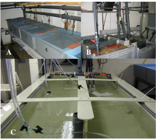
(9) CHAPTER 3 - MATERIAL AND METHODS ............................................................... 34 3.1 - Experimental animals............................................................................................... 35 3.2 – Recirculation system to raise the shrimp................................................................. 36 3.3 – Pre-challenge phase ................................................................................................. 37 3.4 – Shrimp transport ...................................................................................................... 38 3.5 – Challenge facilities .................................................................................................. 38 3.6 – Virus stock preparation and titration ....................................................................... 38 3.7 – Preparation of the virus inoculum ........................................................................... 39 3.8 – Shrimp inoculation .................................................................................................. 39 3.9 – Daily procedures during the challenge experiments................................................ 40 3.10 – Sample collection .................................................................................................. 40 3.11 – Cryosections preparation ....................................................................................... 41 3.12 – WSSV infection detection ..................................................................................... 41 3.13 – Statistical analysis ................................................................................................. 41 3.14 – Experiment 1 ......................................................................................................... 42 3.14.1 – Experimental design ....................................................................................... 42 3.14.2 – Experimental set-up and procedure ................................................................ 44 3.15 – Experiment 2 ......................................................................................................... 45 3.15.1 – Experimental design ....................................................................................... 45 3.15.2 – Experimental procedure and set-up ................................................................ 46 3.16 – Experiment 3 ......................................................................................................... 46 3.16.1- Experimental design......................................................................................... 47 3.16.2 – Experimental procedure and setup ................................................................. 48 CHAPTER 4 – RESULTS ................................................................................................. 51 4.1 – Experiment 1 ........................................................................................................... 51 4.1.1 - Clinical signs..................................................................................................... 51 4.1.2 – Mortality ........................................................................................................... 51 4.1.3 – WSSV detection by Indirect Immunofluorescence (IIF).................................. 53 4.2 - Experiment 2 ............................................................................................................ 54 4.2.1 - Clinical signs..................................................................................................... 54 4.2.2 – Mortality ........................................................................................................... 55 4.2.3 – WSSV detection by Indirect Immunofluorescence (IIF).................................. 56 4.3 - Experiment 3 ............................................................................................................ 57 4.3.1 - Clinical signs..................................................................................................... 57 4.3.2 – Mortality ........................................................................................................... 57 4.3.3 – WSSV detection by Indirect Immunofluorescence (IIF).................................. 59 CHAPTER 5 – DISCUSSION ........................................................................................... 60 5.1 - Challenge model....................................................................................................... 60 5.2 – Experiment 1 ........................................................................................................... 60 5.3 – Experiment 2 ........................................................................................................... 61 5.4 – Experiment 3 ........................................................................................................... 63 5.5 – General discussion................................................................................................... 64 CHAPTER 6 – CONCLUSIONS ...................................................................................... 66 REFERENCES ................................................................................................................... 68. VII.
(10) INTRODUCTION. CHAPTER 1 – INTRODUCTION White spot syndrome virus (WSSV) causes an aggressive and devastating disease (white spot disease, WSD) in shrimp farms throughout Asia, North and South America. Mortalities of 100% can occur within 3-10 days after the onset of disease in grow-out operations. White spot disease constitutes a huge ecological and economical threat for the development of shrimp fisheries and aquaculture. First recorded in Taiwan in 1992 (Chou et al., 1995), it has spread to several shrimp-farming countries in Asia and Latin America (Wang et al., 2000). The disease is characterised by the presence of white spots on the inner surface of the exoskeleton from which the disease name is derived (Lo et al., 1996). Other clinical signs include anorexia, lethargy and reddish discoloration of the body (Otta et al., 1999). WSSV is an enveloped, non-occluded bacilli-form-shaped virus containing a double-stranded DNA. Since the outbreak of white spot disease, shrimp production has decreased significantly in many countries and farmers are facing serious difficulties in continuing production. The resulting economic losses and their impacts are now significantly affecting national economies and the livelihoods of shrimp farmers. Provision of assistance for combating this situation is considered highly appropriate and timely. Such assistance will help secure shrimp aquaculture development, national income through trade (both local and international), and livelihoods of farmers and other service providers (FAO, 2003). In order to face this serious problem, the scientific community promptly answered to gather knowledge on this specific viral disease. Also a considerable number of measures to control WSSV were tested; however with little conclusive results and limited applicability. Of those, temperature manipulation for controlling this specific pathogen appears to be one of the most promissory and potentially applicable in the field. Temperature is one of the most important environmental factors because it can affect an aquatic animals metabolism, oxygen consumption, growth rate, moult cycle, and survival rate directly. Temperature can also affect aquatic animals indirectly when combined with other environmental factors such as salinity and dissolved oxygen. Moreover temperature can have an impact on the development of pathogens and thus. 1.
(11) INTRODUCTION disease in aquatic animals. Studies on the interaction between temperature and crustacean pathogens are however limited. So far there are only three reports on the effect of temperature on WSSV infection in crustaceans (Vidal et al., 2001 and Guan et al., 2003 in penaeid shrimp and Jiravanichpaisal et al., 2004 in freshwater crayfish). The aim of this study was to evaluate the effect of high water temperature (33ºC) on survival of WSSV-infected shrimp (Litopenaeus vannamei), using a highly standardised challenge procedure, with a known infectious dose of white spot syndrome virus. Three experiments were performed. The first experiment aimed to confirm the effectiveness of high water temperature for protecting shrimp against WSSV. For that, four temperature treatments were compared, in which high temperature was applied both before and after the virus inoculation, only before inoculation or only after the inoculation. In a last treatment low temperature (27ºC) was maintained all the time. In the second experiment the objective was to evaluate the protective value of high water temperature, after an initial period of viral replication. The water temperature was raised from 27ºC to 33ºC at different time points, at 12 and 24h after virus inoculation. Knowing that in field conditions, keeping high water temperature for long periods of time will probably be economically unfeasible, the third experiment evaluated the effectiveness of shorter periods of high water temperature exposure. The shrimp were submitted to daily temperature cycles (33ºC/27ºC) with 6, 12 and 18 hours of high water temperature exposure, during five consecutive days.. 2.
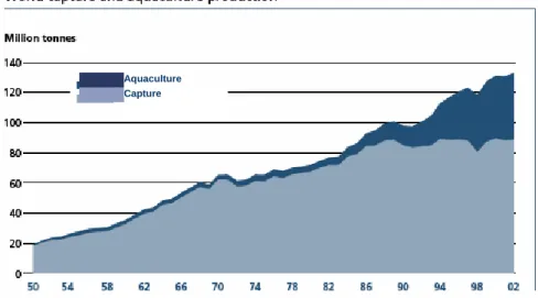
(12) LITERATURE REVIEW. CHAPTER 2 – LITERATURE REVIEW 2.1 – Global aquaculture production According to FAO statistics from 2004, the contribution of aquaculture to global aquatic production continues to grow, increasing from 3.9 percent of total aquatic production by weight in 1970 to 29.9 percent in 2002. Aquaculture continues to grow more rapidly than all other animal food-producing sectors. Worldwide, the sector has grown at an average rate of 8.9 percent per year since 1970, compared with only 1.2 percent for capture fisheries and 2.8 percent for terrestrial farmed meat-production systems over the same period. In 2002, total world aquaculture production (including aquatic plants) was reported to be 51.4 million tonnes (Fig. 1) by quantity and US$ 60.0 billion by value. This represents an annual increase of 6.1 and 2.9 percent in quantity and value respectively, over reported figures for 2000.. Aquaculture Capture. Fig. 1 - Global capture fisheries and aquaculture production data show the increasing importance of aquaculture in the annual global aquatic production (FAO, 2004).. According to data published by FAO (2002), increases in world aquaculture production will be driven by increases in Chinese production, with South Asia, Latin America and the Caribbean and Europe providing smaller increases. Freshwater species and molluscs will dominate aquaculture production. In order to meet growing projected consumption needs in Europe, total production increases in volume are estimated to result primarily from increases in aquaculture production. Indeed, the model estimates. 3.
(13) LITERATURE REVIEW that farmed production will likely double by 2030, exceeding 2.5 million tonnes in 2015 and reaching 4 million tonnes in 2030.. 2.1.1 – Global shrimp production Although cultured crustaceans represented only 5.4 percent of total aquaculture production by weight, they comprised 20.1 percent of total global aquaculture by value in 2002. One of the fastest growing aquaculture production sectors is that of penaeid shrimp. Within this family, the main cultivated species are the giant tiger prawn (Penaeus monodon), the fleshy prawn (Fenneropenaeus chinensis) and the whiteleg shrimp (Litopenaeus vannamei), these three species accounting for over 86% of total shrimp aquaculture production in 2000. Despite being affected by serious disease outbreaks in both Latin America and Asia, the annual rate of growth of the cultured shrimp sector was 6.8 percent (by weight) between 1999 and 2000. Although this had dropped to 0.9 percent during 2002, these growth rates are still relative high compared to other food producing sectors. In recent years, Litopenaeus vannamei has become the leading farm-raised species, representing more than half of the total world production (Fig. 2) (FAO Fishstat database3, 2003). Since a few years China has shifted production towards L. vannamei, producing more than 270 000 metric tonnes in 2002 and an estimated 300 000 metric tonnes (71 percent of the country's total shrimp production) in 2003, which is higher than the current production of the whole of the Americas. Other Asian countries with developing industries for this species include Thailand (120 000 metric tonnes estimated production for 2003), Viet Nam and Indonesia (30 000 metric tonnes estimated for 2003 each). Total production of L. vannamei in Asia was approximately 316 000 metric tonnes in 2002, and it has been estimated that this has increased to nearly 500 000 metric tonnes in 2003, which is worth approximately US$ 4 billion in terms of export income (FAO, 2004).. 4.
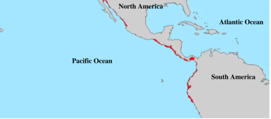
(14) LITERATURE REVIEW. Fig. 2 – Global aquaculture production of Litopenaeus vannamei (FAO Fishstat database3, 2003).. 2.2 – Penaeid shrimp biology 2.2.1 – Habitat and geographical distribution Penaeid shrimp can be found in tropical and subtropical waters around the world, from about 40°N to 40°S latitude. Adult shrimp are rarely found below 180 m and typically inhabit off-shore waters, while juveniles generally occur in protected coastal habitats (Bailey-Brock & Moss, 1992). Litopenaeus vannamei, is native from the pacific coast of America, from Mexico to Peru (Fig. 3), in areas where water temperatures are normally over 20ºC throughout the year (Rosenberry, 2004). This marine shrimp likes muddy bottoms at depths from the shoreline down to about 72 meters (Dore and Frimodt, 1987). It is not currently known whether there is one population or if isolated populations exist, although there appear to be differences between stocks from various areas under culture conditions.. 5.
(15) LITERATURE REVIEW North America Atlantic Ocean. Pacific Ocean South America. Fig. 3 – Geographical distribution of Litopenaeus vannamei. 2.2.2 –Taxonomy Penaeid shrimp belong to the largest phylum in the animal kingdom, the Arthropoda. This group of animals is characterised by the presence of paired appendages and a protective cuticle or exoskeleton that covers the whole animal. The subphylum Crustacea is made up of 42.000, predominantly aquatic species that belong to 10 classes. Within the class Malacostraca, shrimp, together with crayfish, lobsters and crabs, belong to the order Decapoda. Within the suborder Dendrobranchiata, the penaeid shrimp, together with gamba prawns, gamba shrimps, benthesicymid shrimps, rock shrimps and solenocerid shrimps are included in the Superfamily Penaeoidae. The family of the Penaeidae (penaeid shrimp) contains apart from Litopenaeus vannamei many important farmed species such as Penaeus monodon, Litopenaeus stylirostris, Marsupenaeus japonicus, Fenneropenaeus indicus and Fenneropenaeus chinensis.. 6.
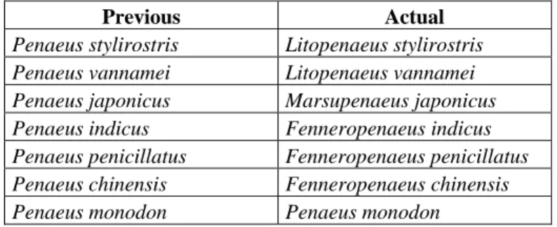
(16) LITERATURE REVIEW Domain = Eucarya Kingdom = Animalia Phylum = Anthropoda Subphylum = Crustacea Class = Malacostraca Subclass = Eumalacostraca Superorder = Eucarida Order = Decapoda Suborder = Dendrobranchiata Super family = Penaeoidea Family = Penaeidae Genus = Litopenaeus Species = vannamei Fig. 4 – Taxonomic classification of Litopenaeus vannamei. Fig. 5 – Drawing of Litopenaeus vannamei. Recently, Pérez Farfante and Kensley (1997) revised the taxonomic classification into genera within the family Penaeidae. The changes for the most important farmed species are shown in the table below. Previous Penaeus stylirostris Penaeus vannamei Penaeus japonicus Penaeus indicus Penaeus penicillatus Penaeus chinensis Penaeus monodon. Actual Litopenaeus stylirostris Litopenaeus vannamei Marsupenaeus japonicus Fenneropenaeus indicus Fenneropenaeus penicillatus Fenneropenaeus chinensis Penaeus monodon. Fig. 6 – Taxonomic changes in important cultured shrimp species (Pérez Farfante & Kensley, 1997). 7.
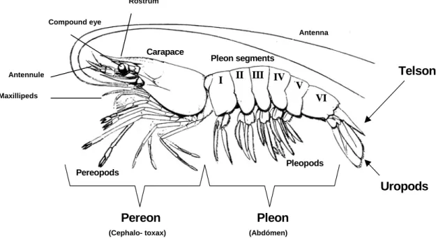
(17) LITERATURE REVIEW. 2.2.3 – Morphology 2.2.3.1 – External morphology Like the other decapod crustaceans, shrimp are bilateral symmetric. The body is protected by an exoskeleton and divided into two regions: the cephalothorax (one unique piece) and the abdomen (several articulated pieces). They are covered by a chitin skeleton more or less calcified (calcium carbonate). This organ is flexible in the abdomen articulation for allowing movement (Morales, 1991). In the head region, antennules and antennae perform sensory functions. The mandibles and the two pairs of maxillae form the jaw-like structures that are involved in food uptake (Solis, 1988). Appendages of the cephalothorax vary in appearance and function. The maxillipeds are the first three pairs of appendages, modified for food handling and the remaining five pairs are the walking legs (pereopods). Five pairs of swimming legs (pleopods) are found on the abdomen (Bell and Lightener, 1988; Baily-Brock and Moss, 1992).. Rostrum Compound eye Antenna. Carapace Antennule. Pleon segments. I. Telson. II III IV. V. Maxillipeds. VI. Pleopods Pereopods. Uropods. Pereon (Cephalo- toxax). Pleon (Abdómen). Fig. 7 - External morphology of shrimp. The body organization of decapod crustaceans is divided into tagmata or specialized regions. These are the pereon, (head and main internal organs), pleon (highly muscularized and specialized for swimming) and telson, or reminiscent tail-like structure. Each tagma possesses specialized appendages, either for feeding and crawling (pereopods) or for swimming and ventilation (pleopods). The uropods of the tail fan are used for escape propulsion.. 8.
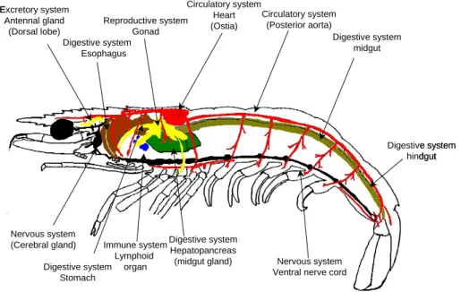
(18) LITERATURE REVIEW. 2.2.3.2 – Internal morphology The internal morphology of the penaeid shrimp is outlined in Figure 7. Penaeids and other arthropods have an open circulatory system and, therefore, the blood and the bloodcells are called haemolymph and haemocytes, respectively. The open spaces in the body are the haemocoel and contain haemolymph. Crustaceans have a muscular heart that is dorsally located in the posterior cephalothorax. It is short and wide, and tapering anteriorly and posteriorly. The blood is pumped by the heart through a complex array of arteries to the haemocoel. The valved haemolymph vessels leave the heart and branch several times before the haemolymph arrives at the sinuses that are scattered throughout the body, where exchange of substances takes place. After passing the gills, the haemolymph returns in the heart by means of three wide non-valved openings. The haemocytes are produced in the haematopoietic tissue. This organ is dispersed in the cephalothorax, but mainly present around the stomach and in the onset of the maxillipeds (Bauchau, 1981; Fox, 2001). The digestive system is divided into a complex, cuticle-lined foregut region; a compact digestive (or midgut) gland at the beginning of the midgut region, followed by a long tubular, simple part; and a cuticle-lined hindgut region, consisting principally of the rectum (Dall, 1967). The stomach and oesophagus are part of the foregut. The stomach is, by divisions, composed of a cardiac and a pyloric region. In the cardiac stomach the cuticle is elaborated to form a complex and intricate gastric mill to grind food. Posterior to the cardiac stomach is located a smaller stomach region, the pyloric stomach which contains a sieve, or filter press, made of cuticular setae (Fox, 2001). In the midgut the hepatopancreas is located. This digestive gland consists of diverticula of the intestine. The spaces between these hepatopancreatic tubules are occupied by haemolymph sinuses. The main functions of the hepatopancreas are the absorption of nutrients, storage of lipids and production of digestive enzymes (Johnson, 1980). The reproductive system in crustaceans is the following. The male has two pairs of modified abdominal appendages on the first and second abdominal segments (the petasma) that deliver sperm to the female's external receptacle (the thelycum) located between the bases of the fifth walking legs.. The gonads (ovaries and testes) are paired tubular structures in the cephalothorax that connect to the exterior by the external sexual. 9.
(19) LITERATURE REVIEW appendages (thelycum and petasma) via paired gonoducts (oviducts and vasa deferentia). (Bailey-Brock & Moss, 1992). The decapod excretory organs are a pair of antennal glands located at the base of the head leading by a duct to the nephridiopores on the second antenna. The antennal gland is a small white pad of tissue just anterior to and lateral to the oesophagus (Fox, 2001). Circulatory system Excretory system Circulatory system Heart Reproductive system Antennal gland (Posterior aorta) (Ostia) Gonad (Dorsal lobe) Digestive system Digestive system midgut Esophagus. Digestive system hindgut. Nervous system (Cerebral gland). Digestive system Immune system Hepatopancreas Lymphoid (midgut gland) Digestive system organ Stomach. Nervous system Ventral nerve cord. Fig. 8 - Diagram of the internal morphology of penaeid shrimp.. 2.2.4 – Penaeid shrimp life cycle Sexes are separated in most cultivated decapods, although occasionally individuals in an intersex hermaphroditic condition are found. In mature decapods mating generally occurs when the female is in a soft-shelled condition (i.e. newly moulted) and results in a deposition of one or more spermatophores in, or close to the genital openings of the female. The spawning occurs directly into the sea in the case of penaeid shrimp, or to the brood chamber beneath the abdomen in other groups. Penaeids eggs hatch a few hours after spawning and each larvae is left to fend for itself as it develops through the nauplius, protozoea and mysis stages before metamorphosing into a post larvae (Fig. 7) (Wickins and Lee, 2002). Their diet ranges from the hereditary yolk sack, during the early naupliar stage, to phytoplankton (microscopic plant organisms) and then to 10.
(20) LITERATURE REVIEW zooplankton (microscopic animals). Finally, at mysis stage and beyond, the shrimp is able to eat a wide variety of organisms. During this period, the larvae drift with the currents. A small percent of them are swept into the bays and estuaries by the currents. Here, the postlarvae remain, through their juvenile stages, until they mature and seek the offshore spawning grounds. It has been estimated that only 1 percent of those spawned in nature actually reach the adult stage (Treece and Yates, 1988).. Fig. 9 – Penaeid shrimp life cycle (Baily-Brock and Moss, 1992).. 2.2.5 – Physiology 2.2.5.1 – Immune system The immune system is commonly divided into two major branches: innate and adaptative immunity. Since vertebrates lack an adaptive immune system in which memory is the hallmark, their defence mechanisms only rely on innate immune responses. Hence, crustaceans cannot readily be vaccinated against particular pathogens. Instead, their defence systems, while effective, tend to be more general and based on haemocytes that can mount phagocytic, cytotoxic and inflammatory responses to invading microbes (Wickins and Lee, 2002). Recently, however, cumulative experimental data from invertebrates provide some specificity and memory might exist in invertebrates (Kurtz, 2005).. 11.
(21) LITERATURE REVIEW Aquatic crustaceans are in intimate contact with their environment, particularly in intensive culture systems, which are enriched with bacteria and viruses. Some of these are pathogenic and many are saprophytic. However, under normal conditions animals maintain a healthy state by defending themselves against potential pathogens (Jiravanichpaisal, 2005). The first line of defence against microbial invasion is the cuticle. It is a physical hard barrier with antimicrobial proprieties, for example it contains inhibitors against enzymatic attack. If it is penetrated, there is an immediate recognition of the non-self material by haemocytes and plasma proteins (Wickins and Lee, 2002). The digestive tract, which is the main route of invasion, is partially lined with chitinous membranes and its hostile environment of acids and enzymes is able to inactivate and digest many virures and bacteria. In most cases the cuticular defences are sufficient to protect against even quite virulent pathogens, which often only produce disease when the integument has been physically damaged. Once pathogens gain entry into the hemocoel of the host, they encounter a complex system of innate defence mechanisms involving cellular and humoral responses (Jiravanichpaisal, 2005). The cellular reactions involve three subpopulations of haemocytes which are responsible for a whole number of reactions: containment of the PO system, phagocytosis, degranulation and release of reactive oxygen intermediates (Song and Hsieh, 1994), and coagulation (Söderhäll and Smith, 1986). The humoral components include the activity of soluble enzymes, either activated in circulating hemolymph, or released by cells that serve to detoxify toxic molecules or inhibit the physiology of invading pathogens (Cardenas and Dankert, 2000). Antimicrobial peptides, proteases and protease inhibitors, as well as lectin-like molecules exist in the white shrimp species Litopenaeus vannamei and L. stylirostris (Gross et al., 2001; Cerenius and Söderhäll, 2004).. 2.2.5.1.1- Haemocytes In crustaceans, the circulating haemocytes play a crucial role in defence against infection, including recognition, phagocytosis, melanization, cytotoxicity and cell–cell communication (Johanson et al., 2000). In decapod crustaceans, these cells can be. 12.
(22) LITERATURE REVIEW divided, according their morphology (presence of cytoplasmic granules) into three types: hyaline, semigranular and granular cells (Bauchau, 1981). The hyaline cells play a major role in phagocytosis (Söderhäll et al., 1986). The semigranular cells take part in encapsulation reactions and have a limited function in phagocytosis. Both granular cells and semigranular cells store the components of prophenoloxidase activating system and are capable of cytotoxic reaction (Smith and Söderhäll, 1983). The semigranular cells are the most sensitive and they are the first to respond to the lipopolysaccharides and β-1,3-glucans by degranulation and then, the components of the proPO system are released (Johansson and Söderhäll, 1985).. 2.2.5.1.2 – The prophenoloxidase activating system (proPO) The primary mediator of the cellular response to injury and disease in invertebrates is the pro-enzyme prophenoloxidase (proPO) activating system (Söderhäll et al., 1994). This system consists of several proteins involved in the immune defence in invertebrates leading to melanin production, cell adhesion, encapsulation, and phagocytosis (Sritunyalucksana, and Söderhäll, 2000), where proPO is released from haemocytes by an active degranulation process that can be stimulated by inflammatory agents such as lipopolysaccharide (LPS) or peptidoglycan (molecules of bacterial cell walls) and ß-1,3glucan (molecules of fungal and yeast cell walls). Once released, ProPO is proteolytically converted, through cleavage of the enzyme at a specific site, to its active form PO, which is the central component of an enzyme cascade that has been identified in crustaceans (Cardenas et al., 2000). The active form of the enzyme then functions to produce antimicrobial effects, wound repair, encapsulation, and phagocytosis.. 2.2.5.1.3 – The coagulation system One of the principal differences between vertebrates and arthropods is the fact that the body fluids in vertebrates are mostly confined to blood and lymphatic vessels, while arthropods have an open circulatory system. Therefore, after wounding, arthropods must produce a matrix that quickly stops the loss of haemolymph, but also aids in trapping. 13.
(23) LITERATURE REVIEW foreign organisms to prevent spreading throughout the haemocoel. Haemolymph clotting is thus an important part of innate immunity and is regulated in many cases by microbial elicitors (Jiravanichpaisal, 2005). In crustaceans, the coagulation system involves plasma clotting protein and a haemocyte-derived transglutaminase. This clotting protein is synthesised in the hepatopancreas and released to the haemolymph. The transglutaminase is synthesised and stored in the haemocytes, and released to the plasma upon the activation of haemocytes. This enzyme covalently crosslinks the clotting protein molecules in the presence of calcium ions to form a soft gel at the wound sites (Bangyeekhun, 2002).. 2.2.5.1.4 – Antimicrobial peptides Antimicrobial peptides are a major component of the innate immune defense system in marine invertebrates. They are defined as molecules less than 10 kDa in mass which show antimicrobial properties (Boman, 1995) and provide an immediate and rapid response to invading microorganisms (Bartlett, 2002). The major classes of antimicrobial peptides include (i) α-helices, (ii) β-sheet and small proteins, (iii) peptides with thio-ether rings, (iv) peptides with an overrepresentation of one or two amino acids, (v) lipopeptides, and (vi) macrocyclic cystine knot peptides (Epand and Vogel, 1999). There is evidence that antimicrobial peptides are widespread in invertebrates (15), especially in tissues such as the gut and respiratory organs in marine invertebrates, where exposure to pathogenic microorganisms is likely (Chisholm and Smith, 1992). These peptides generally act by forming pores in microbial membranes or otherwise disrupting membrane integrity (Tam et al., 2000). The value of antimicrobial peptides in innate immunity lies in their ability to function without either high specificity or memory, and their small size makes them easy to synthesize (Relf et al., 1999). In addition, many antibacterial peptides show remarkable specificity for prokaryotes with low toxicity for eukaryotic cells (Zasloff, 1992). Prominent among crustacean antimicrobial peptides are the penaeidins, which display antifungal and antibacterial properties and were isolated from the haemolymph of the shrimp Litopenaeus vannamei (Destoumieux el al. 1997).. 14.
(24) LITERATURE REVIEW. 2.2.5.1.5 – Non-self recognition system The innate immune system is based on recognition of molecules named pattern recognition receptors that are present on pathogenic microbes. These molecules are structural molecules of pathogens, but not of the host, which are shared by a large group of microbes and are essential for their survival (Medzhitov and Janeway, 2000; Janeway 2001). Example of such molecules are ß-1,3-glucans from fungi, lipopolysaccharide, peptidoglycan and lipoteichoic acid from bacteria, and double-stranded RNA from virus. Therefore, presence of microbial molecules is an indication of an infection, which allows the host to choose a sufficient mechanism to fight against a certain class of pathogens (Medzhitov and Janeway, 2000). The biological function of recognition molecules in innate immune reactions are (i) triggering of proteinase cascades and/or signalling pathways of the defence mechanisms, and (ii) clearance of microbial invaders from the blood system (Bangyeekhun, 2002).. 2.2.5.1.6 – Proteinase inhibitors Proteinase cascades, such as clotting cascades and the proPO system, need to be carefully regulated to prevent excessive activation of endogenous cascades and damage to host tissue (Jiravanichpaisal, 2005). Proteinase inhibitors are present in multiple forms in animals, plants and microorganisms. A number of proteinase inhibitors have been reported from invertebrates. Most of them have a common structural feature as one of well characterised families, such as Kazal, Kunitz, α-macroglobulin, serpin, metalloproteinase inhibitor and cysteine proteinase inhibitor (Bangyeekhun, 2002). In the mechanism of inhibition, that is common among most proteinase inhibitors, the inhibitor molecule combines with the proteinase at the reactive site to block proteolytic activity. Invertebrate proteinase inhibitors can be found in plasma, haemocytes or cuticle. The gross biological function of proteinase inhibitor is to prevent unwanted proteolysis. Two central roles of proteinase inhibitors in invertebrate immunity are defence against microbial proteinases and regulation of endogenous proteinases (Kanost, 1999).. 15.
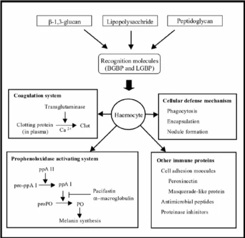
(25) LITERATURE REVIEW These substances play a key role to control and regulate the proPO system, avoiding the deleterious effects of its active components, particularly PO, which can produce highly toxic intermediates (Jiravanichpaisal, 2005).. Fig. 10 – Schematic overview of crayfish defence reactions. In the presence of microbial organisms, the recognition molecules in the plasma participate in binding to the microbial cell wall components. Then the complexes bind to membrane receptors of the haemocytes and consequently activate the defence mechanisms. Haemocytes directly play a role in cellular defence mechanism or release humoral defence molecules, which lead to activation of the prophenoloxidase activating system and coagulation system (Bangyeekhun, 2002).. 2.3 – Shrimp Farming Shrimp farming started more than a century ago in Southeast Asia where farmers raised incidentally wild shrimp crops in tidal fish ponds (Rosenberry, 2004). Modern shrimp farming started in the early 1970s, and today, over fifty countries have shrimp farms. In the Eastern Hemisphere, Thailand, Vietnam, Indonesia, India and China are the leaders, but also Malaysia, Taiwan, Bangladesh, Sri Lanka, The Philippines, Australia. 16.
(26) LITERATURE REVIEW and Myanmar (Burma) have large industries. In the Western Hemisphere, Mexico, Belize, Ecuador and Brazil are the leading producers, and there are shrimp farms in Honduras, Panama, Colombia, Guatemala, Venezuela, Nicaragua and Peru. The shrimp importing nations (United States, Western Europe and Japan) specialize in high-tech "intensive" shrimp farming, but, thus far, their production has been insignificant. In the Middle East, Saudi Arabia and Iran produce the most farmed shrimp (Shrimp News International, 2004). The shrimp farming process can be divided into three main phases, the hatchery, nursery and growout phase.. 2.3.1 – Hatchery The production cycle begins at the hatchery, where the shrimp seed is obtained from the broodstock. In many hatcheries, females with ripe, egg-laden ovaries (gravid females) are brought from the sea for spawning in captivity. As alternative, and due to reasons of price and availability, techniques were developed for inducing maturation of females in captivity. This procedure also allowed the establishment of breeding programs for fast growing, specific pathogen-free and/or resistant stocks. Whether gravid shrimp are captured in the wild or matured in the hatchery, they invariably spawn at night, but with photoperiod manipulation, they can be induced to spawn at any time. Depending on a number of variables (temperature, species, size, wild/captive and number of times previously spawned), they produce between 50,000 and 1,000,000 eggs. After one day, the eggs hatch into nauplii, the first larval stage. Nauplii feed on their egg-yolk reserves for a couple of days. Next they pass through the next two main larval stages, the zoeae (which feed on microalgae and a variety of formulated feeds for three to five days) and mysis (feed on algae, formulated feeds and zooplankton for three or four days). Next they metamorphose into postlarvae. Postlarvae look like adult shrimp and feed on zooplankton, detritus and commercial feeds. Farmers refer to postlarvae as “PLs”, and as each day passes, the stages are numbered PL-1, PL2, and so on. When their gills become branched (PL-13 to PL-17), they can be moved to the nursery or growout farm. From hatching, it takes about 25 days to produce a PL-15.. 17.
(27) LITERATURE REVIEW. 2.3.2 – Nursery In modern semi-intensive farms, a nursery phase (between the hatchery and growout phases) is usually incorporated in the farm design. Nursery ponds may represent between 6 to 15% of all culture area. They are usually made with earthen embankments, and have at sizes that range from 0.04-1 ha. Stocking densities are typically 100-200 juveniles m-2. In South-east Asia, nursery facilities also take the form of concrete tanks, concrete walled ponds with sand bottoms, staked net pens and floating cages (Wickins and Lee, 2002). The PLs are fed with a crumbled diet several times a day, where the protein levels range from 30 to 45%. The nursery phase should not exceed 25 days (Shrimp News International, 2004).. 2.3.3 – Growout The growout operation is stocked with postlarval shrimp and it takes from three to six months to produce a crop of market-sized shrimp. Northern China, the United States and Northern Mexico produce one crop per year, semi-tropical countries produce two crops per year, while farms closer to the equator have produced three crops a year, but rarely (Shrimp News International, 2004). The two most practised production strategies are the extensive and intensive culture, however there are a numerous transitions between them. In extensive shrimp culture, shrimp are stocked at low densities (<25 PLs·m-2) in large ponds or tidal enclosures in which little or no management is exercised or possible. Farmers depend almost entirely on natural conditions in extensive culture. Intensive shrimp culture is carried out in high densities (sometimes >200 PLs·m-2) in intensively managed pens, ponds, tanks and raceways where a high level of investment is required (Rosenberry, 2001). Semiintensive culture falls between these two extremes.. 2.4 – Penaeid shrimp common diseases in farming conditions. 18.
(28) LITERATURE REVIEW The diseases of cultured penaeid shrimp include syndromes with infectious and noninfectious etiologies. Included among the infectious diseases of economic importance to cultured shrimp are those with viral, rickettsial, bacterial, fungal, protistan and metazoan etiologies. A number of non-infectious diseases are also of importance to the industry, and included among these are diseases due to environmental extremes, nutritional imbalances, toxicants, and genetic factors (Lightner and Redman, 1998).. 2.4.1 – Viral diseases Viruses are a group of organisms which must enter a host cell to replicate, since they lack the necessary biochemical machinery to manufacture proteins and metabolize sugars. Some virus also lack the enzymes required for nucleic acid replication, and are dependent on the host cell for these functions (Jiravanichpaisal, 2005). Viral diseases are probably still underestimated in crustaceans, but nevertheless, they emerge as being responsible for serious enzootics or massive pandemics, on a regional scale in shrimp-farming countries. In 1989, 6 viruses were known to affect penaeid shrimp, but by 1997 more than 20 viruses were identified as having affected wild stocks and commercial production. The Office International des Epizooties (OIE) now lists seven viral diseases of shrimp which are considered to be transmissible and of significant socio-economic and/or public health importance (FAO, 2004). These viral diseases are:. •. White Spot Disease (WSSV),. •. Yellow Head disease (YHV),. •. Taura Syndrome Virus (TSV),. •. Spawnerisolated Mortality Virus Disease (SMV),. •. Tetrahedral Baculovirosis (Baculovirus penaei - BP),. •. Spherical Baculovirosis (Penaeus monodon-type baculovirus). •. Infectious Hypodermal and Haematopoietic Necrosis (IHHNV). 19.
(29) LITERATURE REVIEW. 2.4.2 – Bacterial and fungal diseases Bacteria, both gram-positive and gram-negative, are also etiological agents responsible for severe diseases in crustaceans (Söderhäll et al., 1993). These microorganisms act frequently as opportunistic follow-on to viral infection or environmental stress. Similar to bacteria, fungi often infect aquatic organisms as opportunistic invaders, but once established, they are also often fatal and difficult to treat. The most important bacterial and fungal diseases that have socio-economic impact in shrimp farming are: •. Vibriosis – Vibrio spp.;. •. Necrotizing hepatopancreatitis (NHP) - Alfa proteobacteria (new genus);. •. Rickettsial infection - Rickettsia or rickettsia-like microorganisms;. •. Mycobacteriosis. –. Mycobacterium. marinum,. Mycobacterium. fortuitum,. Mycobacterium spp.; •. Larval mycosis – Lagenidium spp., Sirolpidium spp.;. •. Fusarium disease - Fusarium solani, F. moniliforme;. •. Crayfish plague - Aphanomyces astaci.. 2.4.3 – Protozoan diseases Among organisms causing diseases to shrimp, parasites, especially protozoan parasites form an important group. Although several diseases caused by parasites have been noticed in shrimp, often, chronic conditions caused by protozoan play a crucial role in shrimp production. The protozoa affecting shrimp can be grouped as parasites and commensals (Jithendran and Vijayan, 2001). Following are listed the major disease problems caused by the protozoa: •. Protozoan fouling - Peritrichous ciliates such as Zoothamnium, Epistylis, Vorticella and Acinata;. •. Cotton shrimp disease - Microsporeans such as Agmasoma, Ameson and Pleistophora; 20.
(30) LITERATURE REVIEW •. Enterozoic cephaline gregarine infection - Cephaline gregarines such as Nematopsis and Cephalolobus;. •. Invasive protozoan infection - Ciliate protozoa, Paranophrys and Paraoronema, leptomonad-like organisms.. 2.4.4 – Non-infectious and toxic diseases Non-infectious diseases are common in growout farms, as influences of nutritional factors, environmental factors such as temperature extremes and oxygen depletion, toxicity from biotic and abiotic origins, become critical during the lengthy culture period (Jithendran and Vijayan, 2001). The most common non-infectious pathologies are:. • Gas Bubble disease - caused by supersaturation of atmospheric gases, usually nitrogen, but occasionally oxygen;. • Haemocytic enteritis (HE) – caused by toxins released by certain blues-green algae blooms; •. Black gill disease – is associated to the presence of excessive levels of toxic substances such as nitrite, ammonia, heavy metals, crude oils in the culture water;. •. Soft shell syndrome - caused by sudden fluctuation in water salinity, high soil pH, highly reducing conditions in soil, low organic matter in soil, low phosphate content and pesticide pollution in water, nutritional deficiency and insufficient water exchange;. •. Muscle necrosis - is associated with poor environmental conditions such as low oxygen levels, and salinity or temperature shock.. 2.5 - White Spot Syndrome Virus White Spot Disease (WSD) is a pandemic disease of shrimp caused by a virus commonly known as White Spot Syndrome Virus (WSSV). First recorded in Taiwan in. 21.
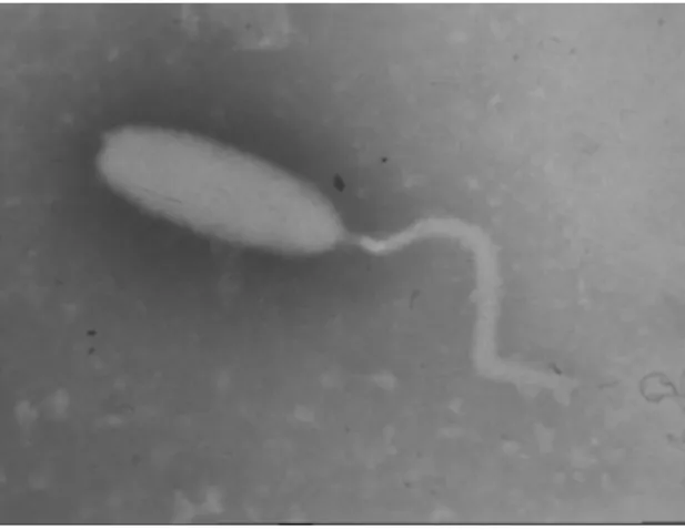
(31) LITERATURE REVIEW 1992 (Chou et al., 1995), the first major outbreaks were first detected in Marsupenaeus japonicus and Fenneropenaeus chinensis in Japan and China in 1993 (Nakano et al., 1994; Zhan et al., 1998) and in the following 18 months the outbreak spread to the majority of the shrimp farming countries in Asia. WSSV has a wide host range among decapod crustaceans (Lo et al., 1996), and is potentially lethal to most of the commercially cultivated penaeid shrimp species (OIE, 2003). Of all shrimp virus, WSSV has the largest impact on shrimp culture and remains a major problem up to the present day (Rosenberry, 2004).. 2.5.1 – Taxonomy Based on extensive phylogenetic analyses, and also on primary genomic structure and composition as well as the distinct morphology of the virion, the International Committee on Taxonomy for Virus (ICTV) approved a proposal in 2002 to accommodate WSSV in a new virus family called Nimaviridae, referring to the threadlike polar extension on the virus particle (Nima: Latin for thread). This virus family consists of a single genus (Whispovirus) and contains White spot syndrome virus I as its sole species so far (Mayo, 2002). Probably all WSSV isolates identified thus far are variants of the same species.. 2.5.2 – Morphology Electron microscopical studies on thin sections and viral suspensions obtained from infected shrimp revealed that the virion of WSSV is a large, ovoidal particle of about 275 nm in length and 120 nm in width, with a tail-like appendage at one end (Duran et al., 1997). It is formed by a road-shaped nucleocapsid with a tight-fitting capsid layer, surrounded by a loose-fitting trilaminar envelop, which consists mainly of the WSSV encoded proteins VP28 and VP19 (van Hulten et al., 2000). VP28 is most likely located on the surface of the virus particle and plays a key role in the virus infection (van Hulten et al., 2001b). Isolated nucleocapsids have a cross-hatched appearance and size of about 300 x 70 nm. The nucleocapsid is formed by stacks of rings (about 14 in total), which. 22.
(32) LITERATURE REVIEW are in turn formed by regular spaced globular subunits of about 8 nm in diameter, arranged in two parallel rows (Durand et al., 1997).. Fig. 11 – Image of a WSSV virion obtained by electron microscopy (source: Wageningen University, Laboratory of Virology). 2.5.3 – Genome The virions of WSSV contain a circular, supercoiled, double-stranded DNA genome, originally estimated to be 300 kilobase pairs (kb) (Wang et al., 2000). The genome contains 292,967 nucleotides encompassing 184 major open reading frames (ORFs). Of these, only 6% of the ORFs have putative homologues in databases, mainly representing genes encoding enzymes for nucleotide metabolism, DNA replication, and protein modification. The remaining ORFs are mostly unassigned, except for five, which encode structural virion proteins. Unique features of WSSV are the presence of a very long ORF of 18,234 nucleotides, with unknown function, a collagen-like ORF, and nine regions, dispersed along the genome, each containing a variable number of 250-bp tandem repeats (van Hulten, et al., 2001a). Between different geographic isolates of WSSV, a few restriction fragment length polymorphisms (RFLPs) were reported, indicating the presence of some genomic variation (Wang et al., 2000).. 23.
(33) LITERATURE REVIEW. 2.5.4 – Epidemiology This virus can be transmitted to benthic crustaceans and other fauna through different feeding pathways such as filter feeding, detritus feeding, and predation (Mortensen, 1993; Vijayan, et al., 2005). The transmission can occur horizontally either per os by predation on diseased individuals, but also by virus particles present in the water. Infection by the latter is thought to occur primarily through the gills, but may occur via other body surfaces as well (Chou et al., 1998). No penaeid shrimp species are known to be resistant to WSSV infection (Lightner, 1996). When viruses pass into the digestive tracts of other invertebrates (bivalves, polychaete worms), they can persist in the alimentary canal, potentially making the animal a passive carrier or vector of the virus. When these passive carriers are consumed by the shrimp, they can potentially infect the shrimp with WSSV (Vijayan et al., 2005). Also, a large number of other wild animals have been reported to be potential carrier of WSSV. These were not only shrimp, prawn and crab species but also planktonic organisms and insect larvae (Flegel and Alday-Sanz 1998). Hence, the passage of the viral pathogen to shrimp broodstock in the hatchery through feeding of infected prey items is a realistic possibility (Vijayan et al., 2005). Once the broodstock is infected, the virus may also be transmitted from mother to offspring, although it is not a clear whether the WSSV virions are present inside the shrimp eggs (Peng et al., 2001). Frequent disease outbreaks in the shrimp farms of India and Asia lead to the offloading of dead and decayed shrimps carrying a heavy load of this virus into the coastal ecosystem. Horizontal transmission of WSSV from the affected shrimp farms to the neighbouring ecosystem has created a realistic scenario in which the receiving ecosystem carries the WSSV load in the form of live or dead tissues, dead and decomposed tissues and free virions (Mortensen, 1993). In addition, contrary to the common belief that free virus cannot survive in natural waters more than 24 hours, WSSV virions can remain infective in decaying tissues or in detritus for up to 4 days, (Bondad-Reantaso et al., 2001).. 24.
(34) LITERATURE REVIEW. 2.5.5 – Cytopathology and histopathology Histopathology of the WSSV infection is characterized by the presence of cells with hypertrophied nuclei showing eosinophilic intranuclear inclusions and marginated chromatin, as WSSV DNA replication and virion morphogenesis take place (Durand et al., 1997; Wang et al., 2000). Inclusion bodies inside the nuclei are markedly distinct and. bigger. than. the. cowdry. A-type. inclusions. characteristic. of. IHHNV. (Wongteerasupaya et al., 1995). Nuclei of infected cells progressively become basophilic and hypertrophied because of the accumulation of intranuclear virions (Chang et al., 1996; Lo et al., 1996; Durand et al., 1996; 1997; Wang et al., 1998; Otta et al., 1999; Takahashi et al., 2000). In the late stage of infection, cells become degenerated, displaying cariorhexis and cellular disintegration which lead to the formation of necrotic areas characterized by vacuolization (Karunasagar et al., 1997; Kasornchandra et al., 1998). WSSV targets tissues of ectodermal and mesodermal origin, such as epithelial and connective tissues of epidermis, stomach, gills, antennal gland, lymphoid organ and haemocytes, muscle, haematopoietic and nervous tissue, eye-stalk, heart, gonads, etc. (Chang et al., 1996; Durand et al., 1996; 1997; Mohan et al., 1998; Rajendran et al., 1999). No haemocytic infiltration can be seen in areas of necrotic tissues (Park et al., 1998; Flegel, 2001).. 2.5.6 – Replication cycle Although during the last decade, intensive efforts were undertaken for detection and characterization of in vivo WSSV infection in shrimp (Maeda, 2004) little is known about the molecular mechanisms underlying the WSSV life cycle and mode of infection. Based on data from Ecobedo-Bonilla et al., (2005), when Litopenaeus vannamei were infected with WSSV, the first infected positive cells were found at 12 hours post inoculation. This data indicates that the virus replication time may not be longer than 12 hours.. 25.
(35) LITERATURE REVIEW. 2.5.7 – General clinical signs Infected animals show lethargic behaviour, such as lack of appetite and slow movement, and reddish to pink body discoloration. Characteristics for the WSSV infected shrimp are white spots on the exoskeleton. These spots are the result of calcified deposits that range in size from a few mm to 1 cm or more in diameter (Chou et al., 1995). However, in case of acute (experimental) infections the only signs of WSSV infection observed are lethargy and lack of appetite. White spots are also not evident in species like L. vannamei, even in normal farming conditions.. 2.5.8 –Diagnostic methods The earliest diagnostic methods developed for virus included the traditional methods of morphological pathology (direct light microscopy, histopathology, and electron microscopy), as well as enhancement and bioassay methods. While tissue culture is considered to be a standard tool in medical and veterinary diagnostic labs, it has never been developed as a useable, routine diagnostic tool for shrimp pathogens. As well, there are few antibody-based diagnostic tests available for the penaeid viral diseases (Lightner and Redman, 1998). PCR or RT-PCR methods are available for several of these viruses and some are in routine use by certain sectors of the industry. For others, specific DNA probes tagged with non-radioactive labels provide highly specific detection methods for application in dot blot formats with haemolymph or tissue extracts, and with routine histological sections using in situ hybridization (Lightner, 1996; Lightner and Redman, 1998).. 26.
(36) LITERATURE REVIEW. 2.5.8.1 – PCR (Polymerase Chain Reaction) The polymerase chain reaction (PCR) is a method for amplification of a specific DNA sequence of interest. PCR will allow a short stretch of DNA (usually fewer than 3000 bp) to be amplified to about a million fold. The particular stretch of DNA to be amplified, called the target sequence, is identified by a specific pair of DNA primers, oligonucleotides usually about 20 nucleotides in length. The PCR product is amplified from the DNA template using a heat-stable DNA polymerase and using an automated thermal cycler. This device promotes the reaction through 30 or more cycles of denaturing, annealing of primers, and polymerization. After amplification by PCR, the products are separated by polyacrylamide gel electrophoresis and are directly visualized after staining with ethidium bromide. In recent years, PCR has been used to detect WSSV in a very specific and sensitive manner (Lo et al., 1996; Nunan and Lightner, 1997; Kim et al., 1998). Nested or twostep PCR has the advantage of increasing the level of sensitivity over singlestep PCR. Nested PCR consists in the reamplification of the PCR product obtained in a single-step PCR reaction by using an aliquot of this first reaction as a template in a second round of amplification (Peinado-Guevara and Lopéz-Meyer, 2005). Frequently, when shrimps show clinical signs of the disease such as lethargy, reduction of food consumption, reddish coloration and white spots on the exoskeleton, WSSV is easily detected by single-step PCR (Lo et al., 1998). However, at low viral loads, WSSV is latent without causing disease symptoms in the shrimps, and can only be detected by nested PCR (Lo et al., 1996, 1998; Kim et al., 1998).. 2.5.8.2 – IIF (Indirect immunofluorescence) A number of antibody-based diagnostic methods have been developed and are described in the literature (Loh et al., 1998; Poulos et al., 2001; Shih et al., 2001) as a confirmation tool for virus infection in shrimp species. One of the antibody-based assays that is being used for detecting WSSV in shrimp is indirect immunofluorescence (Poulos et al., 2001; Wang et al., 2002; Ecobedo-Bonilla et al., 2005; Rahman et al., 2005). This technique detects specific antigens in tissues. The. 27.
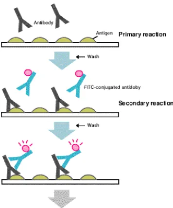
(37) LITERATURE REVIEW specific secondary antibodies are labelled with a compound (fluorescein isothiocyanate) that makes them glow an apple-green colour when observed microscopically under ultraviolet or blue light. In the specific case of the detection of WSSV in Litopenaeus vannamei tissues, tissues from the pereon are embedded in methylcellulose and frozen at –20°C. Cryosections (5 to 6 µm) are made and fixed in absolute methanol at –20°C, washed with white phosphate buffered solution at 1% (WPBS), incubated for 1 h at 37°C with 2 mg ml–1 of the monoclonal antibody 8B7 specific for VP28 (Poulos et al., 2001), washed and incubated for 1 h at 37°C with 0.02 mg ml–1 of fluorescein isothiocyanate (FITC)labeled goat anti-mouse antibody (F-2761, Molecular Probes) in PBS, washed with PBS, rinsed in deionised water, dried and mounted. Slides are analyzed by fluorescence microscopy (Escobedo-Bonilla et al., 2005). Fig. 12 – General principle of Indirect Immunofluorescence technique.. Samples are added on a substrate. slide for primary reaction of antibodies (primary antibodies) and antigens. After washing, conjugated antibodies (secondary antibodies) are added to make complexes of antigens - antibodies - conjugated antibodies. After washing, the fluorescence from FITC is observed by fluorescent microscope (MBL, 2005).. 28.
(38) LITERATURE REVIEW. 2.5.8.3 – Other methods Several other diagnostic methods have been described for WSSV detection: •. Light and electron microscopy (Chou et al., 1995; Wongteerasupaya et al., 1995; Lightner, 1996; Durand et al., 1997). •. In-situ DNA hybridisation ((Chang et al., 1996; Durand et al., 1996). •. Dark-Field Microscopic Observation ( Momoyama et al., 1995). •. Miniarray (Quere et al., 2002),. •. Observation of tissues subjected to fixation or negative staining (Inouye et al.,1994),. •. Reverse passive latex agglutination (Okumura et al., 2005). •. Bioassay (Nunan et al., 1998). •. Rapid staining Hematoxylin and Phloxine/Eosin (H&E) (Sheehan and Hrapchak, 1980).. 2.5.9 – Tested strategies for WSSV control Different approaches knew already some success to control WSSV, including (i) higher or lower than normal water temperatures (Vidal et al., 2001; Guan et al., 2003; Jiravanichpaisal et al., 2004), (ii) treatment with the immunostimulants peptidoglycan, lipopolysaccharide and β-1,3 glucan (Itami et al., 1998; Takahashi et al., 2000; Chang et al., 2003), (iii) vaccination with formalin inactivated bacteria over expressing WSSV proteins, siRNA and WSSV envelope proteins VP19 and VP28 (Namikoshi et al., 2004; Witteveldt et al., 2004; Musthaq et al., 2005; Westenberg et al., 2005) (iv) treatment with egg yolk antibodies (IgY) against WSSV (Kim et al., 2004) (v) feeding antiviral fucoidan, a sulfated polysaccharide extracted from Sargassum polycystum supplemented diet (Chotigeat et al., 2004) and (vi) treatment with cidofovir (antiviral) and a diet supplemented with Spirulina platensis (Rahman et al., 2005).. 29.
(39) LITERATURE REVIEW. 2.5.9.1 – WSSV control with temperature treatment Few reports are available about the influence of temperature on viral diseases in aquatic animals (Amend, 1970; Dorson and Torchy, 1981; Castric and Kinkelin, 1984; Oseko et al., 1988; Sano et al., 1993; Kobayashi et al., 1999) and so far, only three reports on the WSSV infectivity in crustaceans are available. Vidal et al. (2001) have reported that hyperthermia was able to protect Litopenaeus vannamei from WSSV disease after challenge with WSSV. In Marsupenaeus japonicus that protection also occurs at low temperature (Guan et al., 2003). Jiravanichpaisal et al. (2004) reported that protection of WSSV infected freshwater crayfish occur at low temperatures. In the study of Vidal et al. (2005) juveniles of the pacific white shrimp were infected with WSSV by oral and intramuscular route. Both the shrimp inoculated by oral and intramuscular route were divided in two groups, one maintained at ambient temperature (25.8ºC) and the other kept at higher temperature (32.3ºC). The results demonstrated a high degree of protection of the groups kept at high water temperature, as survival was always above 80% against 100% mortality obtained in those maintained at lower temperature. Guan et al. (2003), tested the influence of four temperature levels (15, 23, 28 and 33ºC) on survival of WSSV infected Marsupenaeus japonicus. After virus injection, the four shrimp groups were maintained for 19 days at those temperature levels. The results demonstrate that WSSV infection can by controlled either by low and high temperature levels. Jiravanichpaisal et al. (2004), used two species of freshwater crayfish, Pacifastacus leniusculus and Astacus astacus, for testing the effect of low water temperature on the development of WSSV infection. Crayfish were exposed to different temperatures (4, 12, 22ºC) after WSSV injection or oral exposure and the mortalities were recorded during 45 days. The results showed that the infection could be blocked at lower temperatures (4 and 12ºC), while at high temperature 100% mortality was reached. It was also observed that mortality could be delayed transferring moribund individuals to lower temperature.. 30.
(40) LITERATURE REVIEW. 2.5.10 – Socio-economic impact of WSSV. WSSV disease is responsible for direct losses of billions US$ per year in Asia and Latin America. For example, in Ecuador US$ 280 millions were lost in the first six months of its first appearance in 1999. In the export sector, shrimp exports fell from 115 000 metric tonnes (mt) in 1998 to 38 000 mt in 2000, and have only recovered slightly to 47 000 mt in 2002. This equates to a total direct loss of some 267 000 metric tonnes of shrimp worth nearly US$ 1.8 thousand million between 1999 and mid-2003. Although similar problems have occurred throughout Central and South America, Brazil and Venezuela remained several years free of WSSV due to a rapid and effective closure of their borders to all crustacean imports in 1999. However, recently, on 20 January 2005 the first occurrence of WSSV in Brazil was reported in Litopenaeus vannamei. After initial loses, United States also managed to eradicate WSSV from its shrimp culture industry in 1997 through the implementation of biosecurity measures, including the use of all SPF broodstock, although there are reports of its recent re-emergence in Hawaii in 2004 (Briggs et al., 2004). Estimates for Asia include losses of over US$ 250 million for 1993 (continuing every year) in Mainland China, loosing 120 000 metric tonnes of production of F. chinensis, M. japonicus and P. monodon to WSSV (Jiang, 2000). In addition to direct effects on production, the impacts of diseases are particularly felt by small-scale farmers who, especially in Asia, represent the backbone of many coastal communities. Their very livelihoods are threatened through reduced food availability, loss of income and employment, social disturbance and increased vulnerability. Crop losses to disease for this sector of society may determine whether or not those families are below the UN poverty threshold (Fegan et al., 2001). In Mainland China, for example, the WSSV epidemic in 1993 affected the lives of 1 million people, and has continued to have effects to this day (Jiang, 2000). Similar effects have been noted from Latin American countries. In Ecuador for example, within the first year of the WSSV epidemic in 1999, the disease also lead to the loss of 26 000 jobs (13 percent of the labour force), the closure of 74 %t of the hatcheries, a 68 percent reduction in sales and production for feed mills and packing plants, 64 percent layoffs at feed mills and a total of 150 000 jobs lost in the shrimp farming industry (Alday de Graindorge and Griffith, 2000).. 31.
Imagem

+7
Documentos relacionados
Toit terrasse avec piscine individuelle chauffée, jets d’hydromassages et grand solarium en bois.. Garage en box
de produção de São Paulo, notadamente concentrada no ramo da eletrônica de consumo9 — fenômeno de algum modo também observado no Nordeste que, a partir de um perfil
gra~ de quinta à oitava série, e apenas para os funcionários da universidade. Para os alunos de primeira à quarta série as aulas são diárias e eles são agrupados em
Dentre os sintomas relatados pelos atletas do presente estudo, 78,6% foram referentes ao TGI superior, padrão de desconforto similar a alguns achados prévios
deve ser definido o número da porta que será utilizada por cada uma das três caches do driver: a cache de comandos SQL, a cache de relações e a cache de tuplas. Você pode
Folhas com lâmina lanceolada a estreitamente oblonga, base subcordada, ápice agudo a atenuado, 9–15,6 × 1,8–3,5 cm, membranácea a cartácea, margem plana, 10–14
Sob essa perspectiva, a presente investigação visa compreender como o uso de informações espaciais no desenho e implementação de esquemas de Pagamento por
(Haderlie & Clark 1959, p. We receritly described the subspecies L.. We received some preserved snails of L. Jorge Petersen, kindly informed. Among their roots
