Submitted9 May 2016
Accepted 22 November 2016
Published3 January 2017
Corresponding author
Soo Jeong Han, [email protected]
Academic editor
Tifei Yuan
Additional Information and Declarations can be found on page 10
DOI10.7717/peerj.2818
Copyright
2017 Kim and Han
Distributed under
Creative Commons CC-BY 4.0
OPEN ACCESS
A simple rat model of mild traumatic
brain injury: a device to reproduce
anatomical and neurological changes of
mild traumatic brain injury
Ho Jeong Kim1and Soo Jeong Han2
1Department of Rehabilitation Medicine, Seonam Hospital, Ewha Womans University Medical Center, Seoul,
Republic of Korea
2Department of Rehabilitation Medicine, School of Medicine, Ewha Womans University, Seoul,
Republic of Korea
ABSTRACT
Mild traumatic brain injury typically involves temporary impairment of neurological function. Previous studies used water pressure or rotational injury for designing the device to make a rat a mild traumatic brain injury model. The objective of this study was to make a simple model of causing mild traumatic brain injury in rats. The device consisted of a free-fall impactor that was targeted onto the rat skull. The weight (175 g) was freely dropped 30 cm to rat’s skull bregma. We installed a safety device made of acrylic panel. To confirm a mild traumatic brain injury in 36 Sprague-Dawley rats, we performed magnetic resonance imaging (MRI) of the brain within 24 h after injury. We evaluated behavior and chemical changes in rats before and after mild traumatic brain injury. The brain MRI did not show high or low signal intensity in 34 rats. The mobility on grid floor was decreased after mild traumatic brain injury. The absolute number of foot-fault and foot-fault ratio were decreased after mild traumatic brain injury. However, the difference of the ratio was a less than absolute number of foot-fault. These results show that the device is capable of reproducing mild traumatic brain injury in rats. Our device can reduce the potential to cause brain hemorrhage and reflect the mechanism of real mild traumatic brain injury compared with existing methods and behaviors. This model can be useful in exploring physiology and management of mild traumatic brain injury.
SubjectsNeuroscience, Neurology
Keywords Concussion, Rat, Device, Methodology
INTRODUCTION
and problems with attention and memory can be some of its symptoms (D’Hemecourt, 2011;Ruff, 2011;Sherer, Yablon & Nakase-Richardson, 2009). In the long run, MTBI can cause other post-concussive symptoms such as long-standing somatic symptoms, cognitive symptoms and affective symptoms (Holm et al., 2005). MTBI has shown a high rate of incidence, but it is difficult to detect the symptoms of one. To reveal the damaging mechanism and investigate a therapeutic method, previous studies on MTBI have made use of rat models. The problem is that a rat model is made through a very complicated process of anesthesia and surgery, such as craniotomy followed by the insertion of a plastic injury tube or single impact therapy or hydraulic induction of concussion (Sakurai et al., 2012;Redell et al., 2013;Dixon et al., 1987;Dixon et al., 1991). A recent study focused on tailoring their rat models of MTBI by considering the characteristics of MTBI, namely high-velocity and head acceleration (Kane et al., 2012). However, damaging mechanisms such as a shock to the head or surviving the impact from a fall cannot induce an MTBI alone. For another study, shocks were delivered to the craniums of rats equipped with helmet disks, but it was complicated to have the helmet disks put on (Xu et al., 2016). In the case of a method suggested byTang et al. (1997), including an MTBI was comparatively simple and it did not cause skull fractures. However, their method caused brain edema that lasted about 48 h. The purpose of this study was to develop a tool that could artificially induce an MTBI in a safe and simple way and make the simulated MTBI equal in its damaging mechanism to a real MTBI. To confirm whether an MTBI had occurred, a behavioral test was conducted on experimental rats. Moreover, the tool was inspected for safety with magnetic resonance imaging (MRI) scans and blood tests. The safety inspection was focused on critical injuries such as a skull fracture or cerebral hemorrhage and stress that affected homeostasis.
MATERIALS AND METHODS
Animal groupsForty-four adult male Sprague-Dawley rats (200–250 g, 7 weeks-old) were divided into two groups: sham injury (n=8), and mild traumatic brain injury (n=36). The animals were maintained on a 12-h light/12-h dark cycle and at constant temperature range of 21–24◦
C. Food and water were available ad libitum. All manipulation and experimental procedures on rats were approved by the local Ethics Committee (Ewha Medical Research Institute, No ESM 14-0252).
Mild traumatic brain injury procedure
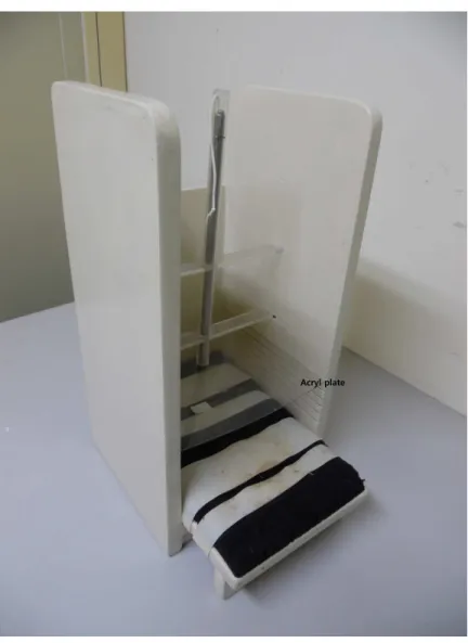
Figure 1 Device for mild traumatic brain injury using a rat as the subject.The components of the de-vice are a vertical guide tube for the dropped weight and an acryl panel to absorb impact.
Neurologic evaluation
Grid-walking and foot-fault test
Before the official test, the rats were pre-trained on a metal grid three times a day for three days. Grid-walking and foot-fault test were performed just after the MTBI. The apparatus consisted of an elevated 52×40 cm2metal grid with grid cell of 3×3 cm2(Barbosa et al.,
Rota rod
Before the official test, rats were adapted to Rota rod three times a day for three days. The tests were performed before and 10 min after Grid-walking and foot-fault test. The specifications were for a Rota rod treadmill of a metal roller of diameter 40 mm, speed tachometer of 15 rpm. The rod was divided into five equal segments at 9 cm intervals. A rat was placed on the roller, and the time the rat stayed on it was measured (Tiwari et al., 2015).
Magnetic resonance imaging and blood sampling
Magnetic resonance imaging (MRI) of mild traumatic brain injured group was performed 24 h after MTBI. The MRI confirmed presence of the skull fracture, brain hemorrhage and diffuse axonal injury. MRI scans were carried out with a four-element phased-array animal-dedicated with a 5-cm inner diameter surface coil (Chenguang Medical Technology, Co., Ltd., Shanghai, China). T2 weighted images were taken using a standard spin echo sequence (TE 22 ms; TR 650 ms; slice thickness 3.00 mm; matrix scan 512; FOV 100.00 mm). In addition, subclavian veins were punctured for blood samples to measure electrolytes, plasma glucose, and plasma calcium before as well as 5 and 20 min after MTBI. Electrolyte levels were measured using the EasyElectrolytes (Medica Corporation, Bedford, MA, USA). Plasma glucose and calcium levels were measured using BS-400 (Mindray, Shenzhen, China).
Immunohistochemical assessment
One day after MTBI, 6 rats (three rats in the MTBI and three rats in the sham injury group) were euthanized and the brains were removed and fixed by immersion in 10% neutral buffered formalin solution. Three sections per group were then congruent compared to each other. Coronal sections at 4µm thickness were prepared for planes having the level primary motor cortex with hippocampus. For immunohistochemical analysis, the sections were treated with primary antibody against glial fibrillary acidic protein (GFAP, rabbit polyclonal antibody 1:5,000 dilution, Abcam Ab7260) (Abcam, Cambridge, UK) at room temperature for 60 min. The sections were washed 3 times in TBS for 5 min each and incubated with conjugated secondary antibodies (Dako EnVision +System-HRP labeled anti-rabbit) (Dako, Glostrup, Denmark). The sections were then washed three times with TBS and incubated with diaminobenzidine (DAB) for 3–5 min. The region of interest (ROI) was the primary motor cortex (layers I–III) and CA1 of hippocampus. Integral intensity of GFAP expressions was measured using computer-assisted image analysis program (AnalySIS, Soft Imaging System, GmbH, Münster, Germany). Images were captured from two areas within the motor cortex and hippocampus. The software automatically converted all immune-labeled elements beyond the threshold range into pure red pixels and converted the rest of the image into pure blue pixels. The software then calculated the density of pure red pixels for statistical analysis.
Statistical analysis
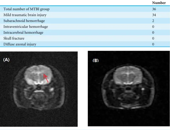
Table 1 Anatomical change after the mild traumatic brain injury (MTBI).
Number
Total number of MTBI group 36
Mild traumatic brain injury 34
Subarachnoid hemorrhage 2
Intraventricular hemorrhage 0
Intracerebral hemorrhage 0
Skull fracture 0
Diffuse axonal injury 0
Figure 2 Magnetic resonance imaging after a mild traumatic brain injury.Arrows represent subarach-noid hemorrhage (A). There was no significant cerebral hemorrhage, intracranial hemorrhage, or diffuse axonal injury in 34 rats (B).
NY, USA) andp-values under 0.05 were considered significant. To analyze the change between before and after sham injury, a Wilcoxon singed rank test was used.
RESULTS
Animal characteristics in magnetic resonance imaging
A total of 36 rats were applied to the concussion model. The MRI findings of 34 rats appeared normal. Only two rats had small amount of subarachnoid hemorrhage, which was not a fatal injury. There was no intracerebral hemorrhage, skull fracture, diffuse axonal injury or death (Table 1andFig. 2). The device took just 1–2 min to organize mild traumatic brain injury.
Neurologic findings
Grid-walking and foot fault test
Thirty-four rats, appeared normal on the MRI, but showed a significant decrease in the total number of footsteps in the foot fault test (p-value < 0.001). The mean in the number of the footsteps was 40.12±10.96 before MTBI, and 17.50±14.50 after MTBI.
Table 2 Comparison of grid walking test parameters before and after the mild traumatic brain injury with sham injury.
Total foot
step (number/min)
Foot fault
step (number/min)
Foot fault ratio
Latency (s)
MTBI
Before MTBI 40.12±10.96 2.21±1.61 0.07±0.09 0.94±1.41
After MTBI 17.50±14.50 0.59±0.74 0.04±0.06 5.26±11.39 p-value <0.001 <0.001 0.01 0.02
Sham injury
Before sham injury 35.50±16.03 1.00±1.07 0.02±0.03 1.00±1.60 After sham injury 31.88±14.38 0.38±0.74 0.02±0.02 0.75±1.75
p-value 0.31 0.13 0.59 0.72
Notes.
Values are mean±standard deviation.
MTBI, Mild traumatic brain injury; s, Second; Min, Minute.
to 0.59±0.74 per min (p-value < 0.001) with the decreased error rate of 0.04±0.06 on the total number of footsteps (p-value=0.01). Latency before MTBI was 0.94±1.41, but it was prolonged to 5.26±11.39 s after MTBI with statistical significance (p-value= 0.02). In the sham injury group, there were no significant changes of the total number of footsteps, the number of foot fault step, foot fault ratios and latency to move (Table 2).
Rota rod test
For the Rota rod evaluation, the rats maintained rolling with balance for 10.00±11.21 s before MTBI. They were able to keep rolling for 13.97±16.09 s after MTBI, thus there was no significant difference in Rota rod results between before and after MTBI. In the sham injury group, animals stayed on the roller for 25.13±16.62 s before and 34.13±11.66 s after the sham injury (p-value > 0.05).
Blood biochemical findings
There is no significant difference between before and after MTBI and the sham injury group in blood test. The sodium level showed no significant difference between before and after MTBI and for the sham injury (before MTBI 138.34±3.33 mmol/L; after MTBI 138.45 ±1.88 mmol/L; before sham injury 138.38±0.67 mmol/L; after sham injury 138.64±0.98 mmol/L). Similarly, for the potassium level changes, there were no significant changes (before MTBI 5.38±0.17 mmol/L; after MTBI 4.98± 0.11 mmol/L; before sham injury 4.74±0.36 mmol/L; after MTBI 4.41±0.54 mmol/L). A decrease in serum glucose level was detected from 210.77±49.33 mg/dL before MTBI to 196.00±42.04 mg/dL after MTBI, but this result was not statistically significant. Also, a calcium level change in the MTBI group was also detected from 1.17±0.27 to 1.21± 0.17 mmol/L, but that was not statistically significant as well. The serum glucose and calcium levels had not changed in the sham injury group (Table 3).
Expression of GFAP immunoreactivity
Table 3 Comparison of serum parameters before and after the mild traumatic brain injury with sham injury.
Sodium (mmol/L)
Potassium (mmol/L)
Glucose (mg/dL)
Calcium (mmol/L)
MTBI
Before MTBI 138.34±3.33 5.38±0.17 210.77±49.33 1.17±0.27
After MTBI 138.45±1.88 4.98±0.11 196.00±42.04 1.21±0.17
p-value 0.87 0.65 0.06 0.51
Sham injury
Before sham injury 138.38±0.67 4.74±0.36 191.38±16.69 0.95±0.27 After sham injury 138.34±0.98 4.41±0.54 188.00±16.23 0.98±0.25
p-value 0.50 0.13 0.18 0.58
Notes.
Values are mean±standard deviation.
MTBI, Mild traumatic brain injury; s, Second; Min, Minute.
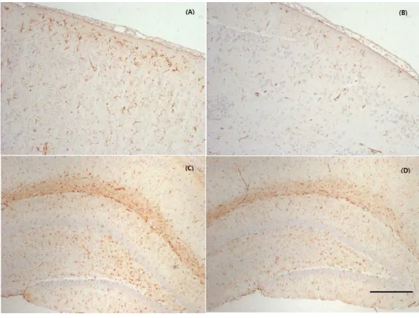
Figure 3 Representative photographs of the cerebral cortex and hippocampus samples showing GFAP
expression.MTBI led to a slightly increased GFAP expression in the cortex (A) and hippocampus (C)
primary motor cortex hippocampus 0
200 400 600 800 1000 1200 1400
Sham injury
MTBI
Inte
g
ra
l
inte
ns
ity
(
μ
m2
)
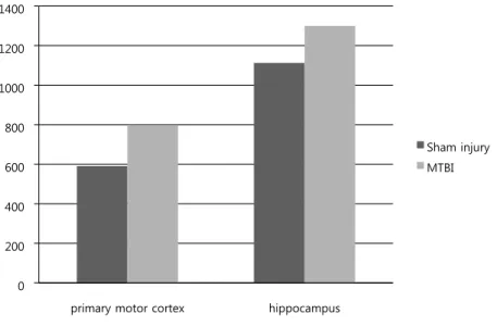
Figure 4 GFAP immunoreactivity analysis.Immunohistochemical analysis of GFAP in primary motor cortex and hippocampus is increased in the mid traumatic brain injury (MTBI) group compared to the sham injury group.
increased immunoreactivity of GFAP in the cerebral cortex around injured bregma and hippocampus. The integral intensity of GFAP expression in the MTBI group (798.03 ± 93.42 in cortex, 1299.35±154.31 in hippocampus) increased in comparison with control group (590.28±137.20 in cortex, 1112.42±123.27) in hippocampus (Fig. 4). The GFAP immunoreactivity showed that increased expression in the cortex and hippocampus in the MTBI group, which was higher than in the control group, but this was not significant.
DISCUSSION
imply that method can be safer than other methods; in addition, our MTBI model can be conducted within 1 or 2 min and does not need an incision and surgery, unlike previous studies (Sakurai et al., 2012; Redell et al., 2013; Dixon et al., 1987; Dixon et al., 1991).
The grid-walking/foot-fault test is known to assay for sensorimotor coordination in neurological diseases such as a cerebral infarction, cortex injury, and Parkinson’s disease that may be affected by motor ability (Zhang et al., 2002;Barth, Jones & Schallert, 1990;
Shanina et al., 2006;Chao et al., 2012). In our study, with the injury method developed herein, MTBI characteristics were seen in rats with a behavioral delay in the grid-walking and foot fault test. In several cases, no movements were observed in the initial one minute of the test. A delay in latency results from a temporary unconsciousness that occurs after an MTBI, or may be due to post-concussive symptoms such as a headache, dizziness, or irritability. From induction of MTBI, rats became slow in movement on the metal grid, considerably reducing the number of their steps in addition to latency, along with many rats lying almost motionless. These seem to be an aspect of alterations in plasticity and activation and from hypometabolism as in the study ofShrey, Griesbach & Giza (2011). Reductions in foot-fault steps and foot-fault error rates do not result from improvement in sensorimotor coordination after an MTBI but are more likely to be caused by a reduction in real movements.
The Rota rod test was to examine balance impairments. The test detected the problems of balance and postural equilibriums subsequent to MTBI (Guskiewicz, 2011). In this study, there was no statistical significant difference before and after MTBI regarding the Rota rod rolling duration. It was probably because the Rota rod test was conducted after the grid-walking and foot-fault test, and that the rats recovered from an MTBI faster, as they were less injured than those in the other studies. It should be noted that the rats were fidgety after getting an MTBI and tended to have difficulty rolling, but these measurements could not be conducted.
By histological analysis, we could also reveal a mild injury at the cellular level. Immuno-histochemical analyses confirmed increased GFAP expression, which indicative of a mild astrocytic response to injury (increased astrocytic activation) to injury. However, this was not significant as the number of stained rats was small and the time of sacrifice was too early.Giza & Hovda (2014)reported that later time points (four days to one month) showed maximal staining for gliosis (GFAP-glial fibrillary acidic protein).
According to the report ofGiza & Hovda (2001)glycolysis reaches its peak 6 min after the induction of MTBI, and hyperglycolysis ends 20 min later, and for 24 h afterwards, the cerebral glucose metabolism slows down. In this study, a blood test was conducted 20 min after the induction of MTBI, and thus hyperglycolysis could not be observed. In addition, although a change occurs in the electrical charge of the membrane, the imbalance of electrolytes in real blood was not observed due to homeostasis.
This study has several limitations. First, hyperglycolysis could not be analyzed because the blood test was conducted 20 min after the induction of MTBI and as not to influence behavioral measurements. Second, the number of experimental rats is considered to be small for the study. It is possible, of course, to generalize the rat models of MTBI with the use of parametric statistics, yet a larger number of experimental rats might be helpful in raising the validity and reliability of the findings of this study. Third, the above results only confirmed the early effect of the MTBI model. The long-term effect on behavior, learning and memory was not revealed. A long-term study would be required to prove the severity of MTBI by the device and its chronic effect. Fourth, with the small number of test animals in the sham injury group, it was hard to compare the measurements with MTBI group directly. This study allowed testing a rat model of MTBI and to demonstrate its safety and simplic-ity. The traumatic brain injuries induced in this study were much milder than those in other studies. This type of modeling of MTBI is expected to allow studying pathophysiology or progression in the injury model as 50% of patients with MTBI do not visit a hospital on the perception of having only mild symptoms, thus making it difficult to assess their cases at early stages of post trauma. Furthermore, due to its characteristics, this type of model can be applied and planned for studies of repetitive MTBI.
ACKNOWLEDGEMENTS
The authors wish to thank Ms. Mina Hong for support conducting the trials.
ADDITIONAL INFORMATION AND DECLARATIONS
Funding
This research was supported by Basic Science Research Program through the National Research Foundation of Korea (NRF) funded by the Ministry of Education (NRF-2013R1A1A2059711) and Research Grant of Alumni Chapters of Medical College of Ewha Womans University in 2015. The funders had no role in study design, data collection and analysis, decision to publish, or preparation of the manuscript.
Grant Disclosures
The following grant information was disclosed by the authors:
National Research Foundation of Korea (NRF): NRF-2013R1A1A2059711. Alumni Chapters of Medical College of Ewha Womans University.
Competing Interests
Author Contributions
• Ho Jeong Kim conceived and designed the experiments, performed the experiments, analyzed the data, wrote the paper, prepared figures and/or tables, reviewed drafts of the paper.
• Soo Jeong Han conceived and designed the experiments, performed the experiments, analyzed the data, contributed reagents/materials/analysis tools, wrote the paper, reviewed drafts of the paper.
Animal Ethics
The following information was supplied relating to ethical approvals (i.e., approving body and any reference numbers):
Ethics Committee of Ewha Medical Research Institute Approval No; ESM 14-0252.
Data Availability
The following information was supplied regarding data availability: The raw data has been supplied asData S1–S4.
Supplemental Information
Supplemental information for this article can be found online athttp://dx.doi.org/10.7717/ peerj.2818#supplemental-information.
REFERENCES
Barbosa EH, Vallim JH, Lachat JJ, De Castro VL. 2015.Assessments of motor
abnor-malities on the grid-walking and foot-fault tests from undernutrition in wistar rats. Journal of Motor Behavior29:1–8DOI 10.1080/00222895.2015.1024824.
Barth TM, Jones TA, Schallert T. 1990.Functional subdivisions of the rat somatic
senso-rimotor cortex.Behavioral Brain Research39:73–95
DOI 10.1016/0166-4328(90)90122-U.
Chao OY, Pum ME, Li JS, Huston JP. 2012.The grid-walking test: assessment
of sensorimotor deficits after moderate or severe dopamine depletion by 6-hydroxydopamine lesions in the dorsal striatum and medial forebrain bundle. Neuroscience202:318–325DOI 10.1016/j.neuroscience.2011.11.016.
Clifton GL, Ziegler MG, Grossman RG. 1981.Circulating catecholamines and
sympa-thetic activity after head injury.Neurosurgery 8(1):10–14
DOI 10.1227/00006123-198101000-00003.
Collins MW, Grindel SH, Lovell MR, Dede DE, Moser DJ, Phalin BR, Nogle S, Wasik M, Cordry D, Daugherty KM, Sears SF, Nicolette G, Indelicato P, McKeag DB.
1999.Relationship between concussion and neuropsychological performance in
college football players.Journal of the American Medical Association282:964–970
DOI 10.1001/jama.282.10.964.
D’Hemecourt P. 2011.Subacute symptoms of sports-related concussion:
outpa-tient management and return to play.Clinics in Sports Medicine30:63–72
Dixon CE, Clifton GL, Lighthall JW, Yaghmai AA, Hayes RL. 1991.A controlled cortical impact model of traumatic brain injury in the rat.Journal of Neuroscience Methods
39:253–262DOI 10.1016/0165-0270(91)90104-8.
Dixon CE, Lyeth BG, Povlishock JT, Findling RL, Hamm RJ, Marmarou A, Young HF,
Hayes RL. 1987.A fluid percussion model of experimental brain injury in the rat.
Journal of Neurosurgery67:110–119 DOI 10.3171/jns.1987.67.1.0110.
Domínguez DC, Raparla M. 2014.Neurometabolic aspects of sports-related concussion.
Seminars in Speech and Language 35(3):159–165DOI 10.1055/s-0034-1384677.
Giza CC, Hovda DA. 2001.The neurometabolic cascade of concussion.Journal of Athletic
Training 36(3):228–235.
Giza CC, Hovda DA. 2014.The new neurometabolic cascade of concussion.Neurosurgery
75(4):24–33DOI 10.1227/NEU.0000000000000505.
Guskiewicz KM. 2011.Balance assessment in the management of sport-related
concus-sion.Clinics in Sports Medicine30:89–102DOI 10.1016/j.csm.2010.09.004.
Harmon KG, Drezner JA, Gammons M, Guskiewicz KM, Halstead M, Herring SA,
Kutcher JS, Pana A, Putukian M, Roberts WO. 2013.American medical society
for sports medicine position statement: concussion in sport.British Journal of Sports Medicine47:15–26DOI 10.1136/bjsports-2012-091941.
Holm L, Cassidy JD, Carroll LJ, Borg J. 2005.Summary of the WHO collaborating
centre for neurotrauma task force on mild traumatic brain injury.Journal of Rehabilitation Medicine37:137–141DOI 10.1080/16501970510027321.
Kane MJ, Angoa-Pérez M, Briggs DI, Viano DC, Kreipke CW, Kuhn DM. 2012.A
mouse model of human repetitive mild traumatic brain injury.Journal of Neuro-science Methods203(1):41–49DOI 10.1016/j.jneumeth.2011.09.003.
Redell JB, Moore AN, Grill RJ, Johnson D, Zhao J, Liu Y, Dash PK. 2013.Analysis of
functional pathways altered after mild traumatic brain injury.Journal of Neuro-trauma30(9):752–764DOI 10.1089/neu.2012.2437.
Ruff RM. 2011.Mild traumatic brain injury and neural recovery: rethinking the debate.
NeuroRehabilitation28:167–180.
Sakurai A, Atkins CM, Alonso OF, Bramlett HM, Dietrich WD. 2012.Mild
hyperther-mia worsens the neuropathological damage associated with mild traumatic brain injury in rats.Journal of Neurotrauma29(2):313–321DOI 10.1089/neu.2011.2152.
Shanina EV, Schallert T, Witte OW, Redecker C. 2006.Behavioral recovery from
unilat-eral photothrombotic infarcts of the forelimb sensorimotor cortex in rats: role of the contralateral cortex.Neuroscience139:1495–1506
DOI 10.1016/j.neuroscience.2006.01.016.
Sherer M, Yablon SA, Nakase-Richardson R. 2009.Patterns of recovery of
post-traumatic confusional state in neurorehabilitation admissions after post-traumatic brain injury.Archives of Physical Medicine and Rehabilitation90:1749–1754
DOI 10.1016/j.apmr.2009.05.011.
Shrey DW, Griesbach GS, Giza CC. 2011.The pathophysiology of concussions in
youth.Physical Medicine & Rehabilitation Clinics of North America22(4):577–602
Tang YP, Noda Y, Hasegawa T, Nabeshima T. 1997.A concussive like brain injury model in mice (I): impairment in learning and memory.Journal of Neurotrauma
14:851– 862DOI 10.1089/neu.1997.14.851.
Tiwari HS, Tripathi AK, Mishra DP, Kalita J, Misra UK. 2015.A study of ER stress in
rat model of cerebral venous sinus thrombosis.Neuroscience Letters589:121–125
DOI 10.1016/j.neulet.2015.01.038.
Xu L, Nguyen JV, Lehar M, Menon A, Rha E, Arena J, Ryu J, Marsh-Armstrong N,
Marmarou CR, Koliatsos VE. 2016.Repetitive mild traumatic brain injury with
impact acceleration in the mouse: multifocal axonopathy, neuroinflammation, and neurodegeneration in the visual system.Experimental Neurology275:436–449
DOI 10.1016/j.expneurol.2014.11.004.
Zhang L, Schallert T, Zhang ZG, Jiang Q, Arniego P, Li Q, Chopp M. 2002.A test for
detecting long-term sensorimotor dysfunction in the mouse after focal cerebral is-chemia.Journal of Neuroscience Methods117:207–214