• Nenhum resultado encontrado

Arq. NeuroPsiquiatr. vol.73 número4

N/A
N/A
Protected

Academic year: 2018

Share "Arq. NeuroPsiquiatr. vol.73 número4"

Copied!
8
0
0

Texto

(1)

342

DOI: 10.1590/0004-282X20150006 ARTICLE

Proteomics analysis after traumatic brain

injury in rats: the search for potential

biomarkers

Análise proteômica após lesão cerebral traumática em ratos: a busca por biomarcadores

Jun Ding1,*, Zhengang Ding2,*, Fang Yuan1, Jianyi Guo1, Hao Chen1, Wenwei Gao1, Ren Wang1, Yi Gu1, Jiong Chen1, Yan Guo1, Hengli Tian1

Traumatic brain injury (TBI) is the leading cause of trau-matic death and disability worldwide1. he main causes of

TBI are motor vehicle accidents (50%), falls (21%), assaults and violence (12%), sports and recreation (10%), and other (7%)2. TBI patients impose a tremendous burden on their

families and society, and increase demands on the healthcare system. In order to achieve favorable outcomes, rapid diagno-sis and treatment are important.

TBI triggers complex changes to the central nervous sys-tem (CNS). A better understanding of its complex pathobi-ology is required to further our ability to evaluate and care for brain injury patients. However, an understanding of

the mechanisms and biomarkers of TBI remains elusive.

Biomarkers relecting the biological response to injury or dis -ease have proven useful for diagnosing many disorders, in-cluding responses to injury, cancer, heart failure, infection, and genetic disorders (Table 1)3. Many biomarkers of TBI

have been identiied, although they are not widely used clini

-cally. hese markers include S-100ß, neuron speciic enolase (NSE), glial ibrillary acid protein (GFAP), and myelin basic protein (MBP). Although these proteins are still being as

-sessed, they appear to lack either the sensitivity or speciicity (except GFAP) to be used efectively alone4. However,

com-binations of these markers can provide valuable information

1Shanghai Jiaotong University affiliated Sixth People’s Hospital, Department of Neurosurgery, Shanghai, 200233, China;

2Chinese Medicine Hospital of Huangdao District (Jiaonan), Department of Neurosurgery, Shandong province, 266406, China.

Correspondence: Heng-Li Tian; Shanghai Jiaotong University Sixth People’s Hospital, Department of Neurosurgery; Shanghai 200233, China; E-mail: tianhlsh@126.com

Conflict of interest: There is no conflict of interest to declare. * These authors contributed equally to this work.

Support: National Nature and Science Foundation of China (grant no. 81271383, 81471245), the Shanghai Science and Technique Committee (grant no. 13411951401) and the Shanghai Municipal Health Bureau project (grant no. 20114242).

Received 15 July 2014; Received in final form 07 November 2014; Accepted 27 November 2014. ABSTRACT

Many studies of protein expression after traumatic brain injury (TBI) have identified biomarkers for diagnosing or determining the prog-nosis of TBI. In this study, we searched for additional protein markers of TBI using a fluid perfusion impact device to model TBI in S-D rats. Two-dimensional gel electrophoresis and mass spectrometry were used to identify differentially expressed proteins. After proteomic analy-sis, we detected 405 and 371 protein spots within a pH range of 3-10 from sham-treated and contused brain cortex, respectively. Eighty protein spots were differentially expressed in the two groups and 20 of these proteins were identified. This study validated the established biomarkers of TBI and identified potential biomarkers that could be examined in future work.

Keywords: traumatic brain injury, biomarkers, proteomics, two-dimensional gel electrophoresis, mass spectrometry.

RESUMO

Muitos estudos de expressão proteica após lesão cerebral traumática (LCT) identificam biomarcadores para determinação diagnóstica ou prognóstica do LCT. No presente estudo, foram investigados marcadores proteicos adicionais de LCT, através de um aparelho de impacto no fluxo e perfusão em ratos S-D. Eletroforese bidimensional em gel e espectrometria de massa foram utilizadas para identificar diferen-tes proteínas expressas. Após a análise proteômica, detectamos marcas de proteínas 405 e 371, com pH variando entre 3-10 no córtex de ratos sham e naqueles com contusão cerebral, respectivamente. Oitenta marcas proteicas foram expressas nos dois grupos e 20 destas proteínas foram identificados. Este estudo validou o estabelecimento de biomarcadores de LCT e identificou potencial biomarcadores que poderão ser estudados em estudos futuros.

(2)

and efectively diagnose and predict the outcome of TBI5.

Given their potential utility for determining the diagnosis

and prognosis of brain injuries, their clinical utility should be explored further.

Proteomic analysis is a powerful tool for the global evalu -ation of protein expression and has been applied widely in the analysis of disease6. Analyses of the TBI proteome can

aid in our understanding of the association between protein changes and brain injury, and several studies have already

ap-plied proteomics to the identiication of biomarkers for TBI7.

Proteomic analysis is a discovery-based method that invariably identiies many proteins that difer in abun -dance between control and experimental samples. TBI studies use samples such as brain tissue (often from

ani-mals), cerebrospinal luid (CSF), and blood for proteomic analysis. hese samples contain information related to the CNS. Of the proteins found in CSF samples, 76% were unique to that bioluid. By contrast, plasma and serum pro -teins have wide, dynamic concentration ranges and it is

of-ten diicult to discover a disease-speciic biomarker with -in the background of blood-borne housekeep-ing prote-ins7.

Consequently, the ideal samples for proteomic analysis are

brain tissue and CSF.

METHOD

Animal model

Forty male Sprague-Dawley rats weighing 250-300 g

were randomized into sham-operated or injury groups

(n = 20/group). Rats were acclimated in a humidiied room

and maintained on a standard pellet diet at the Animal Center of Shanghai Jiao Tong University School of Medicine

for 10 days before the experiment. he temperature in both

the feeding and operating rooms was maintained at

ap-proximately 25 to 28°C. A luid perfusion impact device

was used to model TBI in the rats, as described previous-ly8. All of the surgical, injury, and animal care protocols

de-scribed below were approved by the Scientiic and Ethics Committee of Shanghai Jiaotong University ailiated Sixth People’s Hospital (Permit no. SYXK20130608). he rats in

the TBI group were anesthetized with pentobarbital (3.5%, 40 mg/kg), shaved, and then placed in a stereotaxic frame. Surgery was performed as described by Sullivan and col-leagues9. Briely, a 6-mm ipsilateral craniotomy tangential to

the bregma and the sagittal suture was made, leaving the un-derlying dura mater intact. A 5-mm-wide impactor tip was adjusted so that it just touched the exposed dura mater and

was secured over the right parietal cortex. he next day, the

animals were anesthetized, intubated, and then placed

un-der a percussion brain injury device. A moun-derate

luid-percussion pulse (2.0 ± 0.2 atmospheres) was delivered to the right parietal cortex. Sham-operated rats underwent all of

the surgical manipulations, but without the luid-percussion

pulse, and were monitored under anesthesia for 30 min after the sham operation. All of the animals were kept in the same environment after surgery for 48 hours.

Specimens

he sham-operated and TBI animals were anesthetized 48 h postoperatively and sacriiced by decapitation. Immediately

following decapitation, the brain was removed and the cortex samples were dissected rapidly over dry ice. All of the samples were washed with ice-cold saline before freezing to reduce brain contamination with blood proteins. Samples were then stored in liquid nitrogen for further processing. Injured cortex samples were dissected to about 2 mm from the injury spot;

sham samples were removed from the same location. he to -tal time from decapitation to snap freezing of the samples was about 5 min for all animals in both groups.

Two-dimensional gel electrophoresis

First, 100 mg of tissue sample was ground into a powder in liquid nitrogen, homogenized in 1 ml of lysis bufer (7 M urea, 2 M thiourea, 4% CHAPS, 30 mM Tris-HCl, protease in -hibitor mixture) on ice, and sonicated (10×10-s pulses) on ice.

he homogenate was centrifuged at 12,000 rpm for 30 min at 4°C. he protein was precipitated with cold acetone at -20°C for 2 h and dissolved with rehydration bufer (8 M urea, 2 M thiourea, 4% CHAPS, 100 mM DTT, 2% ampholyte). Protein

concentrations were determined using the Bradford

meth-od (Bio-Rad). Immobilized pH gradient (IPG) strips (18 cm,

pH 4-7, non-linear; Bio-Rad) were rehydrated passively

us-ing 400 μl of rehydration bufer for 12 h at 17°C. Isoelectric focusing electrophoresis (IEF) was performed on an IEF cell (Bio-Rad). he strips were equilibrated in equilibration buf

-fer (25 mM Tris-HCl (pH 8.8), 6 M urea, 20% glycerol, 2% SDS, 130 mM DTT) for 15 min and then in the same bufer con

-taining 200 mM iodoacetamide instead of DTT for anoth

-er 15 min. he two-dimensional gel electrophoresis (2-DE)

separation was performed on 12% sodium dodecyl

sulfate-polyacrylamide gel electrophoresis (SDS-PAGE) gels. he gels

were stained using Coomassie Brilliant Blue R-350 (Merck),

according to the supplier’s protocol.

Table 1. Biochemical markers of injury in various organ systems.

Organ Markers

Heart Troponin, creatine phosphokinase (CPK)-MB Liver Aspartate aminotransferase (AST), alanine aminotransferase (ALT), alkaline phosphatase,

γ-glutamyl transpeptidase (GGTP)

Pancreas Lipase, amylase

Muscle CPK-MM

Kidney Blood urea nitrogen (BUN), creatinine (Cr)

(3)

344 Arq Neuropsiquiatr 2015;73(4):342-349

In-gel tryptic digestion and protein identification by mass spectrometry

In-gel tryptic digestion and protein identiication by mass

spectrometry (MS) were performed as described elsewhere6.

Briely, protein spots of interest were excised and destained. In-gel digestion was performed with 0.01 μg/μl trypsin (Promega) for 20 h at 37°C. he tryptic peptides were extracted from the gel and dried by centrifugal lyophilization. he peptide mix

-tures were redissolved in 0.5% triluoroacetic acid (TFA) and analyzed on an AB4700 Proteomics Analyzer (Bruker Daltonics Inc.). Peptide mass maps were acquired in positive relection

mode, averaging 1,500 laser shots per matrix-assisted laser

desorption/ionization time-of-light (MALDI-TOF) spectrum and 3,000 shots per TOF/TOF spectrum.

Data analysis

After gel staining, the protein spots were detected,

quanti-ied, and matched using PD-Quest 2D analysis software (Bio-Rad). Each sample was run in triplicate. Protein identiicated

by mass spectrometry (MS), and the MS/MS data were as-signed using a MASCOT search against the NCBInr database. Redundant proteins that appeared in the database under dif-ferent names and accession numbers were eliminated. If more

than one protein was identiied in one spot, the single protein

with the highest protein score was selected from the

multi-protein family. Statistical calculations were performed by SPSS statistical software (version 16.0; SPSS, Inc.). Comparison be -tween two groups was performed by Wilcoxon two-sample

test. Statistical signiicance was deined as p < 0.05.

RESULTS

Differentially expressed proteins in the sham-operated and TBI groups

Contused brain cortex from TBI rats and brain cortex from sham-operated rats were analyzed in triplicate

us-ing 2-DE. Coomassie stainus-ing of the gels detected 405 and

371 protein spots within a pH range from 3-10 from the

sham-operated and contused cortex, respectively (Figure 1). Statistically, 80 protein spots were diferentially expressed in the sham-operated and TBI rats brain cortices (p < 0.05).

Mass spectrum identification of differentially expressed proteins

he diferentially expressed protein spots were subject

-ed to MS/MS analysis. Twenty proteins were identii-ed from

the 80 spots (Table 2). Of these, some were expressed more strongly in the sham-operated group (spot numbers: 49, 70, 109, 158, 231, 248, 254, 289 and 369), and others were ex-pressed more strongly in the TBI group (spot numbers: 11, 31, 55, 97, 171, 300, 307, 358, 365, 387 and 388). MS/MS

analy-sis showed these spots had diferent MASCOT scores and se

-quence coverage. he predicted molecular masses/pI values for the 20 spots are listed below, and it the position of the corresponding spot on the 2-DE gel well (Table 2, Figure 2).

Of the 20 identiied proteins, NSE, GFAP, S100B, αII-spectrin, and MAP2 have been studied as biomarkers

of TBI (proteomic data not shown in Table 2). Tumor bio-markers such as 14-3-3 protein have not been studied in TBI.

Proteins associated with cell apoptosis were also identiied, such as GAPDH and RhoB. he other identiied proteins have

not been examined in relation to TBI and little is known re-garding their function, such as nebulin-related anchoring

protein and zinc inger protein 180.

DISCUSSION

his study used proteomic analyses to identify 20 pro

-teins that were diferentially expressed in the sham-operat

-ed and TBI groups of rats. hese includ-ed proteins previously identiied as biomarkers of TBI, such as NSE, GFAP, S100B, αII-spectrin, and MAP2. Others had not been identiied

in TBI patients or animals, including the tumor indicators

M2-PK and 14-3-3 protein. he functions of some of the pro

-teins that were identiied after TBI have not been established.

NSE (spot no. 11) is a biomarker of TBI and has been stud-ied in infants, children, and adults, and in mild and severe

(4)

345

Jun Ding e

t al

. P

o

tential biomark

er

s o

f

TBI

Table 2. Proteins identified by MALDI-TOF/TOF.

Spot

no. Protein name

a Gene name Theoretical

molecular massb

Theoretical pIb

Matched Peptidesc

Coverage (%)

Protein

scored Peptides identified

e TBI/ control ratio MW p (TBI group vs

Sham group)

31 Rho-related GTP-binding

protein (RhoB) RhobArhb 22,109 5.10 28 52 50 K.WVPEVK.H 3.259728 83 0.0104

49 M2 Pyruvate Kinase (M2-PK) Pkm2 52,864 7.96 51 84 102 K.KGATLK.I 0.488157 74 0.0319

55 leucine-, glutamate- and

lysine-rich protein 1 LEKR1 80,817 8.35 95 83 51 K.LELDIEK.E 3.597232 71 0.0082

70 H(+)-transporting ATP

synthase ATP5A1 54,518 8.24 44 67 98 R.ILGADTSVDLEETGR.V 0.021411 67 < 0.0001

109 tubulin beta-4B chain Tubb4b 49,769 4.79 41 67 79 K.AGTATGQIVAVIGAVVDV

QFDEGLPPILNALEVQGR.E 0.000315 54 < 0.0001

158

TRM5 tRNA methyltransferase 5

homolog

Trmt5 56,703 8.45 31 57 48 K.VYWNPR.L 0.345823 42 0.0177

171

glyceraldehyde-3- phosphate dehydrogenase

(GAPDH)

Gapdh 35,805 8.14 25 66 60 K.IVSNASCTTNCLAPLAK.V 2.470165 41 0.0201

248 nebulin-related anchoring

protein Nrap 91,867 9.25 60 57 59 R.YTEDGGQQRGK.G 0.480612 31 0.0396

254 ATP-binding cassette

protein C12 Abcc12 144,124 8.15 51 34 50 R.VVWK.F 0.324121 30 0.0250

289 ras-related protein Rab-39A Rab39a 24,905 8.13 20 66 45 K.SCLLHRFTQGR.F 0.332271 27 0.0282

300

phosphatidylethanolamine-binding protein 1 Pebp1 20,788 5.48 20 84 74 K.DPKFR.E 4.583458 26 < 0.001

358 zero beta-globin MGC72973 15,896 6.81 14 87 63 K.GTFASLSELHCDK.L 2.45697 15 0.0332

365 zinc finger protein 180 Zfp180 55,036 9.57 30 54 47 K.SFGFK.G 7.936295 15 < 0.001

369 thyroid hormone receptor Thr 52,091 6.78 30 34 41 K.LIEENR.E 0.002243 14 < 0.0001

(5)

346 Arq Neuropsiquiatr 2015;73(4):342-349

(6)

TBI. In clinical studies, serum NSE levels have been fre-quently studied. Berger et al. found that NSE was markedly

increased in the CSF after severe TBI10. NSE levels were

sig-niicantly elevated in non-survivors as compared with survi -vors in adults11. Early elevation (≤ 3 days) of NSE secondary to

severe TBI predicts deterioration to brain death. Topolovec-Vranic found that NSE was abnormally elevated in 65% of patients with TBI and predicted a poor outcome at 6 weeks post-injury12. NSE is also a candidate serum marker of

im-pending cerebral hypoxia, which is elevated before the onset of clinical manifestations13. After multiple trauma, elevated

NSE levels have been observed, but systemic NSE increased by similar degrees with and without TBI, limiting its ability to discriminate brain injury magnitude. Reports on correla-tions of serum NSE levels alone with clinical and neurological measures of brain injury magnitude and outcome have been controversial. NSE is not used widely clinically, perhaps be-cause of the above and the lack of a large-scale, multi-center, randomized controlled study.

GFAP (spot no. 307) is a ilament protein found in the astroglial cytoskeleton, is not found outside the CNS. GFAP

could predictably discriminate between severe disability and vegetative state versus good and moderate outcomes as

evaluated by the Glasgow Outcome Scale (GOS). A study of severe TBI patients conirmed its ability to predict mortal -ity, and it was found to discriminate outcome categories of

the GOS and Marshall CT classiication. It also could dis -criminate between patients that had intracranial pressure

(ICP) greater or less than 25 mm Hg, patients that had cere -bral perfusion pressure greater or less than 60 mm Hg, and patients with mean arterial pressure greater or less than

60 mm Hg. GFAP shows good diagnostic potential to predict

outcome after injury, and may also be valuable for diagnos-ing injury magnitude14.

S100B (spot no. 387) is the most well studied proteins for TBI and is considered a promising, non-proprietary brain in-jury biomarker. S100B is most abundant in glial cells of the CNS and peripheral nervous system (Schwann cells). But this marker is that it is not exclusive to the brain, it can be found in other cells such as adipocytes and chondrocytes. A

num-ber of studies have demonstrated S100B’s relationship to in -jury magnitude and outcome in TBI5,14. However, some

stud-ies reported a poor value of S100B as a predictor of outcome after brain injury, particularly mild and pediatric TBI. A poor correlation was found between serum and brain S100B val-ues, suggesting that the serum levels may depend primarily

on the integrity of the blood-brain-barrier and do not relect

the S100B levels in the brain6,15. Despite apparent controver

-sy, S100B still has potential as a brain injury biomarker, and its preclinical and clinical utility should be further explored.

αII-spectrin (spot no. 97) is a major structural compo-nent of the neuron axonal cytoskeleton and a major pro-teolytic substrate for cysteine proteases involved in necrot-ic and apoptotnecrot-ic cell death. Many authors have examined

αII-spectrin after TBI in rats or humans. αII-spectrin levels

in the CSF were shown to increase after TBI in a rat mod -el16. In patients, Brophy found that αII-spectrin breakdown

products (SBDPs) were signiicantly elevated in patients with worse Glasgow Coma Scale (GCS) scores 24 h after in

-jury compared to those whose GCS scores improved17. he

mean CSF levels of SBDPs were signiicantly higher in TBI pa -tients than in controls and in pa-tients who died than in those

who survived, and the SBDP concentration was signiicantly

greater in TBI patients than in controls and was correlated

with the GCS score18. herefore, αII-spectrin is an established

biomarker of TBI.

MAP2 (spot no. 388) is important for microtubule stabil -ity and neural plastic-ity and appears to be among the most vulnerable of the cytoskeletal proteins following neuronal

in-jury. Huh et al. found that MAP2 is an early, sensitive marker

of neuronal damage following TBI. Early after TBI (2 hours)

MAP2 expression decreased than that of control group. But in this study, we found that MAP2 was increased in TBI

group. We speculate that the time of specimen harvesting led

to this diference. In addition, MAP2 is an early blood marker

in ischemic brain injury19. herefore, MAP2 can be used as a

marker for detecting neurotoxic insults, including ischemia and TBI.

here is a growing awareness that RhoB (spot no. 31) is

also important signaling molecules in the CNS. RhoB is a

member of the Rho GTPase family that is dramatically in -duced by brain ischemia or trauma. It seems likely that the increase in RhoB level plays a major role in determining the fate of the neurons, but there is little evidence as to

wheth-er the RhoB induction is beneicial or detrimental to neu

-ronal survival. he increased RhoB expression promotes

neurite outgrowth; but this increase is likely to make a sub-stantial contribution to the neurodegenerative process, and

also promotes caspase 3 activation and DNA fragmentation,

key contributors to cell apoptosis20. RhoB mediates

apopto-sis in neoplastically transformed cells after DNA damage. Of the 20 proteins, GAPDH (spot no. 171) also participates in nuclear events, including transcription, RNA transport, DNA

replication, and cell apoptosis21. herefore, RhoB and GAPDH

may play roles in neuro-cell apoptosis after TBI.

M2-PK (spot no. 49) is a phosphotyrosine-binding protein that has been studied in tumors, but not identiied in TBI. M2-PK expression is critical for rapid growth in cancer cells22.

It has been used in the diagnosis and surveillance of a variety of malignant diseases. With respect to injury, Oehler found

that M2-PK expression and activity were increased in poly -trauma patients compared to controls23. M2-PK is the prod

-uct of the PKM2 gene. PKM2 gene transcription is

activat-ed by hypoxia-inducible factor 1 (HIF-1), i.e., when hypoxia

occurs, such as after TBI, the PKM2 gene produces M2-PK.

Pyruvate kinase (PK) orthologs in many organisms are in -hibited by oxidants. Anastasiou found that the inhibition

(7)

348 Arq Neuropsiquiatr 2015;73(4):342-349

antioxidant responses24. Given the presence of hypoxia after

TBI, M2-PK is a potential biomarker of TBI.

Another protein that has not previously been found after TBI

is ATP-binding cassette protein C12 (spot no. 254). he gene for ATP-binding cassette protein C12 is Abcc12 in rats and ABCC12

in humans. he product of ABCC12 is MRP9, which is expressed

in breast tissue, brain, bone, and ovary25. SUR1, the product of

Abcc8, has been found in the CNS after TBI and has an efect on

progressive hemorrhagic necrosis (PHN)26. Abcc8 and Abcc12

are members of the Abcc superfamily and their products, SUR1

and MRP9, respectively, have been found in brain. herefore, similar to SUR1, MRP9 might be an indicator of TBI.

he 14-3-3 protein (spot no. 387) family plays an impor -tant role in tumorigenesis and development. In addition, 14-3-3 protein is a biomarker of neurodegenerative diseas-es27. In the motoneurons of rats, Namikawa reported that

en-hanced expression of the molecules involved in Ras-Erk sig-naling, such as 14-3-3 protein, is required for peripheral nerve regeneration28. Apoptosis is important in cell death after TBI

and is associated with B-cell lymphoma 2 (Bcl-2). 14-3-3

pro-tein can bind Bcl-2-antagonist cell death (BAD) and then

trigger cell apoptosis29. Consequently, 14-3-3 protein is a

po-tential marker of apoptosis after TBI.

he remaining proteins, including leucine-, glutamate-, and lysine-rich protein 1, H(+)-transporting ATP synthase, nebulin-related anchoring protein, and zinc inger protein 180, have not been studied suiciently and their functions af

-ter TBI have not been established. We can ind no conclu -sive published relationships between these proteins and TBI.

herefore, more studies should target these proteins. This study has several limitations. First, the study ex -amined one time point after TBI (48 h), and the animal model was for moderate TBI. We did not determine pro-tein expression at other times after TBI, nor did we de-termine protein expression in mild or severe TBI models. Second, the 20 proteins identified in the proteomic analy-sis have not been confirmed using Western blotting, im-munohistochemistry, or reverse transcription polymerase

chain reaction (RT-PCR) in animals or patients. Future

work should focus on these limitations and identify pro-tein expression at different time points in mild, moderate, and severe TBI, in animal models or in patients at one or more centers.

In general, this study validated several established

bio-markers of TBI and identiied other potential biobio-markers

that can be evaluated in the future.

References

1. Chiu WT, Huang SJ, Tsai SH, Lin JW, Tsai MD, Lin TJ et al. The impact of time, legislation, and geography on the epidemiology of traumatic brain injury. J Clin Neurosci. 2007;14(10):930-5. http://dx.doi.org/10.1016/j.jocn.2006.08.004

2. Centers for Disease Control and Prevention. TBI outcomes and consequences. [Cited 2005 July]. Available from: http://www.cdc.gov/ node.do/id/0900f3ec8000dbdc/aspectId/A0400027

3. Hergenroeder GW, Redell JB, Moore AN, Dash PK. Biomarkers in the clinical diagnosis and management of traumatic brain injury. Mol Diagn Ther 2008;12(6):345-58. http://dx.doi.org/10.2165/1250444-200812060-00002

4. Bazarian JJ, Zemlan FP, Mookerjee S, Stigbrand T. Serum S-100B and cleaved-tau are poor predictors of long-term outcome after mild traumatic brain injury. Brain Inj 2006;20(7):759-65.

5. Berger RP, Beers SR, Richichi R, Wiesman D, Adelson PD. Serum biomarker concentrations and outcome after pediatric traumatic brain injury. J Neurotraum. 2007;24(12):1793-801. http://dx.doi.org/10.1089/neu.2007.0316

6. Sun W, Xing B, Sun Y, Du X, Lu M, Hao C et al. Proteome analysis of hepatocellular carcinoma by two-dimensional difference gel electrophoresis: novel protein markers in hepatocellular carcinoma tissues. Mol Cell Proteomics. 2007;6(10):1798-808.

7. Ottens AK, Kobeissy FH, Fuller BF, Liu MC, Oli MW, Hayes RL et al. Novel neuroproteomic approaches to studying traumatic brain injury. Prog Brain Res. 2007;161:401-18. http://dx.doi.org/10.1016/S0079-6123(06)61029-7

8. Finnie JW. Animal models of traumatic brain injury: a review. Aust Vet J. 2001;79(9):628-33. http://dx.doi.org/10.1111/j.1751-0813.2001.tb10785.x

9. Sullivan PG, Keller JN, Bussen WL, Scheff SW. Cytochrome c release and caspase activation after traumatic brain injury. Brain Res. 2002;949(1-2):88-96. http://dx.doi.org/10.1016/S0006-8993(02)02968-2

10. Berger RP, Pierce MC, Wisniewski SR, Adelson PD, Clark RS, Ruppel RA et al. Neuron-specific enolase, and S100B in cerebrospinal fluid after severe traumatic brain injury in infants and children. Pediatrics. 2002;109(2):E31.

11. Böhmer AE, Oses JP, Schmidt AP, Perón CS, Krebs CL, Oppitz PP et al. Fibrillary acidic protein levels as outcome predictors in patients with severe traumatic brain injury. Neurosurg. 2011;68(6):1624-31. http://dx.doi.org/10.1227/NEU.0b013e318214a81f

12. Topolovec-Vranic J, Pollmann-Mudryj MA, Ouchterlony D, Klein D, Spence J, Romaschin A et al. The value of serum biomarkers in prediction models of outcome after mild traumatic brain injury. J Trauma. 2011;71(5 Suppl 1):S478-86. http://dx.doi.org/10.1097/TA.0b013e318232fa70

13. Stein DM, Lindell AL, Murdock KR, Kufera JA, Menaker J, Bochicchio GV et al. Use of serum biomarkers to predict cerebral hypoxia after severe traumatic brain injury. J Neurotrauma. 2012;29(6):1140-9. http://dx.doi.org/10.1089/neu.2011.2149

14. Pelinka LE, Kroepfl A, Leixnering M, Buchinger W, Raabe, A, Redl H. GFAP versus S100B in serum after traumatic brain injury: relationship to brain damage and outcome. J Neurotrauma. 2004;21(11):1553-61. http://dx.doi.org/10.1159/000103304

15. Piazza O, Storti MP, Cotena S, Stoppa F, Perrotta D, Esposito G et al. S100B is not a reliable prognostic index in paediatric TBI. Pediatr. Neurosurg. 2007;43(4):258-64.

16. Aikmana J, O’Steen B, Silver X, Torres R, Boslaugh S, Blackband S et al. Alpha-II-spectrin after controlled cortical impact in the immature rat brain. Dev Neurosci. 2006;28(4-5):457-65. http://dx.doi.org/10.1159/000094171

(8)

18. Berger RP, Hayes R, Richichi R, Beers SR, Wang KK. Serum concentrations of ubiquitin C-terminal hydrolase-L1 and alII-spectrin breakdown product 145 kDa correlate with outcome after pediatric TBI. J Neurotrauma. 2012;29(1):162-7. http://dx.doi.org/10.1089/neu.2011.1989

19. Park D, Joo SS, Lee HJ, Choi KC, Kim SU, Kim YB. Microtubule-associated protein 2, an early blood marker of ischemic brain injury. J Neurosci Res. 2012;90(2):461-7. http://dx.doi.org/10.1002/jnr.22769

20. Barberan S, McNair K, Iqbal K, Smith NC, Prendergast GC, Stone TW et al. Altered apoptotic responses in neurons lacking RhoB GTPase. Eur J Neurosci. 2011;34(11):1737-46. http://dx.doi.org/10.1111/j.1460-9568.2011.07891.x

21. Tisdale EJ. Glyceraldehyde-3-phosphate dehydrogenase is phosphorylated by protein kinase Ciota /lambda and plays a role in microtubule dynamics in the early secretory pathway. J Biol Chem. 2002;277(5):3334-41. http://dx.doi.org/10.1074/jbc.M109744200

22. Christofk HR, Heiden MG, Wu N, Asara JM, Cantley LC. Pyruvate kinase M2 is a phosphotyrosine-binding protein. Nature. 2008;452(7184):181-6. http://dx.doi.org/10.1038/nature06667

23. Luo W, Hu H, Chang R, Zhong J, Knabel M, O’Meally R et al. Pyruvate kinase M2 is a PHD3-stimulated coactivator for hypoxia-inducible factor 1. Cell. 2011;145(5):732-44. http://dx.doi.org/10.1016/j.cell.2011.03.054

24. Anastasiou D, Poulogiannis G, Asara JM, Boxer MB, Jiang JK, Shen M et al. Inhibition of pyruvate kinase M2 by reactive oxygen species contributes to cellular antioxidant responses. Science. 2011;334(6060):1278-83. http://dx.doi.org/10.1126/science.1211485

25. Bera TK, Iavarone C, Kumar V, Lee S, Lee B, Pastan I. MRP9, an unusual truncated member of the ABC transporter superfamily, is highly expressed in breast cancer. Proc Natl Acad Sci USA. 2002;99(10):6997-7002. http://dx.doi.org/10.1073/pnas.102187299

26. Simard JM, Tsymbalyuk O, Ivanov A, Ivanova S, Bhatta S, Geng Z et al. Endothelial sulfonylurea receptor 1– regulated NC Ca-ATP channels mediate progressive hemorrhagic necrosis following spinal cord injury. J Clin Invest. 2007;117(8):2105-13. http://dx.doi.org/10.1172/JCI32041

27. Bersano A, Fiorini M, Allaria S, Zanusso G, Fasoli E, Gelati M et al. Detection of CSF 14-3-3 protein in Guillain-Barré syndrome. Neurology. 2006;67(12):2211-6. http://dx.doi.org/10.1212/01.wnl.0000249150.98891.d1

28. Namikawa K, Su Q, Kiryu-Seo S, Kiyama H. Enhanced expression of 14-3-3 family members in injured

motoneurons. Brain Res Mol Brain Res. 1998;55(2):315-20. http://dx.doi.org/10.1016/S0169-328X(98)00012-6

Imagem

Table 1. Biochemical markers of injury in various organ  systems.
Figure 1. Proteomic analysis of sham-operated and traumatic brain injury (TBI) brain cortex using 2-DE.
Table 2. Proteins identified by MALDI-TOF/TOF.
Figure 2. MALDI-TOF/TOF maps for the 20 identified proteins.

Referências

Documentos relacionados

In Audiology, auditory processing and English language proiciency, the upper semicircular canal dehiscence syndrome and auditory training after traumatic brain injury are

El artículo: “ Measurement of intracranial pressure and short-term outcome of patients with traumatic brain injury: a propensity-matched analysis ” por Biselli-Ferreira et al., (2)

Tal circunstância, além de ser vista como penosa para os próprios AO’s (responsáveis pelo transporte e distribuição deste tipo de produtos pelas várias unidades do

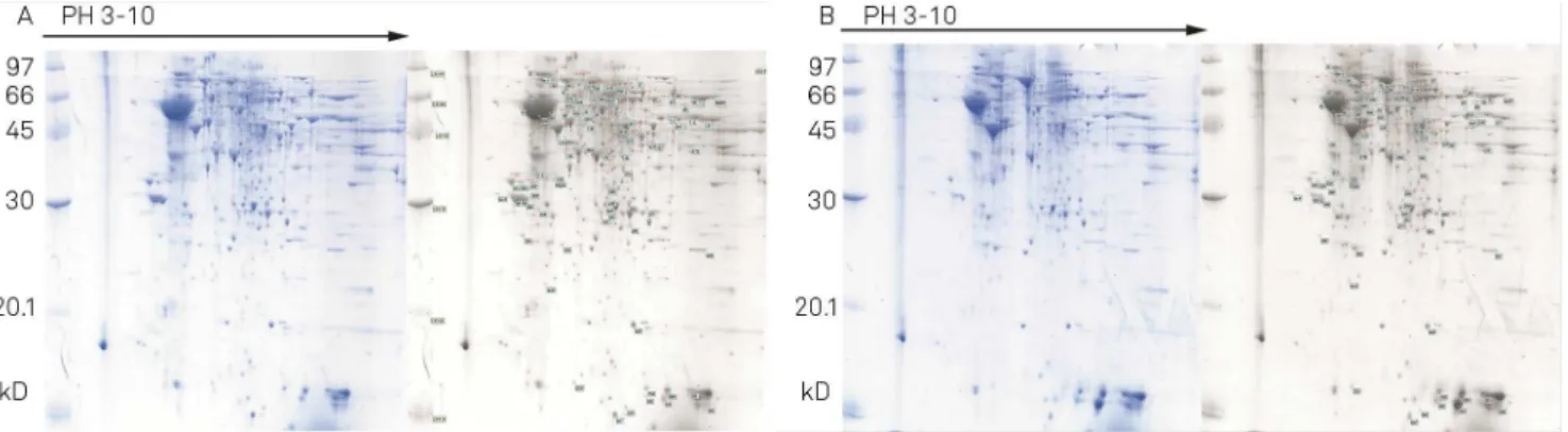
ment applied systemically is effective in pre- venting brain damage in animal models of focal ischemia (14,15), spinal cord ischemia (13), traumatic brain injury (10), and neuro-

Evidence for cellular damage in normal-appearing white matter correlates with injury severity in patients following traumatic brain injury: A magnetic resonance spectroscopy

Objective documentation of traumatic brain injury subsequent to mild head trauma: multimodal brain imaging with MEG, SPECT, and MRI. Head Trauma

Os resultados da avaliação realizada pelos alunos, através do questionário, sinalizaram para uma boa receptividade relativa a metodologia ativa. As questões propostas, apresentadas

We hypothesized that (i) as a group, participants with complicated MTBIs would report greater symptoms and have worse neurocognitive outcomes than those with uncomplicated MTBI,