Arq Neuropsiquiatr 2006;64(1):122-124
1Departamento de Neurologia da Faculdade de Medicina da Universidade de São Paulo (FMUSP), São Paulo SP, Brasil;2Serviço de Neurofisiologia Clínica da Escola Paulista de Medicina da Universidade Federal de São Paulo (EPM/UNIFESP), São Paulo, SP, Brazil.
Received 24 May 2005, received in final form 18 August 2005. Accepted 10 October 2005.
Dr. Ricardo Nitrini - Rua Bartolomeu Feio 560 - 045580-001 São Paulo - Brasil. E-mail: [email protected]
GENERALIZED PERIODIC EEG ACTIVITY
IN TWO CASES OF NEUROSYPHILIS
Renato Anghinah
1, Érica C.S. Camargo
1, Nádia I. Braga
2,
Simone Waksman
1, Ricardo Nitrini
1ABSTRACT - Neurosyphilis is a recognized cause of epileptic seizures and cognitive impairment, but is not usually associated with the finding of generalized periodic activity in the EEG. We report two similar cas-es characterized by pro g rcas-essive cognitive impairment followed by partial complex seizurcas-es, in whom the EEG showed generalized periodic activity. Both cerebrospinal fluid and the response to penicillin therapy confirmed the diagnoses of neurosyphilis in the two cases. The finding of EEG generalized periodic activ-ity in patients with cognitive or behavioral disorders is usually associated with Creutzfeldt-Jakob disease, although there are other conditions, some of them potentially reversible, which may also present this EEG abnormality. Neurosyphilis has tended not to be included among them, and our present findings support the importance of first ruling out neurosyphilis in those patients with cognitive or behavioral disord e r s associated with generalized periodic epileptiform discharges.
KEY WORDS: neurosyphilis, EEG, generalized periodic activity, cognitive impairment.
Atividade periódica generalizada no EEG em dois casos de neurossífilis
RESUMO - N e u rossífilis é uma causa conhecida de crises convulsivas e de comprometimento cognitivo, mas não é associada geralmente a atividade periódica generalizada no eletroencefalograma (EEG). Relatamos dois casos similares caracterizados por declínio cognitivo pro g ressivo seguido de crises parciais complexas, em que o EEG mostra a atividade periódica generalizada. O líquido cefalorraquidiano e uma boa respos-ta à terapia com penicilina confirmaram os diagnósticos de neurossífilis nos dois casos. Achados de ativi-dade periódica generalizada no EEG de pacientes com distúrbios cognitivos ou de comportamento são associados geralmente com a doença de Creutzfeldt-Jakob, embora haja outras circunstâncias, algumas delas potencialmente reversíveis, que podem também apresentar esta anormalidade no EEG. A neurossí-filis tende a não ser incluída entre eles, e nossos achados sustentam a importância de afastar o diagnósti-co de neurossífilis naqueles pacientes diagnósti-com declínio diagnósti-cognitivo ou diagnósti-comportamental associados diagnósti-com as descar-gas periódicas generalizadas no EEG.
PALAVRAS-CHAVE: neurossífilis, EEG, atividade periódica generalizada, comprometimento cognitivo.
Periodic activity in the electro e n c e p h a l o g r a m
(EEG) was initially related to subacute sclero s i n g
panencephalitis
1. Jones and Nevin
2were the first to
describe this EEG pattern in what is now classified as
C reutzfeldt-Jakob disease
3 , 4. EEG periodic activity
con-sists of repetitive stereotyped graphoelements,
main-ly sharp waves or triphasic waves with generalized
distribution; according to the repetition rate it is
clas-sified as short (0.5-4s) or long (>4s) periodic
activi-t y
1 , 4. Short generalized periodic activity has been
cons i d e red the hallmark for the diagnoconsicons of Cre u t z f e l d t
-Jakob disease, although it may be present, mainly as
a transient pattern, in other conditions
3-8.
We now describe two cases of neurosyphilis that
presented with generalized periodic activity.
CASES
Arq Neuropsiquiatr 2006;64(1) 123
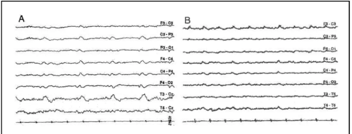
asymmetric with the right pupil greater than the left, and t h e rewas a mild left hemiparesis. Cere b rospinal fluid (CSF) examination revealed 3 leukocytes/mm3(no red blood cells),
69 mg/dL glucose, 33 mg/dL protein with 35% gammaglob-ulin, reagent VDRL and positive Treponema pallidumh e-magglutination assay (TPHA) at a titre of 1:2048. Tests for H I V-1 were negati ve. Magnetic resonance imaging (MRI) showed mild ventricular enlargement and slight widening of the cortical sulci. EEG showed short generalized period-ic activity (slow sharp waves), being more prominent over the right posterior areas, together with disorganization of background activity (Fig 1A). Two days after examination, EEG showed periodic lateralized epilept iform discharg e s (PLEDs) over the right posterior areas. The patient was tre a t-ed with aqueous crystall ine penicill in G, 24 million unit is per day, administered as 4 millions units intravenously every 4 hours for 20 days starting from the second day of admis-sion, with partial improvement. CSF examination at the end of the treatment with penicillin showed 3 leukocytes/mm3,
65 mg/dL glucose, 21 mg/dL protein and reagent VDRL. An EEG done at time of discharge show ed only di sorg a n i z a-tion of the background activity over the right hemisphere . One year later, EEG was normal, and the patient w as still partially dependent and unable to work.
Case 2 – A 48-year-old, right-handed tradesman pre-sented with one month-history of pro g ressive memory de-cline and temporal and spatial disorientation. Four days prior to admission he had suff e red a prolonged partial com-plex seizure. On examination at the time of admission, he was dro w s y, his pupils were asymmetric with the left pupil g reater than the right, both reacting to light, and there was a mild right hemiparesis. CSF examination revealed 42 l e u k o c y t e s / m m3, mainly lymphocytes (no red blood cells),
48 mg/dL glucose, 122 mg/dL protein with 49.6% gamma-globulin, reagent VDRL and positive TPHA at 1:512. Te s t s for HIV-1 were negative. MRI revealed widening of the c o rtical sulci most severe in the frontotemporal regions and a small area consistent with a lacunar infarct in the white matter of the right parietal lobe. EEG showed short
genera-lized periodic activity, most prominent over the left hemi-sphere, against a slow background activity (Fig 1B). Treat-ment with aqueous crystalline penicillin G, 24 million uni-tis per day, administered as 4 millions units intravenously e v e ry 4 hours for 20 days caused partial im provement. In the second EEG, obtained 14 days after admission, no ab-n o rmality was fouab-nd duriab-ng wakefulab-ness. CSF examiab-natioab-n at the end of the treatment with penicillin showed 3 leuko-c y t e s / m m3, 51 mg/dL glucose, 83 mg/dL protein and re a g e n t
VDRL. One year after treatment, the patient was still unable to work.
DISCUSSION
The clinical features, CSF findings with positive
immunological reactions, and the response to
peni-cillin therapy were supportive of the diagnosis of
n e u rosyphilis in both patients. EEG generalized
peri-odic activity was seen as a transient phenomenon in
both cases, and in one case it was rapidly
substitut-ed by PLEDs.
In neurosyphilis, PLEDs have been re p o rted more
f re q u e n t l y
9, while periodic generalized discharg e s
have seldom been described. PLEDs are more
fre-quent in EEG practice than generalized periodic
epi-l e p t i f o rm discharges. As far as we know, the onepi-ly
pre-vious report of generalized periodic activity in
neu-rosyphilis was produced by Radhakrishnan et al., who
described one case in 1984
10.
Pathophysiological mechanisms responsible for
the periodicity in the EEG are unknown. One
hypoth-esis associates this condition with increased GABA
hippocampal activity
1 1, while another assumes that
generalized periodic epileptiform discharges are
cau-sed by disconnection between pre f rontal cort i c a l
areas and ventral tegmental area
12.
124 Arq Neuropsiquiatr 2006;64(1)
The finding of short generalized periodic
epilep-t i f o rmdischarges in paepilep-tienepilep-ts wiepilep-th cogniepilep-tive or
behavioral disorders is usually associated with Cre u t z f e l d t
-Jakob disease, although there are other conditions,
some of them potentially reversible, which may also
present cognitive and behavioral disturbances
asso-ciated with this EEG abnormality (see Bro w n
1 3for a
review). However, neurosyphilis may manifest
peri-odic activity as consequence either of epileptic
activ-ity or stroke caused by the encephalitic process, in
both cases that we are re p o rting a seizure pre c e d e d
the EEG abnormality.
N e u rosyphilis has tended not to be included in
the diff e rential diagnosis of these conditions pre s e
n-ting with EEG periodic activity. However, our
find-ings bear out the need of first ruling out neuro s y p h i l i s
in those patients with cognitive or behavioral
disor-ders associated with generalized periodic
epilepti-f o rm discharges, and point to the key role oepilepti-f
sequen-tial EEG re c o rds tracking the evolution of encephalitic
manifestations. Since tests for syphilis may be no
lon-ger be mandatory in the “routine” blood tests in
pa-tients being evaluated for dementia
1 4, the
impor-tance of including neurosyphilis in the diff e re n t i a l
diagnosis of generalized periodic activity should be
stressed.
REFERENCES
1. Gaches J. Activités periodiques en EEG. Rev EEG Neuro p h y s i o l 1971;1:9-33.
2. Jones DP, Nevin S. Rapidly pro g ressive cerebral degeneration with mental disorder, focal disturbances, and myoclonic epilepsy. J Neurol Neurosurg Psychiatry 1954;17:148-159.
3. Niedermeyer E. Abnormal EEG patterns: epileptic and paroxysmal. In Niedermeyer E, Lopes da Silva F (Eds). Electroencephalography: basic principles, clinical applications, and related fields. Baltimore: Wi l l i a m s & Wilkins, 1993:217-240.
4. Radermecker FJ. Aspects électroencéphalographiques dans trois cas d'encéphalite subaigue. Acta Neurol Psychiatr Belg 1949,49:222-232. 5. Takahashi M, Kubota F, Nishi Y, Miyanaga K. Persistent synchronous
periodic discharges caused by anoxic encephalopathy due to card i o p u l-monary arrest. Clin Electroencephalogr 1993;24:166-172.
6. K u ro d a Y, Ikeda A, Kurohara K, et al. Occurrence of paroxysmal syn-chronous EEG discharges in subcortical arteriosclerotic encephalopa-thy. Intern Med 1993;32:243-246.
7. Isozumi K, Fukuuchi Y, Tanaka K, Nogawa S, Ishihara T, Sakuta R. A MELAS (mitochondrial myopathy, encephalopathy, latic acidosis and s t roke like episodes) mt DNA mutation that induces subacute demen-tia whith mimics Creutzfeldt- Jakob disease. Intern Med 1994;33:543-546.
8. Yemisci M, Gurer G, Saygi S, Ciger A. Generalized periodic epilepti-form discharges: clinical features, neuroradiological evaluation and prognosis in 37 adult patients. Seizure 2003;12:465-472.
9. Camacho-Salas A, Martinez-Salio A, Garcia-Morales I, Vi l l a re j o - G a l e n d e A, de la Pena P. Descargas epileptiformes lateralizadas y periódicas como forma de presentación de neurosífilis. Rev Neurol 2002;35:734-737.
10. Radhakrishnan K, Ashok PP, Sridharan R, El-Mangoush MA. Periodic EEG pattern in meningovascular syphilis. J Neurol Neuro s u rg Psychiatry 1984;47:1360-1361.
11. P a p a t h e o d o ropoulos C, Kostopoulos G.Spontaneous GABA(A)- depen-dent synchronous periodic activity in adult rat ventral hippocampal slices. Neurosci Lett 2002; 8:319:17-20.
12. Peters Y, Barnhardt NE, O`Donnell P. Pre f rontal cortical up states are synchronized with tegmental area activity. Synapse 2004;52:143-152. 13. B rown P. Transmissible human spongiform encephalopathy (infectious
c e rebral amyloidosis): Creutzfeldt-Jakob disease, Gerstmann-Sträussler-Scheinker syndrome, and Kuru. Annu Rev Med 1995;46:57-65. 14. Knopman DS, DeKosky ST, Cummings JL, et al. Report of the quality