AN ALTERNATIVE MANEUVER TO TREAT
GINGIVAL RECESSION
JOSÉ RICARDOKINA1*, THAÍS YUMI UMEDASUZUKI2, EUNICE FUMICO UMEDAKINA3
1.Associate Professor of Department of Surgery and Integrated Clinic, Araçatuba School of Dentistry, São Paulo State University – UNESP;
2.Postgraduate Student of Department of Dental Materials and Prosthodontics, Araçatuba School of Dentistry, São Paulo State University – UNESP;3.Private practice in Araçatuba, SP - Brazil.
* Department of Surgery and Integrated Clinic – UNESP. Rua José Bonifácio, 1193 Araçatuba, São Paulo, Brazil. CEP: 16015-050.
kinajr@hotmail.com
Received:30/10/2014;Accepted:24/11/2014
ABSTRACT
This article reports a clinical case in which was applied autol-ogous bone graft associated with subepithelial connective tis-sue graft, harvested by gingivectomy procedure with technical modifications to increase gingival graft extension, also to be used as guided tissue regeneration, to treat a single gingival recession. After 1 year and 2 months of follow-up, the cover-age of the recession was 4.0 mm, which corresponded to the gain of attached keratinized gingival tissue. An increase in the gingival tissue thickness was observed, without significant probing depth. The procedures applied to treat this case may be biologically and clinically useful to treat gingival recession.
KEYWORDS:Case report, connective tissue, esthetics, gin-gival recession.
1. INTRODUCTION
Gingival recession is defined as apical migration of junctional epithelium, loss of attachment, resorption of alveolar bone, with root surfaces exposure without for-mation of periodontal pocket. It is prevalent in patients with poor or good plaque control, with or without gingi-val margins inflammation being always part of perio-dontal disease process1. Its etiology is a complex
inter-action of bacteria and predisposing risk factors, as: ana-tomical features, iatrogenic process, emotional condi-tions, behaviors (habits), traumatic occlusion, mechani-cal trauma, chemimechani-cal trauma, tobacco consumption and has been found frequently on buccal surfaces than on other aspects of the teeth1,2,3.
Gingival recession in its localized or generalized form, often result in non-esthetic condition and exposed root surfaces are prone to abrasion, caries and hypersen-sitivity1. The management of gingival recession, a
con-sequence of periodontal disease progression is based on a thorough assessment of the etiological factors and de-gree of periodontal tissue involvement1,4. Once the
eti-ology of the condition has been uncovered and addressed, the treatment plan to arrest or reverse the gingival
reces-sion may be established4,5.
The treatment plan will be based on the severity of symptoms as dentinary hipersensibility, the future con-sequence of the lesion as radicular caries and the goal of the patient as esthetic concerns5. In this case, the
regen-erative periodontal procedures as subepithelial connec-tive tissue graft employed as guided tissue regeneration and autogenous bone graft were applied, attempting to gain new clinical attachment, keratinized gingiva, im-prove bone level, and minimize postoperative gingival recession.
2. CASE REPORT
A 22-year-old female patient was referred to the Per-iodontology Clinic, São Paulo State University-UNESP, in January 2013, for evaluation and treatment of single recession in the lower left central incisor. Her complaints were esthetics and dental sensitivity. She was non-smoking, presented good systemic health, did not take any medication in the previous 3 months, had no known allergies and brushed his teeth with a soft-bristle toothbrush using horizontal motions.
The clinical examination revealed a plaque index of 39%,6 a gingival index of 18%,6and probing depth
al-most in all teeth ≤ 2 mm however, by lingual side in posterior teeth in the maxilla, the marginal and papillary gingiva appeared enlarged and prominent with probing deep around 4mm, but without attachment loss. Ap-proximately 3 mm of excessive gingival tissue was ob-served in relation to the cementum-enamel junction. Ra-diographic evaluation showed no bony defect. Only lower left central incisor displayed evident gingival in-flammation with 4 mm of clinical attachment loss (Fig-ure 1).
The lesion was caused by anatomic features (lower left central incisor buccally malpositioned in the arch), associated with traumatic occlusion (Figure 2)1,2,3. The
traumatic occlusion was treated by adjusting centric po-sition and anterior guidance, to distribute anterior teeth contacts on protrusion movement (Figure 3)7. As the
marginal tissue recession of lower left central incisor extended to the mucogingival junction and a clear at-tachment loss and tooth malposition existed, classifica-tion of the gingival recession was consistent with class II according to Miller8. The patient was given a detailed
explanation concerning the procedure, and informed consent was obtained from her. The patient underwent complete root scaling with sonic scaler and oral hygiene instructions10. After 1 month, the plaque index was
22%,6and the gingival index was 8%; thus, a
subepithe-lial connective tissue graft in association with autolo-gous bone graft was proposed, aiming for root coverage of lower left central incisor9,10.
Figure 2.Gingival recession caused by lower left central incisor buc-cally malpositioned in the arch, associated with traumatic occlusion.
Initially, intra and extra oral antisepsis was carried out using 0.12% chlorhexidine digluconate. Following local anesthesia, the exposed root surfaces were submit-ted to physical treatment by root scaling with sonic scal-er. Then, a sulcular incision was made through the buc-cal aspect with a 15C sbuc-calpel blade, preserving the
integ-rity of the papillae.
Figure 3.Adjusting anterior guidance, to distribute anterior teeth con-tacts on protrusion movement.
The incision was initiated in the distal aspect of the lower right lateral incisor and extended to the distal as-pect of the lower left central incisor. A vertical incision was performed in the distal aspect of both teeth. The flap was elevated by carefully full-thickness dissection per-formed with a periosteal elevator. The exposed perio-dontitis-affected root surface was altered chemistrilly with topical application of tetracycline hydrochloride paste for 4 minutes. To the purposes of demineralization and decontamination, to make it a hospitable substrate to support and encourage migration, proliferation, proper phenotypic expression of periodontal connective tissue progenitor cells and attachment11,12. To influence faster
bone graft union and incorporation, the bone surround-ing root of lower left central incisor was decorticated, to expose bone marrow and endosteum (Figure 4)13.
Figure 4.Decortication of the bone-surrounding root of lower left central incisor to expose bone marrow and endosteum.
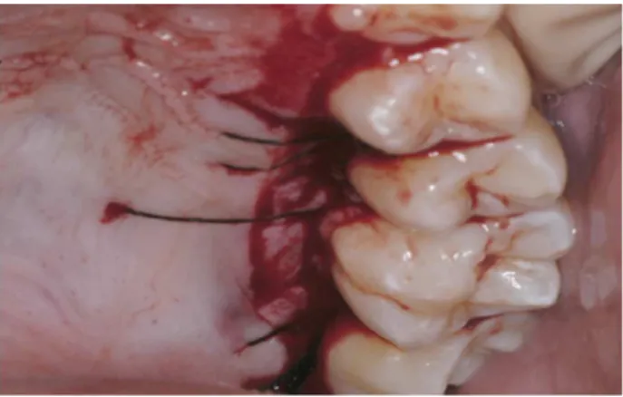
as-sessment revealed a fibrous and consistent tissue, with-out bleeding on probing depth. A gingivectomy proce-dure with technical modifications was planned, to cor-rect gingival contour and improve aesthetics crown lengthening by reducing the amount of gingiva. First, an intra sulcular incision was made through the lingual as-pect with a 15C scalpel blade until achieving the bone crest. This incision was initiated in the distal aspect of the upper left first premolar and extended longitudinally to the distal aspect of the upper left first molar, until ex-posing maxillary tuberosity. A vertical incision was per-formed in the distal aspect of upper left first premolar. The flap was deflected by carefully full-thickness dis-section performed with a periosteal elevator. A straight horizontal releasing incision technique was executed on the level and in the direction of cementoenamel junction, from the distal aspect of upper left second molar until distal aspect of upper left first premolar, to remove ex-cess of gingival tissue which was used as the tissue to be grafted (Fig. 5). After that, the epithelial tissue was re-moved from the graft by acute dissection to obtain only connective tissue. A scheme was performed to enlarge the extension of the graft to cover completely avascular root surface, with its major extension over adjacent vas-cularized tissues. For that reason, the connective tissue graft harvested with the maximum thickness was posi-tioned on a sterilized glass plate and immobilized with a sterile spatula. The graft was split cross-sectionally with a 15C scalpel blade; however, it was not divided com-pletely into two parts. After this procedure, the graft had almost twice the length of the initial graft and a thick-ness around 1.5 mm (Fig. 6). The autologous bone graft was harvested from maxillary tuberosity by using a roungeur (Fig. 7).12
Figure 5. Gingivectomy procedure with technical modifications to harvest connective tissue to be grafted.
The palatal flap was then incised by internal bevel incision and the excess of connective tissue was dissect-ed and the flap was thinndissect-ed. The excess of connective tissue was not removed; it was dislocated and
reposi-tioned coronally, over the crest of alveolar bone. The flap was sutured to hold tissues passively in the position with suspensory 4.0 silk sutures (nonabsorbable organic material: silk black braided, #4-0, cutting needle, P-3, Ethicon, São José dos Campos, SP, Brazil) (Figure 8).
Figure 6.Connective tissue graft harvested by gingivectomy.
Figure 7.The autologous bone graft harvested from maxillary tuberos-ity by using a roungeur.
Figure 8.Donor site sutured.
Figure 9.The bone graft positioned and trimmed on the receptor site.
Figure 10. The subepithelial graft was placed and sutured over the grafted bone with suspensory sutures.
Figure 11.The flap sutured over the grafted site.
The patient was instructed to take analgesic medica-tion (acetaminophen, 750 mg, three times a day for 4 days) and to use mouthrinse with 0,12% chlorhexidine digluconate twice daily for 21 days. The periodontal dressing and all sutures were removed after 10 days (Fig. 12). The patient was followed up weekly and monthly up to the third month.
The healing process was uneventful, and the patient did not report pain or discomfort during the overall postoperative period. During the postoperative follow-up, no sign of necrosis or hemorrhage was observed in the donor and receptor areas.
Figure 12.Postoperative after 10 days.
Figure 13.1 year and 2 months of follow-up.
Figure 14.Donor site 1 year and 2 months of follow-up.
The color of the tissues was nearly homogeneous 3 weeks following the surgical procedure and esthetic im-provements were observed 3 months postoperatively and were maintained during 1 year and 2 months of fol-low-up (Figure 13).
3. DISCUSSION
The etiology of gingival recession is considered mul-tifactorial, where bacteria are essential, but always needs to be associated with predisposing risk factors to develop the disease1,2,3,4. The predisposing risk factor may be an
inherent characteristic associated with an increased rate of a subsequently occurring disease, but does not neces-sarily cause the disease4. The gingival recession may
cause esthetic concern, dental root sensitivity and radic-ular caries predisposition1. To treat gingival recession,
always will be necessary to eliminate or to establish a control in all etiologic factors or improve host local de-fense against the entire etiologic factors, to promote ho-meostasis in diseased areas through a long stated period4.
In this case the etiological factors playing determinant role in the gingival recession development were: bacteria, traumatic occlusion and the tooth buccally malposition-ated; an anatomical feature1,2,3. To establish bacterial
plaque control, the patient underwent complete root scaling and the oral hygiene instructions.
The traumatic occlusion was promoted due buccally malpositioned lower left central incisor. When the man-dible was moved into protrusion, the lower left central incisor was aimed anteriorly and first come to contact with the maxillary upper incisor, promoting deleterious contact in mandibular eccentric movement7. Posterior
centric contact was checked out and an occlusal adjust-ment was carried out, because premature contact in cen-tric position may move the mandibular arch anteriorly, promoting undesirable anterior contact7. In the sequence,
the lingual face of upper incisor was adjusted to distrib-ute anterior teeth contacts on protrusion movement.7
Malposition of lower left central incisor also may induce alterations in the widths of the keratinized and attached gingiva and a thin bony in buccal aspect, decreasing amount of bone marrow, thus, predisposing bone reab-sorption and gingival recession1,2,3. When all etiologic
factors may not be eliminated or controlled, the alterna-tive procedure should be to improve host defense against aggressor agents to establish disease contro4.
In order to improve local resistance in lower left cen-tral incisor, an autogenous bone graft associated with guided tissue regeneration was considered9,10. The
au-togenous bone graft from the maxillary tuberosity is the most viable periodontal bone graft, due presence of hematopoietic tissue and their osteogenic potentials to form the new bone, by processes of osteogenesis, oste-oinduction, and osteoconduction10. Therefore, the bone
graft harvested from maxillary tuberosity was positioned and trimmed on exposed root surface and adjacent de-corticated vascularized tissues. A subepithelial graft was used as barrier membranes to direct the growth of new bone, to augment keratinized attached gingival tissue and to recover the root of lower left central incisor9. An
adequate blood supply from the tissues adjacent to the
graft bed seems to be the single most important factor for the survival of grafted tissue5,9,12,13. Then, a large
extension of the graft to cover completely avascular root surface, with its major extension over adjacent vascular-ized tissues seem to be important. The method applied to harvest a large connective tissue graft in this case was significant to aid an initial stability of the subepithelial graft on vascularized portion of receptor site, which was decorticated, allowing the revascularization and mainte-nance of the amount of connective tissue graft during healing process. The subepithelial graft was placed and sutured over the grafted bone, attempting to exclude gingival epithelium interference, guiding only required tissue regeneration, to recuperate diseased root surface by establishing new resistant tissue development as bone, and keratinized attached gingiva2,3,4,9. The space created
by bone graft may allow easily cells from the periodontal ligament and bone marrow exposed by decortication, to expand blood supply and to populate with mesenchymal stem cells penetration into the bone graft in its early re-parative phase9,10,13. The release of local growth factors,
which would be caused by receptor bed decortication, also has been suggested as one of the factors explaining the hastening of bone graft incorporation process12,13.
These procedures could induce new bone formation and new connective tissue attachment to the avascularized root surface, preventing epithelial migration and the es-tablishment of the long junctional epithelium until the base of the original periodontal defect4,12,14. The gain of
keratinized attached gingival tissue and possible bone neoformation, may induce supplementary resistance against the gingival recession recidivism4. Then, the goal
of treatment was not only to eliminate or to control etio-logic factors; application of procedures which could re-cuperate some lost tissues improving its quality and quantity, may also increase host resistance against ag-gressor agents, helping to achieve homeostasis in altered area4. Another aspect regarding to the biologic
mecha-nism that facilitates healing of lost periodontium by using guided tissue regeneration is attributed to stabilization of the root-clot-graft interface by membrane12. In surgical
periodontal therapy when periodontal wounds are closed and sutured, one of the wound margins is an avascular and rigid periodontitis-affected and altered root surface and another wound margin is a soft tissue vascular flap margin12,15. This detail induces a fibrin clot formation
with a fragile initial attachment to the altered root surface, for preventing epithelial down growth and to form a scaffold for development of a cell and collagen fiber attachment mechanism12,15. Then, a fibrin clot adherent to
of the original periodontal defect is expected to occur to prevent infection12,14,15.
Then, the suture also may be considered as an im-portant factor during regenerative attempts, stabilizing and protecting the root-clot-graft interface in earlier pe-riod of wound healing.12For this reason, the flap margin
was sutured positioned coronally to the cemento-enamel junction, in a manner that could be well stabilized against the tooth, limiting the possibilities of gingival margin movement12. The patient is maintained over periodic
plaque control supervision, to keep the area clinically health.
4. CONCLUSION
The present clinical results, after 1 year and 2 months of periodic control, show an adequate mucogingival complex in which the mucogingival tissues can sustain their biomorphological integrity and maintain an endur-ing attachment to the tooth and the underlyendur-ing alveolar bone, allowing to conclude that the procedures applied to treat this case may be biologically and clinically useful to treat gingival recession.
REFERENCES
[1] Kassab MM, Cohen RE. The etiology and prevalence of gingival recession. J Am Dent Assoc. 2003; 134:220-5. [2] Ericsson I, Lindhe J. Recession in sites with inadequate
width of the keratinized gingiva. An experimental study in the dog. J Clin Periodontol. 1984; 11:95-103.
[3] Kennedy JE, Bird WC, Palcanis KG, Dorfman HS. A lon-gitudinal evaluation of varying widths of attached gingiva. J Clin Periodontol. 1985; 12:667-75.
[4] Kina JR, Suzuki TYU, Kina J, Kina M, Kina EFU. Repara-tive phase events on periodontal disease progression: in-terpretation and considerations. Int J Microbiol Res. 2013; 5:439-44.
[5] Oates TW, Robinson M, Gunsolley JC. Surgical Therapies for the Treatment of Gingival Recession. A Systematic Review. Ann Periodontol. 2003; 8:303-20.
[6] Ainamo J, Bay I. Problems and proposal for recording gingivitis and plaque. Int Dent J 1975; 25:229-35.
[7] Dawson PE. Evaluation, Diagnosis, and Treatment of Occlusal Problems. 2nd ed. St. Louis, Mosby. 1989. [8] Miller PD Jr. A classification of marginal tissue recession.
Int J Periodontics Restorative Dent. 1985; 5:8-13.
[9] Langer B, Langer L. Subephitelial connective tissue graft technique for root coverage. J Periodontol. 1985; 56:715-20.
[10]Brunsvold MA, Mellonig JT. Bone grafts and periodontal regeneration. Periodontal. 2000; 1993; 1:80-91.
[11]Lowenguth RA, Blieden TM. Periodontal regeneration: root surface demineration. Periodontol. 2000; 1993; 1:54-58. [12]Polson AM. Periodontal regeneration: current status and
directions. Chicago:Quintessence Books.1994.
[13]Greenstein G, Greenstein B, Cavallaro J, Tarnow D. The role of bone decortications in enhance the results of guided
bone regeneration: a literature review. J Periodontol. 2009; 80:175-89.
[14]Bossahardt DD, Lang NP. The junctional epithelium: from heath to disease. J Dent Res. 2005; 84:9-20.