Iranian Journal of Otorhinolaryngology No.3, Vol.24, Serial No.68, Summer 2012
143 Case Report
A Bilateral Pediculated Palatal Periosteal Connective Tissue Flap
for Coverage of Large Bone Grafts in the Anterior Maxillary Region
Amin Rahpeyma1,*Saeedeh khajeh Ahmadi2, Vahid Reza Hosseini3, Hamidreza Azimi34
Abstract
Introduction:
Coverage of bone grafts is very important in reconstructive surgery. In edentulous alveolar ridges this coverage is particularly important for supporting dental prostheses. Here we present the case of a patient with a large deficient maxillary anterior region that was reconstructed with a bilateral palatal submucosal periosteal connective tissue flap: a soft tissue reserve for upper jaw reconstructive surgeries. The bilateral pediculated palatal periosteal connective tissue flap was used for coverage of a large bone graft in the anterior maxillary region.
Conclusion:
Palatal submucosa can be used as a soft tissue reserve in upper jaw reconstructions.
Keywords:
Bone graft, Reconstructive surgical procedures, Surgical flap
Received date: 8 Nov 2011 Accepted date: 28 Feb 2012
1Department of oral and maxillofacial surgery, Oral and Maxillofacial Diseases Research Center, Faculty of
dentistry, Mashhad University of Medical Sciences, Mashhad, Iran
2Department of oral and maxillofacial pathology, Dental Research Center, Faculty of dentistry, Mashhad
University of Medical Sciences, Mashhad, Iran
3Department of oral and maxillofacial surgery, Dental Research Center, Faculty of dentistry, Mashhad
University of Medical Sciences, Mashhad, Iran
*Corresponding author:
Dental Research Center, Faculty of dentistry, Mashhad University of Medical Sciences, Vakilabad Blvd, Mashhad, Iran P.O. Box: 91735-984
144, Iranian Journal of Otorhinolaryngology No. Introduction
Bone grafting is used for replacement hard tissue that has been lost due
or pathological processes that jaw regions. Soft tissue coverage grafts in the alveolar region is an factor for the success of the graft. this coverage should meet the criteria:
1. Provide complete coverage graft (1,2).
2. Play a role in nourishment (3,4).
3. Provide histological similarity the preexisting soft tissue and the tissue coverage (5,6).
4. Have an appropriate volume
with no need for reshaping procedures the soft tissue (7).
5. Maintain the osteogenic capacity soft tissue coverage (8).
In this case report with detailed notes we present a soft tissue from palatal connective tissue used to cover a 3 × 2 cm2 bone
anterior maxilla, and simultaneously reconstruct hard and soft tissue
vertical and horizontal dimensions.
Case Report
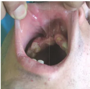
The patient was a 20-year-old had experienced a motor vehicle three months earlier. After primar and treatment of facial fractures referred for reconstruction of anterior maxillary ridge. The defect anterior maxilla extended from surface of the maxillary left lateral to the mesial surface of the maxillary second bicuspid. The edentulous severely deficient in the buccolingual vertical dimensions. Soft tissue the vestibule and previous surgeries reconstruction more difficult (Fig 1). Surgical procedure:
Under general anesthesia an incision made on top of the residual crest the underlying bone, which was
nolaryngology No.3, Vol.24, Serial No.68, Summer 2012
for replacement of lost due to trauma processes that involve the coverage of bone grafts in the alveolar region is an important of the graft. Ideally meet the following
coverage of the bone
nourishment of the graft
similarity between tissue and the new soft
volume (not bulky) reshaping procedures on
osteogenic capacity of the
detailed technical tissue flap created connective tissue that was bone graft in the simultaneously soft tissue in the
mensions.
old male who motor vehicle accident After primary care fractures he was reconstruction of a deficient e defect in the extended from the distal left lateral incisor the maxillary right edentulous ridge was the buccolingual and oft tissue scarring in previous surgeries made reconstruction more difficult (Fig 1).
anesthesia an incision was residual crest to access which was atrophied,
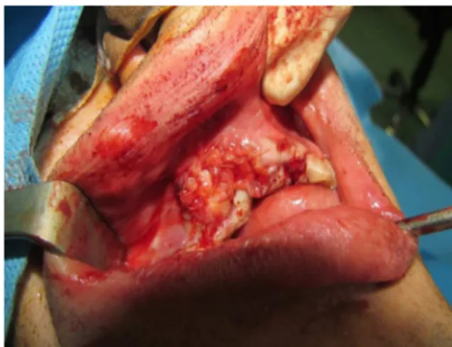
thin, and irregular (Fig obtain a pediculated periosteal tissue flap from the palate, lidocaine with epinephrine subperiostealy in the hard facilitate subperiosteal control of hemostasis.
Fig 1:Photograph showing the m
anterior alveolar ridge defect.
Fig 2:Photograph showing the actual
defect and soft tissue deficiency preparation.
The flap needed to have
and width to reach the vestibular depth cover the bone graft. So
long incision horizontally palate with a 5 mm vertical the scalloped gingival margin from the second molar
Rahpeyma A, et al
irregular (Fig 2). In order to pediculated periosteal connective the palate, we injected 2%
epinephrine (1:80,000) the hard palate region to subperiosteal dissection and
Photograph showing the maxillary anterior alveolar ridge defect.
Photograph showing the actual bone issue deficiency after the bed
preparation.
Bilateral Pediculated Palatal Periosteal Connective Ttissue Flap
Iranian Journal of Otorhinolaryngology No.3, Vol.24, Serial No.68, Summer 2012,145
incisor. Sharp dissection with a no. 15 blade was used to separate the connective tissue from the overlying epithelium. This dissection continued toward the midline. Great caution was needed to avoid perforation or thinning of the overlying mucosa. Blunt dissection with a periosteum, elevated this tissue from the bone.
The most distal part of the flap was incised vertically and brought to the anterior maxillary buccal region underneath the mucosa.
Hemorrhage from the greater palatine artery was controlled by cautery and surgicel® applied for dead space management. The palatal mucosal incision was sutured primarily and the whole procedure was done bilaterally in the palate.
A corticocancellus bone graft (3 × 2 cm2) was obtained from anterior iliac crest with a medial approach and fixed in the recipient site with a titanium mini screw. The height of the bone graft was 4 mm more than the residual bone and augmented the deficient ridge (Fig 3).
Fig 3:Photograph showing the
corticocancellus bone graft fixed in place with a titanium mini screw.
The bone graft was covered with bilateral anteriorly-based soft tissue flaps from the periosteal connective tissue of the hard palate. The edges of the flaps were sutured in the depth of the vestibule and together in
the midline. Sutures were made with 4-0, 5-0 vicryl® (Figs 4–6).
Fig 4:Schematic diagram of the anteriorly based pediculated palatal periosteal connective tissue flap. 1 Bone graft, 2 Titanium screw, 3 Pediculated palatal periosteal connective tissue flap, and 4
Alveolar mucosa.
Fig 5:Palatal view of the bilateral pedicle
flaps.
Fig 6:Photograph of the bilateral palatal
Rahpeyma A, et al
146, Iranian Journal of Otorhinolaryngology No.3, Vol.24, Serial No.68, Summer 2012 Discussion
The use of a pediculated periosteal connective tissue flap of the hard palate is valuable in maxillary reconstructions (9, 10). The flap has a random pattern blood supply, can have a width/ length ratio of up to 1/5 (8), and is used for minor ridge reconstruction of maxillary esthetic regions and simultaneously reconstructs both hard and soft tissue defects (9, 10). The flap also contains periosteum and connective tissue from the hard palate, allowing the bone graft to be covered with osteogenic tissue. The pedicled blood supply derived from the connective tissue periosteal plexus within the flap provides the biological basis for predictable bone graft coverage. The simultaneous hard and soft tissue reconstruction also reduces the number of operations that are needed for the reconstruction and reduces the time until placement of dental implants. This
flap does not need to be covered by mucosa and heals by secondary epithelialization, so the vestibular depth does not decrease, which is an important factor for prosthetic replacement of lost teeth in the most critically esthetic area of the jaw. In this report we have shown that this flap is capable for successfully covering large bone grafts.
Conclusion
Palatal submucosa can be used as a soft tissue reserve in upper jaw reconstructions. The donor site is near the surgical field and has minor morbidity. The surgical technique is simple, quick, and predictable. Simultaneous hard and soft tissue augmentation in the most esthetic area of the jaw is possible using this procedure.
References
1. Sindet-Pedersen S, Enemark H. Comparative study of secondary and late secondary bone-grafting in patients with residual cleft defects. Short-term evaluation. Int J Oral Surg 1985; 14(5): 389-98. 2. Jia YL, James DR, Mars M. Bilateral alveolar bone grafting: A report of 55 consecutively-treated patients. Eur J Orthod 1998; 20(3): 299-307.
3. van der Meij AW, Baart JA, Prahl-Andersen B, Kostense PJ, van der Sijp JR, Tuinzing DB. Outcome of bone grafting in relation to cleft width in unilateral cleft lip and palate patients. Oral Surg Oral Med Oral Pathol Oral Radiol Endod 2003; 96(1): 19-25.
4. Prolo DJ, Rodrigo JJ. Contemporary bone graft physiology and surgery. Clin Orthop Relat Res 1985; 200: 322-42.
5. Miller LL, Kauffmann D, St John D, Wang D, Grant JH, Waite PD. Retrospective review of 99 patients with secondary alveolar cleft repair. J Oral Maxillofac Surg 2010; 68(6): 1283-9.
6. Enemark H, Sindet-Pedersen S, Bundgaard M. Long-term results after secondary bone grafting of alveolar clefts. J Oral Maxillofac Surg 1987; 45(11): 913-9.
7. Sándor GK, Carmichael RP, Brkovic BM. Dental implants placed into alveolar clefts reconstructed with tongue flaps and bone grafts. Oral Surg Oral Med Oral Pathol Oral Radiol Endod 2010; 109(2): e1-7.
8. Sclar AG. Soft tissue and esthetic consideration in implant therapy. Surrey: Quintessence; 2003: 163-85.
9. Feichtinger M, Mossböck R, Kärcher H. Evaluation of bone volume following bone grafting in patients with unilateral clefts of lip, alveolus and palate using a CT-guided three-dimensional navigation system. J Craniomaxillofac Surg 2006; 34(3): 144-9.