ALEXANDRE REIS MACHADO
PHYLOGENY, IDENTIFICATION AND PATHOGENICITY OF THE BOTRYOSPHAERIACEAE ASSOCIATED WITH COLLAR AND ROOT ROT
OF THE BIOFUEL PLANT Jatropha curcas IN BRAZIL, WITH A DESCRIPTION OF NEW SPECIES
Dissertação apresentada à Universidade Federal de Viçosa, como parte das exigências do Programa de Pós-Graduação em Fitopatologia, para obtenção do título de Magister Scientiae.
VIÇOSA
ALEXANDRE REIS MACHADO
PHYLOGENY, IDENTIFICATION AND PATHOGENICITY OF THE BOTRYOSPHAERIACEAE ASSOCIATED WITH COLLAR AND ROOT ROT
OF THE BIOFUEL PLANT Jatropha curcas IN BRAZIL, WITH A DESCRIPTION OF NEW SPECIES
Dissertação apresentada à Universidade Federal de Viçosa, como parte das exigências do Programa de Pós-Graduação em Fitopatologia, para obtenção do título de Magister Scientiae.
APROVADA: 19 de julho de 2012.
____________________________ _____________________________ Prof. Maria Catarina Megumi Kasuya Dr. Harold Charles Evans
____________________________ Prof. Olinto Liparini Pereira
ii “Não há conquistas fáceis. São as estradas sinuosas que levam ao caminho certo. O profissional, em qualquer ofício, alcançará o triunfo a partir de um espírito tenaz, forte, obstinado”.
iii
AGRADECIMENTOS
Primeiramente agradeço à Deus pela saúde, pelas oportunidades que surgiram na minha vida, pelas conquistas, por nunca ter me abandonado nas horas difíceis e por ter colocado as pessoas certas no meu caminho.
Aos meus pais e irmãos por todo apoio durante a minha jornada.
À minha namorada Janiele pelo apoio e companheirismo mesmo nos momentos difíceis. Ao Professor Olinto Liparini Pereira, por ter me orientado, por todo apoio, pela amizade, incentivo, ensinamentos e por toda a dedicação na pesquisa e na formação de seus alunos.
Ao colega de laboratório Danilo pela amizade, ensinamentos e por toda a contribuição na execução deste trabalho.
Aos demais colegas e amigos do Laboratório Patologia de Sementes e Pós-Colheita, Deiziane, André Firmino, Stefania, Athus, Camila e Taís pela excelente convivência, pela ajuda, ensinamentos e amizade.
Aos colegas e amigos da Clínica de Doenças de Plantas pela amizade e auxílio.
À professora da UNINONTES, Adelica Xavier e ao pesquisador da EPAMIG, Nívio Poubel, pela amizade, pelos ensinamentos, por todo apoio e incentivo para que eu seguisse carreira na pesquisa.
Aos colegas de mestrado pela excelente convivência e amizade.
Ao Departamento de Fitopatologia da Universidade Federal de Viçosa, pela oportunidade de realização do curso de Mestrado.
Aos Professores do Departamento de Fitopatologia se empenharam em passar todo o conhecimento e experiência.
iv À empresa NOVABRA pela conceção de material para a realização deste trabalho e por todo apoio de seus técnicos, agrônomos e dirigentes.
Às empresas BIOJAN e SADA BIOENERGIA, por ceder suas áreas para visita e coleta. A todos aqueles que me apoiaram e torceram por mim.
v
BIOGRAFIA
ALEXANDRE REIS MACHADO, filho de Paulo Renato Machado e Mary de Souza Reis Machado, nasceu na cidade de Montes Claros, Minas Gerais, no dia 11 de novembro de 1986.
Realizou os estudos básicos na cidade de Janaúba, no mesmo estado.
Em 2005 iniciou o curso de graduação em Agronomia na Universidade Estadual de Montes Claros, graduando-se em julho de 2010.
vi
SUMÁRIO
RESUMO ... vii
ABSTRACT ... ix
INTRODUÇÃO GERAL ...1
REFERÊNCIAS ...4
ARTIGO ...6
Abstract ...7
INTRODUCTION ...8
MATERIALS AND METHODS ...10
Sample collection and isolation ...10
Morphological studies ...11
DNA extraction, Sequencing and Phylogenetic studies ...11
Pathogenicity tests ...13
RESULTS ...13
Symptomatology and isolations ...13
DNA extraction, PCR amplification and Phylogeny ...14
Taxonomy ...15
Pathogenicity tests ...21
DISCUSSION ...22
ACKNOWLEDGEMENTS ...25
REFERENCES ...25
CONCLUSÃO GERAL ...31
vii
RESUMO
MACHADO, Alexandre Reis, M.Sc., Universidade Federal de Viçosa, julho de 2012.
Phylogeny, identification and pathogenicity of the Botryosphaeriaceae associated with collar and root rot of the biofuel plant Jatropha curcas in Brazil, with a description of new species. Orientador: Olinto Liparini Pereira. Coorientador: Robert Weingart Barreto.
A partir da iniciativa do Governo Federal em introduzir o biodiesel na matriz energética brasileira, surgiu a necessidade de se pesquisar plantas oleaginosas potenciais para produção de matéria-prima para este biocombustível. O pinhão manso (Jatropha curcas) tem se destacado por ser uma planta perene, de fácil manejo, além de produzir
sementes com alto teor de óleo. A grande expansão das áreas de cultivo tem sido acompanhada pelo surgimento de diversas enfermidades, do qual pouco se conhece sobre os reais agentes etiológicos. Atualmente, em diversas áreas do Brasil, tem-se relatado a ocorrência de uma nova doença que não apenas reduz a produtividade, como tem causado a morte das plantas. Esta doença está associada a uma podridão das raízes e do colo das plantas. Alguns patógenos já foram relatados para essa doença, sendo mais frequente a ocorrência de fungos da família Botryosphaeriaceae, grupo conhecido pela dificuldade de separação das espécies utilizando características morfológicas. Assim, o objetivo deste trabalho foi identificar os possíveis agentes etiológicos, investigar a diversidade de Botryosphaeriaceae associado a essa doença, utilizando características morfológicas aliadas às ferramentas moleculares, bem como provar a potogenicidade dos isolados. Foram realizadas coletas nos estados de Minas Gerais e Espírito Santo. Em adição a estes, foram obtidas amostras dos estados do Piauí e São Paulo. Isolados monospóricos foram obtidos e armazenados. Estes tiveram o DNA extraído e as regiões ITS e TEF1-α sequenciadas. A partir dos resultados das análises filogenéticas, foram separados dois a três isolados de cada espécie para a caracterização morfológica. Nove espécies de Botryosphaeriaceae foram identificadas, sendo Lasiodiplodia theobromae, L. parva, L. pseudotheobromae, L. iraniensis, Neoscytalidium dimidiatum, Macrophomina phaseolina e três a serem propostas como novas espécies (Lasiodiplodia
sp.1, Lasiodiplodia sp.2 e Macrophomina sp.1). Todas as espécies distinguiram
morfologicamente e filogeneticamente, com exceção de Macrophomina sp.1 que não
viii Pelos testes de Blotter foram encontrados associados às sementes: Lasiodiplodia theobromae, Macrophomina phaseolina e Macrophomina sp.1. Espécies de
ix
ABSTRACT
MACHADO, Alexandre Reis, M.Sc., Universidade Federal de Viçosa, july, 2012.
Phylogeny, identification and pathogenicity of the Botryosphaeriaceae associated with collar and root rot of the biofuel plant Jatropha curcas in Brazil, with a description of new species. Adviser: Olinto Liparini Pereira. Co-adviser: Robert Weingart Barreto.
The introduction of biodiesel in the Brazilian energy matrix by initiative of the Federal Government, encouraged the search for potential oleaginous crops to supply raw material for biofuel production. The physic nut (Jatropha curcas) has been
highlighted since it is a perennial plant, easy to manage, and produces seeds with a high oil content. The expansion of areas of Jatropha in the world has contributed to the
emergence of various diseases. Currently in Brazil, the occurrence of a new disease that not only reduces the productivity, since it causes death of the plants. This disease is associated with a collar and root rot of plants. A number of pathogens have been associates with this disease, the occurrence of Botryosphaeriaceae fungi being the most frequent, which is a group known to be difficult to delimit species based on morphological characters. Thus, the purposes of this work was to investigate the diversity of Botryosphaeriaceae associated with collar and root rot of J. curcas, with the
aid of morphology and molecular tools and to asses the pathogenicity of the species involved. Samples were colleted in states of Minas Gerais and the Espirito Santo. In addition, samples were obtained from Piauí and São Paulo states. Single spore cultures were obtained and stored. These had their DNA extracted and the ITS and TEF1-α regions were sequenced. From the results of the phylogenetic analyses, two to three isolates of each species were separated for the morphological characterization and pathogenicity tests. With the purpose of investigating the association of the pathogens in seeds, Blotter tests were performed. Nine Botryosphaeriaceae species were identified:
Lasiodiplodia theobromae, L. parva, L. pseudotheobromae, L. iraniensis, Neoscytalidium dimidiatum, Macrophomina phaseolina and three to be proposed as new
species (Lasiodiplodia sp.1, Lasiodiplodia sp.2 and Macrophomina sp.1). All species
were distinguished morphologically and phylogenetically, except Macrophomina sp.1
that failed sporulate in culture.Currently, only Lasiodiplodia theobromae, Lasiodiplodia
sp.1 and Neoscytalidium dimidiatum have proven to be pathogenic. Lasiodiplodia theobromae, Macrophomina phaseolina and Macrophomina sp.1 were associated with
x are often referred to as endophytes and latent or opportunistic pathogens, because manifestation of the disease is directly linked with host stress. The great expansion of
Jatropha areas in the world have contributed to the emergence of several diseases,
INTRODUÇÃO GERAL
A partir da iniciativa do Governo Federal em introduzir o biodiesel na matriz energética brasileira, surgiu a necessidade de se pesquisar plantas oleaginosas com potencial para produção de matéria-prima para este biocombustível. Dentre as várias culturas com potencial para esse fim, o pinhão manso (Jatropha curcas) tem se
destacado especialmente em áreas com déficit hídrico recorrente.
O pinhão manso pertence à família Euphorbiaceae, com provável origem na
América Central, mas já se encontra amplamente distribuído na América do Sul, África e Ásia (Heller 1996). É uma cultura perene, de fácil manejo (Saturnino et al. 2005) e que não requer alto investimento financeiro durante o seu cultivo, tornando-se uma opção para a agricultura familiar (Arruda et al. 2004).
Produz sementes com alto teor de óleo, aproximadamente 47,25% (Akintayo 2004) e com excelentes propriedades, sendo que, em mistura de até 50% com o óleo diesel, pode ser utilizado em motores sem qualquer modificação (Pramanik 2003), contribuindo para a redução da emissão de gases poluentes produzidos pela queima de combustíveis fósseis. Além disso, o seu cultivo seria uma forma de se estocar carbono da atmosfera (Harinder e Makkar 2009).
Além das diversas vantagens de se cultivar o pinhão manso, a maioria dos trabalhos o consideram uma cultura resistente à pragas e doenças (Saturnino et al. 2005), e isso têm contribuído muito para o aumento da área plantada no Brasil. Entretanto, essa grande expansão das áreas de cultivo tem sido acompanhada pelo surgimento de diversas enfermidades, do qual pouco se conhece sobre os reais agentes etiológicos.
A maior parte das doenças descritas na cultura do pinhão manso são causadas por fungos, com destaque para: Glomerella cingulata (Stoneman) Spauld. & H.
Schrenk, Psathyrella subcorticalis Speg., Schizophyllum alneum (L.) Kuntze, Aecidium cnidoscoli Henn., Ramulariopsis cnidoscoli Speg., Uromyces jatrophicola
2 P. Syd.) E.J. Butler & Bisby, Passalora ajrekari (Syd. & P. Syd.) U. Braun (Freire e
Parente 2006), Phakopsora arthuriana Buriticá & J.F. Hennen (Hennen et al. 2005), Cochliobolus spicifer R.R. Nelson (Mendes et al. 1998), Cercospora jatrophicola
(Speg.) Chupp, Cercospora jatrophigena U. Braun, Pseudocercospora jatrophae-curcas (J.M.Yen) Deighton, Pseudocercospora jatrophae (G.F. Atk.) A.K. Das &
Chattopadh. e Pseudocercospora jatropharum (Speg.) U. Braun (Crous e Braun 2003)
e Elsinoë jatrophae Bitanc. & Jenkins (Bitancourt e Jenkins 1951).
A grande maioria dos patógenos citados são causadores de manchas foliares e outras doenças que ainda não representam uma limitação à produtividade do pinhão manso. Entretanto, em diversas áreas do Brasil, tem-se relatado a ocorrência de uma nova doença que não apenas é capaz de reduzir a produtividade, como tem causado a morte súbita de plantas, inviabilizado áreas de cultivo. Esta doença está associada a uma podridão do colo e radicular, nas quais os primeiros sintomas são a murcha repentina e amarelecimento das folhas, que em estágio mais avançado caem, finalizando com a morte da planta. Existem trabalhos de várias regiões do mundo que associam alguns patógenos a esses sintomas, a exemplo de: Nectria haematococca
Berk.& Br. [Haematonectria haematococca (Berk. & Broome) Samuels & Nirenberg]
e seu anamorfo Fusarium solani (Mart.) Sacc. na China (Yue-kai et al. 2011),
Clitocybe tabescens (Scop.) Bres. nos EUA (USDA 1960) e
Phytophthorapalmivoravar.palmivora(E.J. Butler) E.J. Butler (Erwin e Ribeiro
1996) em diversos países.
A primeira constatação da referida doença no Brasil e comprovação de sua etiologia foi feita por Pereira et al. (2009) no estado de São Paulo. O agente etiológico foi identificado morfologicamente como Lasiodiplodia theobromae e a doença foi
considerada como de baixa incidência ou esporádica (Pereira et al. 2009).
Entretanto, recentemente, cultivos de pinhão-manso têm sido abandonados em diferentes regiões do país por conta da podridão do colo associado também ao surgimento de uma nova sintomatologia de podridão radicular. Adicionalmente, a identificação somente morfológica de L. theobromae por Pereira et al. (2009) é
limitada, uma vez que a taxonomia de Lasiodiplodia (e outros Botryosphaeriaceae) foi
3 et al. 2008; Abdollahzadeh et al. 2010) e também pelo fato de nenhum estudo sistemático de coleta de materiais de diferentes regiões do Brasil ter sido feito até o momento.
O controle da doença pode ser futuramente alcançado pelo uso de variedades ou porta-enxertos resistentes. Entretanto, tal possibilidade encontra forte entrave fitopatológico pelo desconhecimento do complexo de espécies envolvidas na referida sintomatologia no país. Em qualquer cultura, o conhecimento dos agentes etiológicos associados a doenças de plantas é pré-requisito crucial para o estudo de possíveis práticas de manejo visando o controle dessas doenças de plantas.
4
REFERÊNCIAS
Abdollazadeh J, Javadi A, Mohammadi Goltapeh E, Zare R, Phillips AJL (2010) Phylogeny and morphology of four new species of Lasiodiplodia from Iran.
Persoonia 25: 1-10.
Akintayo ET (2004) Characteristics and composition of Parkia biglobosa and Jatropha curcas oils and cakes. Bioresource Technology 92: 307–310.
Alves A, Crous PW, Correia A, Phillips AJL (2008) Morphological and molecular data reveal cryptic species in Lasiodiplodia theobromae. Fungal Diversity 28: 1–
13.
Arruda FP De, Beltrão NEM, Andrade AP De, Pereira WE, Severino LS (2004) Cultivo de Pinhão-manso (Jatropha curcas L.) como alternativa para o
semi-árido nordestino. Revista Brasileira de Oleaginosas e Fibrosas 8: 789-799.
Bitancourt AA, Jenkins AE (1951) Estudos sôbre as Myriangiales. II. Vinte novas espécies de Elsinoaceas neotropicais. Arquivos do Instituto Biológico 20: 1-28. Crous PW, Braun U (2003) Mycosphaerella and its anamorphs: 1. Names published in
Cercospora and Passalora. CBS Biodiversity Series 1: 1-569.
Erwin DC, Ribeiro OK (1996) Phytophthora Diseases Worldwide. APS Press, St.
Paul, Minnesota, 562 p.
Freire FCO, Parente, GB (2006) As doenças das Jatrofas (Jatropha curcas L. e J. podagrica Hook.) no estado do Ceará. Embrapa, Comunicado Técnico 120, 4p.
Harinder PS, Makkar KB (2009) Jatropha curcas, a promising crop for the generation
of biodiesel and value-added coproducts. European Journal of Lipid Science and Technology 111: 773–787.
Heller J (1996) Physic nut (Jatropha curcas)– Promoting the conservation and use of
underutilized and neglected crops. Rome, IPGRI, 66p.
5 Mendes MAS, Silva VL, Dianese JC, Ferreira MASV, Santos CEN, Gomes Neto E, Urben AF, Castro C (1998) Fungos em plantas no Brasil. Brasília. Embrapa Cenargen. 555p.
Pereira OL, Dutra DC, Dias LAS (2009) Lasiodiplodia theobromae is the causal agent
of a damaging root and collar rot disease on the biofuel plant Jatropha curcas in
Brazil. Australasian Plant Disease Notes 4: 120-123.
Phillips S (1975) A new record of Pestalotiopsis versicolor on the leaves of Jatropha curcas. Indian Phytopathology 28: 546.
Pramanik K (2003) Properties and use of Jatropha curcas oil and diesel fuel blends in
compression ignition engine. Renewable Energy 28: 239–248.
Saturnino HM, Pacheco DD, Kakida J, Tominaga N, Gonçalves NP (2005) Cultura do pinhão-manso (Jatropha curcas L.). Informe Agropecuário 26: 44-78.
USDA (1960) Index of plant diseases in the United States. U.S.D.A. Agriculture Handbook 165: 1-531.
Viégas AP (1961) Índice de fungos da América do Sul. Campinas. Instituto Agronômico, 919p.
Yue-Kai W, Guo-Teng O, Jin-Yong Y (2011) First report of Nectria haematococca causing root rot disease of physic nut (Jatropha curcas) in China.
6
ARTIGO
According to the guidelines of Fungal Diversity
Phylogeny, identification and pathogenicity of the Botryosphaeriaceae associated with collar and root rot of the biofuel plant Jatropha curcas in Brazil, with a
7
Phylogeny, identification and pathogenicity of the Botryosphaeriaceae associated with collar and root rot of the biofuel plant Jatropha curcas in Brazil,
with a description of new species
Alexandre Reis Machado1
e-mail: [email protected] Danilo Batista Pinho1
e-mail: [email protected] Olinto Liparini Pereira1
e-mail: [email protected]
1 Universidade Federal de Viçosa - Departamento de Fitopatologia - 36570-000, Brazil Abstract:
The expansion of areas of Jatropha in the world, have contributed to the
emergence of various diseases. Currently in Brazil, the occurrence of a new disease has been reported that not only reduces the productivity, but also causes the death of plants. This disease is associated with a collar and rot root of plants. From morphological and phylogenetic studies nine species of Botryosphaeriaceae were identified. These include Lasiodiplodia theobromae, Lasiodiplodia parva, Lasiodiplodia pseudotheobromae, Lasiodiplodia iraniensis, Neoscytalidium dimidiatum, Macrophomina phaseolina and three to be proposed as new species
(Lasiodiplodia sp.1, Lasiodiplodia sp.2 and Macrophomina sp.1). Of these Lasiodiplodia theobromae, Macrophomina phaseolina and Macrophomina sp.1 were
encountered in seeds. Until now, only Lasiodiplodia theobromae, Lasiodiplodia sp.1
and Neoscytalidium dimidiatum have proven to be pathogenic. The results show that
root rot of physic nutis not associated with a single pathogen. Perhaps the emergence of this disease has been favored by stress conditions that the plant has encountered in the field. This study provides new information for future studies of disease management, quarantine programs and especially the development of resistant varieties for the collar and root rot disease of J. curcas.
8 INTRODUCTION
Jatropha curcas L., popularly known as Physic nut, is a plant of the family
Euphorbiaceae, that currently has been cultivated widely in the world with the main purpose supplying raw material for biofuel production. Among oil plants, this species has gained importance for being considered as easy to cultivation (Saturnino et al. 2005), which produces seeds with high oil content (47.25%) (Akintayo 2004) and with excellent fuel properties (Pramanik 2003).
Several research papers have also described J. curcas as resistant to pests and
diseases, and this has been an additional factor that encouraged its cultivation. However, the great expansion of cultivated areas has been accompanied by the emergence of various diseases of unknown etiology.
Recently in Brazil, a new disease that not only is able to reduce productivity, but also causes the sudden death of plants whas been observed. This disease is associated with root and collar rot, in which, the first symptoms are wilt and yellowing of the leaves, ending with leaf fall and death of the plant. According to Pereira et al. (2009), this disease is associated with Lasiodiplodia theobromae (Pat.) Griffon &
Maubl. in Brazil, as also reported by Latha et al. (2009) in India.
Lasiodiplodia theobromae is a fungus of the family Botryosphaeriaceae
(Botryosphaeriales, Ascomycetes), that has great phytopathological importance. Currently, 1060 hosts species are reported for L. theobromae (Farr and Rossman
2012). In recent years, this fungus has been the target of many controversial taxonomic studies. Denman et al. (2000) proposed the synonymy of the genus
Lasiodiplodia with Diplodia and Dothiorella. However, several other studies showed
that they are different genera (Phillips et al. 2005; Burgess et al. 2006; Alves et al. 2008; De Wet et al. 2008).
Others Botryosphaeriaceae have been reported causing the same disease in J. curcas, such as Macrophomina phaseolina (Patel et al. 2008), pathogen of great
phytopathological importance, due to its wide host range and ability of survivability by sclerotia (Dhingra and Sinclair 1978), and Neoscytalidium dimidiatum
9 form Scytalidium-like synanamorphs in the aerial mycelia and Fusicoccum-like
conidia in the pycnidia which has recently been reported in Brazil (Machado et al. in press).
Members of the family Botryosphaeriaceae occur in a wide range of hosts, including Gymnosperms and Angiosperms, and can be found as saprophytic, parasites or endophytes. The survival of these fungi as endophytes can represents a great danger and a problem for quarantine barriers, due to possibility of these being introduced into new environments through asymptomatic propagative material. Due to the emergence of unfavorable conditions for the plant, the pathogen can induce the symptoms and cause extensive losses (Slippers and Wingfield 2007). The identification and description of the new species of fungus was based on an analysis of morphological characters, but to undertake this task without a polyphasic approach, can underestimate the true diversity of the species (Taylor et al. 2000).
The species L. theobromae was distinguished from others basically on conidia
and paraphyses morphology. However, in recent years, various species have emerged from phylogenetic studies, showing the existence of a species complex (Pavlic et al. 2004; Burgess et al. 2006; Damm et al. 2007; Alves et al. 2008; Pavlic et al. 2008; Abdollahzadeh et al. 2010; Begoude et al. 2010; Urbez-Torres et al. 2012).Thus, the importance of combining and applying morphological and molecular tools to this group of fungi is clear.
In view of the importance that physic nut has acquired in the world, the lack of studies related to the etiology of root and collar rot disease is critical for future plant breeding programs, as well as to assist the establishment of phytosanitary measures and quarantine, in order to avoid its spread.
The purposes of this work were to investigate the diversity of Botryosphaeriaceae associated with collar and root rot of J. curcas, with aid of
10 MATERIALS AND METHODS
Sample collection and isolation
Field surveys were carried out during October 2010 and January-February 2011 in J. curcas plantations, with the purpose of finding and collecting plants with
symptoms of wilt, leaf fall and yellowing due to root or collar rot. The areas visited belong to the States of Minas Gerais (Biojan; SADA Bioenergia; EPAMIG-URENM) and Espírito Santo (NOVABRA). In addition, samples from the States of Piauí and São Paulo were also obtained from symptomatic plants. The samples were sent to the Laboratório de Patologia de Sementes e de Pós-colheita (Departamento de Fitopatologia, Universidade Federal de Viçosa) where they were first examined for the possible presence of fungal fruiting structures. Longitudinal sections of the stem and roots were made manually for observation of vascular necrosis, from which small fragments of areas of transition between the healthy tissue and the symptomatic tissue were obtained for fungal isolations. These fragments were disinfected in 70% ethanol for 1 min, followed by 1% sodium hypochlorite for 3 min and washed in sterile distilled water. Later, these were placed in Petri dishes with Potato Dextrose Agar (PDA - Acumedia®) and incubated at 25°C.
The isolates with dark mycelium and lack sporulation, typical of
Botryosphaeriaceae, were grown in Petri dishes with 2% Water Agar (WA - Agar
Agar, type I Himedia®) overlaid with double-sterilized maize straw and incubated at 25°C in 12 hours light-dark regime for 3-4 weeks for induction of sporulation. From these, single-spore cultures was obtained and stored in tubes on PDA at 10°C.
With the purpose of detecting these fungi in seeds, Blotter tests were performed as described by Alfenas et al. (2007). The isolates obtained from seeds, were processed in the same manner as described for the other isolates.
11
Morphological studies
The isolates were grown on Petri dishes containing 2% WA overlaid with double-sterilized twigs of Pinus and corn straw and incubated at 25°C with a
photoperiod of 12 hours to induce the formation of fruit bodies and sporulation. Sections of the fruiting bodies were manually made and mounted in lactophenol.
Thirty measurements of all relevant morphological characters (conidia, paraphyses and conidiogenous cells) were made using a light microscope OLYMPUS CX31 for identification of the species. The images were obtained with an OLYMPUS BX 51 light microscope fitted with a digital camera (OLYMPUS EVOLT330).
DNA extraction, Sequencing and Phylogenetic studies
Single spore isolates were grown on PDA at 25 °C for one week. Approximately 40 mg of fungus mycelia were scraped from the agar surface and placed in a sterile 1.5 mL microcentrifuge tube. The extraction was processed by freezing with liquid nitrogen and grinding into a fine powder using a microcentrifuge tube pestle. The crushing continued to add 100 µL of Nuclei Lysis Solution of the Wizard® Genomic DNA Purification Kit (Promega Corporation, WI, U.S.A.). After, an additional 500 µL of the previous solution was added. The extraction continued as described by Pinho et al. (in press).
PCR reactions were set-up using the following ingredients for each 25 µL reaction: 12.5 µL of Dream Taq TM PCR Master Mix 2X (MBI Fermentas, Vilnius, Lithuania), 1 µL of 10 µM of each forward and reverse primer synthesised by Invitrogen (Carlsbad, U.S.A), 1 µL of dimethyl sulfoxide (DMSO, Sigma–Aldrich, St. Louis, MO, U.S.A.), 5 µL of 100× (10 mg/mL) Bovine Serum Albumin (BSA, Sigma– Aldrich, St. Louis, MO, U.S.A.), 2 µL of genomic DNA (25 ng/µl), and nuclease-free water to complete the total volume.
(5’-12
TGCGGTGGTATCGACAAGCGT-3’) and EF2R
(5’-AGCATGTTGTCGCCGTTGAAG-3’) (Jacobs et al. 2004), EF1-688F (5’- CGGTCACTTGATCTACAAGTGC-3’) (Alves et al. 2008) and EF1-986R (5’-TACTTGAAGGAACCCTTACC-3’) (Carbone et al. 1999) for partial TEF1-α. The thermal cycle consisted of 95 ºC for 5 min, followed by 35 cycles of 94 ºC for 1 min (denaturation), 55 ºC for 1 min (for TEF1-α) or 52 ºC for 1 min (for ITS) (annealing), 72 ºC for 2 min (elongation), and 72 ºC for 10 min (final extension). PCR products were analyzed by 2 % agarose electrophoresis gels stained with GelRed™ (Biotium Inc., Hayward, CA, U.S.A.) in a 1× TAE buffer and visualized under UV light to check for amplification size and purity. PCR products were purified and sequenced by Macrogen Inc., Korea (http://www.macrogen.com). The nucleotide sequences were edited with the DNA Dragon software (Hepperle 2011). All sequences were checked manually and nucleotides with ambiguous positions were clarified using both primer direction sequences. New sequences were deposited in GenBank (http://www.ncbi.nlm.nih.gov). Sequences of ITS and TEF1-α of additional species were retrieved from GenBank (Table 1).
Consensus regions were compared against GenBank’s database using their Mega BLAST program. The closest hit sequences were then downloaded in FASTA format and aligned using the multiple sequence alignment program MUSCLE® (Edgar 2004), built in MEGA v. 5 software (Tamura et al. 2011). Alignments were checked and manual adjustments were made when necessary. All the ambiguously aligned regions within dataset were excluded from the analyses. Gaps (insertions/deletions) were treated as missing data. The resulting alignment was deposited into TreeBASE (http://www.treebase.org/).
13 performed using MrBayes v.3.1.1 (Ronquist and Huelsenbeck, 2003). In MrBayes, data were partitioned by locus and the parameters of the nucleotide substitution models for each partition were set as described above. Four MCMC chains were run simultaneously, starting from random trees for 10 000 000 generations. Trees were sampled every 1000th generation for a total of 10 000 trees. The first 2 500 trees were discarded as the burn-in phase of each analysis. Posterior probabilities (Rannala & Yang, 1996) were determined from a majority-rule consensus tree generated with the remaining 7 500 trees. Trees were visualized in FigTree (Rambaut 2009) and exported to graphics programs. The species Spencermartinsia viticola CBS117009 were used as
an outgroup in these analyses.
Pathogenicity tests
Five J. curcas plants of six months old obtained from disinfested seeds, were
utilized in the pathogenicity test for each fungal species isolated. The plants were grow in pots with 5kg of sterilized soil, maintained in a greenhouse, and watered once a week until the appearance of symptoms. For inoculation, one representative isolate of each species was grown in a Petri dish with PDA for seven days at 25°C. Six millimeters diameter disks of bark (from the collar regions of healthy plants) were removed with a sterile cork borer and replaced with 6 mm diameter disks containing mycelia from the margins of the growing culture, just below a portion of moistened cotton was placed and subsequently covered with parafilm. On control plants, only PDA plugs were placed on wounded stems. After 2 weeks, the parafilme and cotton were removed. The inoculated plants were maintained in a greenhouse at 25°C for 60 days. From the symptomatic inoculated plants, the fungi were reisolated in culture.
RESULTS
Symptomatology and isolations
14 the appearance of black fungal structures in the bark of the plant could be observed. Upon being removed from the soil, plant roots were found to be rotted and the vascular system were necrotic, ranging from light brown to black. Due to loss of support, the plants were often already toppled by wind action (Fig 1) or invaded by termites.
A total of 55 plants were sent to the laboratory and from these, 35 isolates belonging to Botryosphaeriaceae were obtained. Of these, 20 isolates belong to
Neoscytalidium, 13 to Lasiodiplodia and 2 to Macrophomina. In addition to these
fungal genera, some Fusarium spp., Colletotrichum spp. and Chaetomium spp., were
also isolated. However, due to the prevalence and diversity of species of Botryosphaeriaceae associated with root and collar rot of J. curcas, the other genera
were not included in this work. The Neoscytalidium was not detected in the State of
the Espírito Santo. In the State of Piauí was detected only Neoscytalidium. In São
Paulo State only one isolate of Lasiodiplodia was obtained. However, in Minas Gerais
State, it was possible to detect all genera. From seeds from Minas Gerais and Espírito Santo, five isolates of Macrophomina and one isolate of Lasiodiplodia were obtained. Neoscytalidium was not associated with seeds.
DNA extraction, PCR amplification and Phylogeny
15 The combined analyzes of ITS and TEF1-α dataset included 52 taxa and contained 718 characters with 193 parsimony-informative, 237 variable and 481 conserved.
From the phylogenetic analysis, it was possible to identify nine different species of Botryosphaeriaceae: one species of Neoscytalidium, two species of Macrophomina and six species of Lasiodiplodia. The consensus tree generated with
Bayesian analyses is shown (Fig 2).
Taxonomy
The isolates previously identified as Lasiodiplodia had common
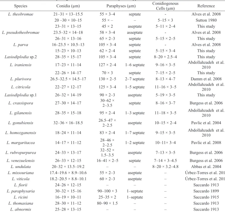
characteristics, such as the presence of paraphyses within the pycnidial conidiomata, conidia initially hyaline and aseptate, but becoming brown and 1-septate with age, with the formation of longitudinal striations due the deposition of melanin granules on the inner surface of the wall. Based on phylogenetic analysis, followed by the morphological descriptions, six species were delimited, in which four are known species and two are to be proposed as new. Dimensions of each Lasiodiplodia species
are available in Table 2.
Lasiodiplodia theobromae (Pat.) Griffon & Maubl., Bull. trimest. Soc. Mycol. Fr. 25:
57 (1909) (Fig 3)
Mycelium immersed or superficial, branched, septate, dark brown. Conidiomata pycnidial, stromatic, superficial, separate, globose, dark brown, uni- or
multilocular, often covered by aerial mycelium, formed superficially on twigs of Pinus
or corn straw in culture. Wall dark brown, thick-walled textura angularis, paler and thinner towards the conidiogenous region. Conidiophores absent. Conidiogenous cells
holoblastic, determinate, discrete, cylindrical, hyaline, smooth and thin-walled, formed from cells lining the inner pycnidial walls, 5‒11 × 2‒4 μm. Paraphyses hyaline,
cylindrical, aseptate, not branched, ends rounded, up to 45 μm long, 2 μm wide.
Conidia acrogenous, aseptate, ellipsoid to ovoid, hyaline when young, later becoming
16
Habitat: On Jatropha curcas
Known distribution: São Paulo, Espírito Santo and Minas Gerais States, Brazil
Material utilized for morphological description: BRAZIL, Colatina, Espírito Santo,
collar and root rot of Jatropha curcas, 2011, A. R. Machado & O. L. Pereira, Isolate
114. Other isolates examined are listed in Table 1.
Lasiodiplodia parva A.J.L. Phillips, A. Alves & Crous, Fungal Diversity 28:9 (2008)
(Fig 4)
Mycelium immersed or superficial, branched, septate, dark brown. Conidiomata pycnidial, stromatic, superficial, separate, globose, dark brown,
unilocular, often covered by aerial mycelium, formed superficially on twigs of Pinus
or corn straw in culture. Wall dark brown, thick-walled textura angularis, paler and thinner towards the conidiogenous region. Conidiophores absent. Conidiogenous cells
holoblastic, determinate, discrete, cylindrical, hyaline, smooth and thin-walled, formed from cells lining the inner pycnidial walls, 5‒15 × 2‒5 μm. Paraphyses hyaline,
cylindrical, septate, occasionally branched, ends rounded, up to 62 μm long, 2‒4 μm wide. Conidia acrogenous, aseptate, ellipsoid to ovoid hyaline when young, later
becoming medianly one-septate, dark brown, thick-walled, ellipsoid, frequently with rounded apex, truncate base, 15‒23 × 10‒13 μm and longitudinal striations.
Habitat: On Jatropha curcas
Known distribution: Espírito Santo and Minas Gerais States, Brazil
Material utilized for morphological description: BRAZIL, Colatina, Espírito Santo,
collar and root rot of Jatropha curcas, 2011, A. R. Machado & O. L. Pereira, Isolate
17
Lasiodiplodia pseudotheobromae A.J.L. Phillips, A. Alves & Crous, Fungal Diversity
28:9 (2008) (Fig 5)
Mycelium immersed or superficial, branched, septate, dark brown. Conidiomata pycnidial, stromatic, superficial, separate, globose, dark brown,
unilocular, often covered by aerial mycelium, formed superficially on twigs of pinus or corn straw in culture. Wall dark brown, thick-walled textura angularis, paler and thinner towards the conidiogenous region. Conidiophores absent. Conidiogenous cells
holoblastic, determinate, discrete, cylindrical, hyaline, smooth and thin-walled, formed from cells lining the inner pycnidial walls, 5‒15 × 2‒5 μm. Paraphyses hyaline,
cylindrical, septate, not branched, ends rounded, up to 65 μm long, 2‒3 μm wide.
Conidia acrogenous, aseptate, ellipsoid, hyaline when young, later becoming medianly
one-septate, dark brown, thick-walled, ellipsoid, frequently with rounded apex, truncate base, 26‒31 × 13‒16 μm and longitudinal striations.
.
Habitat: On Jatropha curcas
Known distribution: Espírito Santo State, Brazil
Material utilized for morphological description: BRAZIL, Colatina, Espírito Santo,
Collar and root rot of Jatropha curcas, 2011, A. R. Machado & O. L. Pereira, Isolate
163.
Lasiodiplodia iraniensis Abdollahzadeh, Zare & A.J.L. Phillips, Persoonia 25:8
(2010) (Fig 6)
Mycelium immersed or superficial, branched, septate, dark brown. Conidiomata pycnidial, stromatic, superficial, separate, globose, dark brown,
unilocular, often covered by aerial mycelium, formed superficially on twigs of pinus or corn straw in culture. Wall dark brown, thick-walled textura angularis, paler and thinner towards the conidiogenous region. Conidiophores absent. Conidiogenous cells
18 cylindrical, septate, occasionally branched, ends rounded, up to 70 μm long, 3 μm wide. Conidia acrogenous, aseptate, subglobose to subcylindrical, hyaline when
young, later becoming medianly one-septate, dark brown, thick-walled, ovoid to subcylindrical, frequently with rounded apex, truncate base, 22‒26 × 14‒17μm and longitudinal striations.
Habitat: On Jatropha curcas
Known distribution: Espírito Santo State, Brazil
Material utilized for morphological description: BRAZIL, Colatina, Espírito Santo,
Collar and root rot of Jatropha curcas, 2011, A. R. Machado & O. L. Pereira, Isolate
67.
Lasiodiplodia sp. 1 (To be proposed as new species) (Fig 7)
Mycelium immersed or superficial, branched, septate, dark brown. Conidiomata pycnidial, stromatic, superficial, separate, globose, dark brown, uni- or
multilocular, often covered by aerial mycelium formed superficially on twigs of pinus or corn straw in culture. Wall dark brown, thick-walled textura angularis, paler and thinner towards the conidiogenous region. Conidiophores absent. Conidiogenous cells
holoblastic, determinate, discrete, cylindrical, hyaline, smooth and thin-walled, formed from cells lining the inner pycnidial walls, 5‒19 × 3‒5 μm. Paraphyses hyaline,
cylindrical, aseptate, not branched, ends rounded, up to 90 μm long, 2‒3 μm wide.
Conidia acrogenous, aseptate, ellipsoid, hyaline when young, later becoming dark
brown, thick-walled, ellipsoid to obpyriform, frequently with rounded apex, truncate base, forming up to two septa, 26‒32 × 14‒19 μm and longitudinal striations.
Habitat: On Jatropha curcas
Known distribution: Espírito Santo State, Brazil
Material utilized for morphological description: BRAZIL, Colatina, Espírito Santo,
Collar and root rot of Jatropha curcas, 2011, A. R. Machado & O. L. Pereira, Isolate
19
Lasiodiplodia sp. 2(To be proposed as new species) (Fig 8)
Mycelium immersed or superficial, branched, septate, dark brown. Conidiomata pycnidial, stromatic, immersed or superficial, separate or aggregated,
globose, dark brown, uni- or multilocular, often covered by aerial mycelium, formed superficially on twigs of pinus or corn straw in culture. Wall dark brown, thick-walled textura angularis, paler and thinner towards the conidiogenous region. Conidiophores
absent. Conidiogenous cells holoblastic, determinate, discrete, cylindrical, hyaline,
smooth and thin-walled, formed from cells lining the inner pycnidial walls, 8‒20 × 2.5‒4 μm. Paraphyses hyaline, cylindrical, septate, not branched, ends rounded, up to
105 μm long, 3‒4 μm wide. Conidia acrogenous, up to three-septate, ellipsoid to
ovoid, hyaline when young, later becoming medianly one-septate, dark brown, thick-walled, ellipsoid, frequently with rounded apex, truncate base, 28‒35 × 15‒17 μm and longitudinal striations.
Habitat: On Jatropha curcas
Known distribution: Espírito Santo and Minas Gerais States, Brazil
Material utilized for morphological description: BRAZIL, Colatina, Espírito Santo,
Collar and root rot of Jatropha curcas, 2011, A. R. Machado & O. L. Pereira, Isolate
116. Other isolates examined are listed in Table 1.
The isolates previously identified as Neoscytalidium, had common
characteristics: the formation of chains of arthroconidia on the aerial mycelium and on twigs of Pinus or corn straw in culture, pycnidial conidiomata, immersed in or
superficially on a stroma, with release of Fusicoccum-like conidia.
From the phylogenetic analysis, as well as from the morphology, it was possible to separate one Neoscytalidium species. The species could be identified as Neoscytalidium dimidiatum (Penz.) Crous & Slippers. Dimensions of the species are
20 Neoscytalidium dimidiatum (Penz.) Crous & Slippers Stud. Mycol., 55:244 (2006)
(Fig 9) The aerial mycelia formed chains of 0‒1 septate arthroconidia, oblong to
globose, initially hyaline becoming brown thick-walled with age, 4‒12 × 2.5‒8 µm.
Pycnidia dark, globose, base up to 250 µm and a neck up to 810 µm, immersed in or
superficially on a stroma. Conidiogenous cells holoblastic, lageniform to ampulliform,
hyaline, 6‒10 × 1.5‒2.5 µm. Conidia hyaline, ellipsoid to nearly fusiform, 8‒12 × 4‒5
µm. Dark septate conidia not observed.
Habitat: On Jatropha curcas
Known distribution: Piauí and Minas Gerais States, Brazil
Material utilized for morphological description: BRAZIL, Piauí, Collar and root rot of Jatropha curcas, 2010, A. R. Machado & O. L. Pereira, Isolate 77. Other isolates
examined are listed in Table 1.
The isolates previously identified as Macrophomina had common
characteristics of the genus, such as the formation of dark mycelia and sclerotia on PDA. On twigs of Pinus in culture the formation of pycnidial conidiomata was
observed, with release of hyaline conidia with apical mucoid appendages. Only a single isolate sporulated in culture.
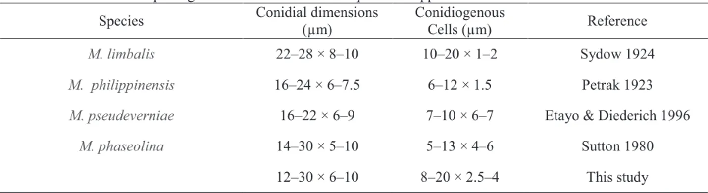
Based on morphology and the phylogenetic analyses, it was possible to identify two Macrophomina species. Macrophomina phaseolina was found only on
roots, however, on seeds a new phylogenetic species was found. Dimensions of species are given in Table 4.
Macrophomina phaseolina (Tassi) Goid., Annali della Sperimentazione Agaria, 1:
457 (1947) (Fig 10)
Mycelium superficial or immersed, light to dark brown, branched, septate. Conidiomata pycnidial, separate, globose, dark brown, unilocular, thick-walled. Conidiophores absent. Conidiogenous cells holoblastic, phialidic, determinate,
21
Conidia hyaline, aseptate, obtuse at each end, fusiform, thin-walled, smooth, with
apical mucoid appendages. Brown, 1-septate mature conidia were present.
Habitat: On Jatropha curcas
Known distribution: Espírito Santo and Minas Gerais States, Brazil
Material utilized for morphological description: BRAZIL, Nova Porteirinha, Minas
Gerais, Collar and root rot of Jatropha curcas, 2011, A. R. Machado & O. L. Pereira,
Isolate 70. Other isolates examined are listed in Table 1.
Macrophomina sp.1(To be proposed as new species)
It was not possible to induce sporulation of the isolates obtained from seeds. However, the phylogenetic studies show that it is a possible new species, due to the formation of a monophyletic group with in the clade, and well supported by posterior probability.
Habitat: On Jatropha curcas
Known distribution: Espírito Santo and Minas Gerais States, Brazil
Material utilized for morphological description: BRAZIL, Jaíba, Minas Gerais, from
seeds of Jatropha curcas, 2011, A. R. Machado & O. L. Pereira, Isolate 113. Other
isolates examined are listed in Table 1.
Pathogenicity tests
Sixty days after inoculation, the plants were evaluated for presence or absence of symptoms. The fungus Neoscytalidium dimidiatum reproduced the
symptoms observed in the field, which caused collar rot that progressed to the roots. From the lesion, it was possible to isolate and retrieve the inoculated fungus (Fig.11 a– c). The species Lasiodiplodia theobromae and Lasiodiplodia sp.1 caused localized
22 produce symptoms in the conditions under which the test was conducted. For the newly found species Lasiodiplodia sp. 2, L. iraniensis and L. pseudotheobromae, the
pathogenicity tests are on going.
DISCUSSION
In this work, through phylogenetic analysis supported by morphological studies, nine species of Botryosphaeriaceae were identified associated with the collar rot disease of J. curcas. Among these, two new species of Lasiodiplodia and one of Macrophomina will be proposed.
As with previous studies (Damm et al. 2007; Alves et al. 2008; Abdollahzadeh et al. 2010), the Lasiodiplodia species, except for Lasiodiplodia sp.2.,
cannot be distinguished based only on sequences of ITS, however when TEF1-α is also included in the analysis, the species are clearly separated and with strong support. This occurs because the newly described taxa belong to a complex of species within what was previously known as L. theobromae.
Utilizing only the TEF1-α sequences for phylogenetic analysis, it was possible to distinguish the species in separate groups, with a similar topology to the combined data, but without sufficient support to propose new species (data not shown).
Allied to the phylogenetic analyses, several studies have used morphological parameters, such as conidial morphology, size, shape and septation of paraphyses, growth and pigment production in culture to differentiate Lasiodiplodia species
(Pavlic et al. 2004; Burgess et al. 2006; Damm et al. 2007; Alves et al. 2008; Pavlic et al. 2008; Abdollahzadeh et al. 2010; Begoude et al. 2010; Urbez-Torres et al. 2012). However, Abdollahzadeh et al. (2010) make case against differentiating species on paraphyses septation, due to the fact that this parameter can give erroneous results and should be interpreted with caution. For example, paraphyses can be aseptate when young and become septate with age. In this study, L. pseudotheobromae produced
23 In the material of L. parva studied by Alves et al. (2008), the paraphyses are
longer than the isolates from the same species retrieved in the present study. But, as in the above situation, the dimensions of the conidia are the same.
Alves et al. (2008) also used pigment production in culture at 35 ° C and growth at 10 ° C for the differentiation of L. parva and L. pseudotheobromae from L. theobromae. However, Abdollahzadeh et al. (2010) concludes that this parameter has
limited value in species determination. Thus, this parameter was not used in the present study.
The conidia of L. iraniensis were larger than reported for Abdollahzadeh et
al. (2010). However, they still differ from L. theobromae because they were often
smaller and can be sepatared from L. parva because the conidial shape were often
subglobose to subcylindrical.
Lasiodiplodia sp.1 is phylogenetically closer to L. citricola Abdollahzadeh,
Javadi & A.J.L. Phillips, but conidia are longer and wider than those of L. citricola. In
addition, conidia up to two-septate and obpyriform were observed. Paraphyses and conidiogenous cells are smaller than in Lasiodiplodia. sp.1.
Lasiodiplodia sp.2 is phylogenetically distant from the other species and can
be distinguished solely with ITS sequences. The dimensions of the paraphyses are similar to those of L. parva, however, the conidia are larger and hyaline and up to
three-septate.
L. theobromae analyzed in this study had the same dimensions of conidia and
paraphyses as cited by Sutton (1980) and Alves et al. (2008).
These results show the great diversity of Lasiodiplodia species associated
with J. curcas. This is the first description of the occurrence of L. pseudotheobromae, L. parva and L. iraniensis in Brazil, and also from this host. As shown by Begoude et
al. (2010), these fungi have a wide geographical distribution and a broad host range that has been increased substantially with these studies.
The isolates of Neoscytalidium dimidiatum formed one well supported
monophyletic group, being different from N. novaehollandiae in contrast to the results
24 dimensions of conidia, arthroconidia and conidiogenous cells similar to the same species in previous studies and of the species N. novaehollandiaePavlic, T.I. Burgess
& M.J. Wingf.. Pavlic et al. (2008) reached the same conclusion, but according to these authors, the presence of Dichomera-like conidia in N. novaehollandiae is the
main character that differentiates this species from N. dimidiatum.
The fungus Neoscytalidium dimidiatum is cosmopolitan, occurring in a wide
range of hosts, which includes apple, banana, citrus, fig, grapevine, potato, walnut, yam and mango, sometimes causing canker and rot or branch wilt (Sutton 1980; Polizzi et al. 2009; Ray et al. 2010). Recently, it has been described causing collar and rot root of J. curcas in Brazil (Machado et al. in press).
In this study it was observed that this fungus is present in regions with a semi-arid climate (North of Minas Gerais and Piauí) and was not found in more humid regions (Espírito Santo). It is known that climatic variations can influence the distribution of different Botryosphaeriaceae (Úrbez-Torres et al. 2006; Begoude et al. 2010). But, for Neoscytalidium, more studies should be conducted with a broader
survey to confirm this hypothesis.
Two species of Macrophomina are reported in this paper. Macrophomina phaseolina formed a monophyletic group with other sequences from the same species.
The conidia had dimensions similar to those described by Sutton (1980), however, the conidiogenous cells were longer and narrower.
Based on the phylogenetic analysis, we obtained a new species of
Macrophomina isolated from seeds, but to confirm this information, a morphological
differentiation from the other species previously reported will be required. So far, these isolates did not sporulate in culture using the method tested.
This study shows that the Botryosphaeriaceae species isolated exhibit different levels of pathogenicity on J. curcas. Lasiodiplodia theobromae and N. dimidiatum reproduced the symptoms observed in the field, and can be considered as
primary pathogens. Other species of Lasiodiplodia and Macrophomina tested, possibly
25 since factors such as the age of the plant, inoculation method or environmental conditions can influence the inoculations results. Future studies should be realized with the purposes of investigating the possible existence of the severity levels between species and to test the pathogenicity of isolates in plants under environmental stress.
Botryosphaeriaceae species occur in a wide range of hosts and environments, and are often referred to as endophytes, latent or opportunistic pathogens, because the manifestation of the disease is directly linked with the occurrence of stress of the host (Slippers and Wingfield 2007).
The great expansion of Jatropha areas in the world has contributed to the
emergence of several diseases for which, so far, the etiological agents are unknown. This study provides new information for future studies of disease management, quarantine programs and especially the development of resistant varieties for collar and root rot of J. curcas.
ACKNOWLEDGEMENTS
This work forms part of a research project submitted as a M.Sc. dissertation to the Departamento de Fitopatologia/Universidade Federal de Viçosa by A. R. Machado. The authors thanks Conselho Nacional de Desenvolvimento Científico e Tecnológico –CNPq, and the Coordenação de Aperfeiçoamento de Pessoal de Nível Superior –CAPES and Fundação de Amparo a Pesquisa do Estado de Minas Gerais – FAPEMIG for the financial support.
REFERENCES
Abbas SQ, Sutton BC, Ghaffar A, Abbas A (2004) Reassessment of Sphaeropsis undulata Berk. & Curt. Pakistan Journal of Botany 36: 209–218.
Abdollahzadeh J, Jvadi A, Mohammadi-Goltapeh E, Zare R, Phillips AJL (2010) Phylogeny and morphology of four new species of Lasiodiplodia from Iran.
26 Akintayo ET (2004) Characteristics and composition of Parkia biglobosa and
Jatropha curcas oils and cakes. Bioresource Technology 92: 307–310.
Alfenas AC, Ferreira FA, Mafia RG, Gonçalves RC (2007) Isolamento de fungos fitopatogênicos. In: Alfenas AC, Mafia RG (Eds) Métodos em Fitopatologia. UFV, Viçosa, pp 53−90.
Alves A, Crous PW, Correia A, Phillips AJL (2008) Morphological and molecular data reveal cryptic species in Lasiodiplodia theobromae. Fungal Diversity 28: 1–
13.
Begoude BAD, Slippers B, Wingfield MJ, Roux J (2010) Botryosphaeriaceae associated with Terminalia catappa in Cameroon, South Africa and Madagascar.
Mycological Progress 9(1): 101-123.
Burgess TI, Barber PA, Mohali S, Pegg G, de Beer W, Wingfield M.J (2006) Three new Lasiodiplodia spp. from the tropics, recognized based on DNA sequence
comparisons and morphology. Mycologia 98: 423-435.
Carbone I, Anderson JB, Kohn LM (1999) A method for designing primer sets for the speciation studies in filamentous ascomycetes. Mycologia 91:553–556
Crous PW, Slippers B, Wingfield MJ, Rheeder J, Marasas WFO, Phillips AJL, Alves A, Burgess T, Barber P, Groenewald JZ (2006) Phylogenetic lineages in the Botryosphaeriaceae.Studies in Mycology 55: 235-253.
Damm U, Crous PW, Fourie PH (2007) Botryosphaeriaceae as potential pathogens of
Prunus in South Africa, with descriptions of Diplodia africana and Lasiodiplodia plurivora sp. nov. Mycologia 99: 664–680.
De Wet J, Slippers B, Preisig O, Wingfield BD, Wingfield MJ (2008) Phylogeny of Botryosphaeriaceae reveals patterns of host association. Molecular Phylogenetics and Evolution 46:116–126.
Denman S, Crous PW, Taylor JE, Kang JC, Pascoe I, Wingfield MJ (2000) An overview of the taxonomic history of Botryosphaeria, and a re-evaluation of its
27 Dhingra OD, Sinclair JB (1978) Biology and pathology of
Macrophomina phaseolina. Imprensa Universitária, Universidade Federal de
Viçosa, Viçosa, Brasil. 166 pp.
Edgar RC (2004) MUSCLE: multiple sequence alignment with high accuracy and high throughput. Nucleic Acids Research 32: 1792–1797.
Etayo J, Diederich P (1996) Lichenicolous fungi from the western Pyrenees, France and Spain. II. More deuteromycetes. Mycotaxon 60: 419-420.
Farr DF, Rossman AY (2012) Fungal Databases, Systematic Mycology and Microbiology Laboratory, ARS, USDA. http://nt.ars-grin.gov/fungaldatabases/. Acessed 10 July 2012.
Hepperle D (2011) DNA Dragon 1.4.1 – DNA sequence contig assembler software. http://www.dna-dragon.com/. Acessed 15 November 2010.
Jacobs K, Bergdahl DR, Wingfield MJ, Halik S, Seifert KA, Bright DE, Wingfield BD (2004) Leptographium wingfieldii introduced into North America and found
associated with exotic Tomicus piniperda and native bark beetles. Mycological
Research 108: 411–418.
Latha P, Prakasam V, Kamalakannan A, Gopalakrishnan C, Raguchander T, Paramathma M, Samiyappan R (2009) First report of Lasiodiplodia theobromae
(Pat.) Griffon & Maubl causing root rot and collar rot disease of physic nut (Jatropha curcas L.) in India. Australasian Plant Disease Notes 4: 19-20.
Machado AR, Pinho DB, Dutra DC, Pereira OL (In press) Collar and root rot caused by Neoscytalidium dimidiatum in the biofuel plant Jatropha curcas. Plant
Disease.
Nattrass RM (1933) A new species of Hendersonula (H. toruloidea) on deciduous
trees in Egypt. Transactions of the British Mycological Society18: 189–198. Patel DS, Patel SI, Patel RL (2008) A new report on root rot of Jaropha curcas caused
by Macrophomina phaseolina from Gujarat, India. Journal of Mycology and
28 Pavlic D, Slippers B, Coutinho TA, Gryzenhout M, Wingfield MJ (2004)
Lasiodiplodiagonubiensis sp. nov., a new Botryosphaeria anamorph from native Syzygium cordatum in South Africa. Studies in Mycology 50: 313-322.
Pavlic D, Wingfield MJ, Barber P, Slippers B, Hardy GESJ, Burgess TI (2008) Seven new species of the Botryosphaeriaceae from baobab and other native trees in Western Australia. Mycologia 100: 851–866.
Pereira OL, Dutra DC, Dias LAS (2009) Lasiodiplodia theobromae is the causal agent
of a damaging root and collar rot disease on the biofuel plant Jatropha curcas in
Brazil. Australasian Plant Disease Notes 4: 120-123.
Petrak F (1923) Mycologische Notizen V. No. 200. Über die Pseudosphaeriaceen v.H. und ihre Bedeutung für die spezielle Systematik der Pyrenomyzeten. Annales Mycologici21: 30–69.
Phillips AJL, Alves A, Correia A, Luque J (2005) Two new species of Botryosphaeria
with brown, 1-septate ascospores and Dothiorella anamorphs. Mycologia 97:
513-529.
Pinho DB, Firmino AL, Pereira OL, Ferreira Junior WG (In press) Meliola centellae
sp. nov., first record of a black mildew on a member of the Apiaceae and its phylogenetic relationship. Mycotaxon.
Polizzi G, Aiello D, Vitale A (2009) First report of shoot blight, canker, and gummosis caused by Neoscytalidium dimidiatum on citrus in Italy. Plant Disease93: 1215.
Posada D, Buckley TR (2004) Model selection and model averaging in phylogenetics: advantages of Akaike information criterion and Bayesian approaches over likelihood ratio tests. Systematic Biology 53:793−808.
Pramanik K (2003) Properties and use of Jatropha curcas oil and diesel fuel blends in
compression ignition engine. Renewable Energy 28: 239–248.
Rambaut A (2009) FigTree 1.2.2. Available at:
29 Rannala B, Yang Z (1996) Probability distribution of molecular evolutionary trees: a new method of phylogenetic inference. Journal of Molecular Evolution 43: 304−311.
Ray JD, Burgess T, Lanoiselet VM (2010) First record of Neoscytalidium dimidiatum
and N. novaehollandiae on Mangifera indica and N. dimidiatum on Ficus carica
in Australia.Australasian Plant Disease Notes 5: 48‒50.
Ronquist F, Heulsenbeck JP (2003) MrBayes 3: Bayesian phylogenetic inference under mixed models. Bioinformatics 19: 1572–1574.
Saccardo PA (1899) Sylloge Fungorum omnium hucusque cognitorum 14: 938. Saccardo PA (1913) Sylloge Fungorum omnium hucusque cognitorum 22: 1012. Saccardo PA (1915) Fungi ex insula Melita (Malta) lecti a Doct. Caruana-Gatto et
Doct. G. Borg annis MCMXIII et MCMIV. Nuovo Giornale botanico italiano 22: 61.
Saturnino HM, Pacheco DD, Kakida J, Tominaga N, Gonçalves NP (2005) Cultura do pinhão-manso (Jatropha curcas L.). Informe Agropecuário, 26: 44-78.
Slippers B, Wingfield MJ (2007) Botryosphaeriaceae as endophytes and latent pathogens of woody plants: diversity, ecology and impact. Fungal Biology Review 21: 90–106.
Sutton BC (1980) The Coelomycetes: Fungi imperfecti with acervuli, pycnidia and stromata. Commonwealth Mycological Institute, Kew, UK.
Sydow H (1924) Beschreibungen neuer südafrikanischer Pilze IV. Annales Mycologici 22: 430-431.
30 Taylor JW, Jacobson DJ, Kroken S, Kasuga T, Geiser DM, Hibbett DS, Fisher MC (2000) Phylogenetic species recognition and species concepts in fungi. Fungal Genetics and Biology 31:21–32.
Úrbez-Torres JR, Leavitt GM, Voegel TM, Gubler WD (2006) Identification and distribution of Botryosphaeria spp. associated with grapevine cankers in
California. Plant Disease 90:1490–1503.
Urbez-Torres JR, Peduto F, Striegler RK, Urrea-Romero KE, Rupe JC, Cartwright RD, Gubler WD (2012) Characterization of fungal pathogens associated with grapevine trunk diseases in Arkansas and Missouri. Fungal Diversity 52:169– 189.
31
CONCLUSÃO GERAL
Foram identificadas nove espécies de Botryosphaeriaceae associados a
32 ANEXOS
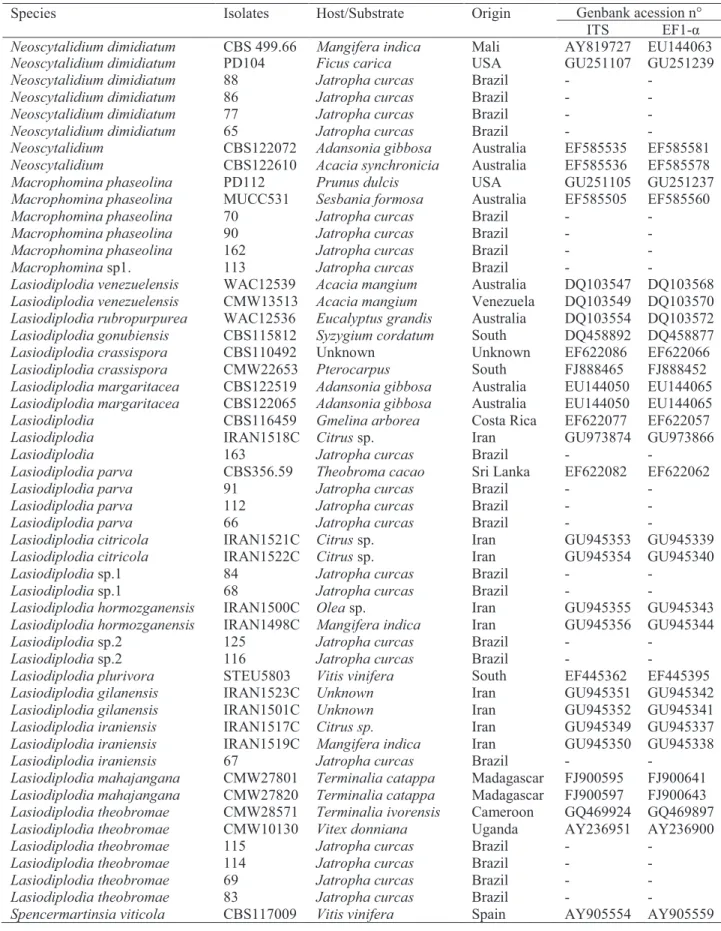
Table 1 Isolates from GenBank and this study used in the phylogenetic analyses.
Species Isolates Host/Substrate Origin Genbank acession n°
ITS EF1-α Neoscytalidium dimidiatum CBS 499.66 Mangifera indica Mali AY819727 EU144063
Neoscytalidium dimidiatum PD104 Ficus carica USA GU251107 GU251239
Neoscytalidium dimidiatum 88 Jatropha curcas Brazil - -
Neoscytalidium dimidiatum 86 Jatropha curcas Brazil - -
Neoscytalidium dimidiatum 77 Jatropha curcas Brazil - -
Neoscytalidium dimidiatum 65 Jatropha curcas Brazil - -
Neoscytalidium CBS122072 Adansonia gibbosa Australia EF585535 EF585581
Neoscytalidium CBS122610 Acacia synchronicia Australia EF585536 EF585578
Macrophomina phaseolina PD112 Prunus dulcis USA GU251105 GU251237
Macrophomina phaseolina MUCC531 Sesbania formosa Australia EF585505 EF585560
Macrophomina phaseolina 70 Jatropha curcas Brazil - -
Macrophomina phaseolina 90 Jatropha curcas Brazil - -
Macrophomina phaseolina 162 Jatropha curcas Brazil - -
Macrophomina sp1. 113 Jatropha curcas Brazil - -
Lasiodiplodia venezuelensis WAC12539 Acacia mangium Australia DQ103547 DQ103568 Lasiodiplodia venezuelensis CMW13513 Acacia mangium Venezuela DQ103549 DQ103570 Lasiodiplodia rubropurpurea WAC12536 Eucalyptus grandis Australia DQ103554 DQ103572 Lasiodiplodia gonubiensis CBS115812 Syzygium cordatum South DQ458892 DQ458877
Lasiodiplodia crassispora CBS110492 Unknown Unknown EF622086 EF622066
Lasiodiplodia crassispora CMW22653 Pterocarpus South FJ888465 FJ888452 Lasiodiplodia margaritacea CBS122519 Adansonia gibbosa Australia EU144050 EU144065 Lasiodiplodia margaritacea CBS122065 Adansonia gibbosa Australia EU144050 EU144065
Lasiodiplodia CBS116459 Gmelina arborea Costa Rica EF622077 EF622057
Lasiodiplodia IRAN1518C Citrus sp. Iran GU973874 GU973866
Lasiodiplodia 163 Jatropha curcas Brazil - -
Lasiodiplodia parva CBS356.59 Theobroma cacao Sri Lanka EF622082 EF622062
Lasiodiplodia parva 91 Jatropha curcas Brazil - -
Lasiodiplodia parva 112 Jatropha curcas Brazil - -
Lasiodiplodia parva 66 Jatropha curcas Brazil - -
Lasiodiplodia citricola IRAN1521C Citrus sp. Iran GU945353 GU945339
Lasiodiplodia citricola IRAN1522C Citrus sp. Iran GU945354 GU945340
Lasiodiplodia sp.1 84 Jatropha curcas Brazil - -
Lasiodiplodia sp.1 68 Jatropha curcas Brazil - -
Lasiodiplodia hormozganensis IRAN1500C Olea sp. Iran GU945355 GU945343
Lasiodiplodia hormozganensis IRAN1498C Mangifera indica Iran GU945356 GU945344
Lasiodiplodia sp.2 125 Jatropha curcas Brazil - -
Lasiodiplodia sp.2 116 Jatropha curcas Brazil - -
Lasiodiplodia plurivora STEU5803 Vitis vinifera South EF445362 EF445395
Lasiodiplodia gilanensis IRAN1523C Unknown Iran GU945351 GU945342
Lasiodiplodia gilanensis IRAN1501C Unknown Iran GU945352 GU945341
Lasiodiplodia iraniensis IRAN1517C Citrus sp. Iran GU945349 GU945337
Lasiodiplodia iraniensis IRAN1519C Mangifera indica Iran GU945350 GU945338
Lasiodiplodia iraniensis 67 Jatropha curcas Brazil - -
Lasiodiplodia mahajangana CMW27801 Terminalia catappa Madagascar FJ900595 FJ900641
Lasiodiplodia mahajangana CMW27820 Terminalia catappa Madagascar FJ900597 FJ900643 Lasiodiplodia theobromae CMW28571 Terminalia ivorensis Cameroon GQ469924 GQ469897
Lasiodiplodia theobromae CMW10130 Vitex donniana Uganda AY236951 AY236900
Lasiodiplodia theobromae 115 Jatropha curcas Brazil - -
Lasiodiplodia theobromae 114 Jatropha curcas Brazil - -
Lasiodiplodia theobromae 69 Jatropha curcas Brazil - -
Lasiodiplodia theobromae 83 Jatropha curcas Brazil - -