1994, 62(4):1465. Infect. Immun.
Moraes, L R Travassos and J D Lopes
A P Vicentini, J L Gesztesi, M F Franco, W de Souza, J Z de
leads to enhancement of fungal pathogenesis.
laminin through surface glycoprotein gp43
Binding of Paracoccidioides brasiliensis to
http://iai.asm.org/content/62/4/1465
Updated information and services can be found at:
These include:
CONTENT ALERTS
more»
cite this article),
Receive: RSS Feeds, eTOCs, free email alerts (when new articles
http://journals.asm.org/site/misc/reprints.xhtml
Information about commercial reprint orders:
http://journals.asm.org/site/subscriptions/
To subscribe to to another ASM Journal go to:
on January 22, 2014 by UNESP - Universidade Estadual Paulista
http://iai.asm.org/
Downloaded from
on January 22, 2014 by UNESP - Universidade Estadual Paulista
http://iai.asm.org/
INFEcrIONANDIMMUNITY,Apr. 1994,p. 1465-1469 0019-9567/94/$04.00+0
CopyrightX) 1994, AmericanSocietyforMicrobiology
Binding of
Paracoccidioides brasiliensis
to
Laminin
through
Surface
Glycoprotein gp43
Leads
to
Enhancement
of Fungal Pathogenesis
ADRIANA P. VICENTINI,' JEAN-LUC GESZTESI,' MARCELO F.FRANCO,2 WANDERLEY DESOUZA,3
JANEZ. DEMORAES,' LUIZR.TRAVASSOS,' AND
JOSE'
DANIELLOPES1*Disciplina de Biologia Celular, Escola Paulista de Medicina, 04023-062 Sdo Paulo, SP,' Departamento de Patologia, Faculdade de Medicina, UNESP, 18600 Botucatu, SP,2and Laborat6rio de Ultraestrutura CelulareMicroscopia
Eletr6nica, Institutode
Bioflsica
Carlos Chagas Filho, UFRJ, 21941,Riode Janeiro, RJ,3BrazilReceived20September 1993/Returned for modification 15 October 1993/Accepted 7January 1994
Extracellular matrix protein laminin binds specifically to yeast forms of Paracoccidioides brasiliensis and enhances adhesion of the fungus to the surface of epithelial Madin-Darby canine kidney cells in vitro. Immunoblotting of fungal extracts showed that thegp43 glycoprotein is responsible for adhesion. Thiswas confirmedby bindingassaysusing purified gp43,withaKdof 3.7 nM. ThecoatingofP.brasiliensisyeast forms
with laminin beforeinjectioninto hamstertesticlesenhanced thefungus virulence, resultingina fasterand more severegranulomatousdisease.Theseresultsindicatethatinteraction offungiwith extracellularmatrix elements may constitute a basis for the evolution of fungal infection toward regional spreading and dissemination.
Paracoccidioidomycosis isasystemic mycosis caused by the
dimorphic fungus Paracoccidioides brasiliensis withaspectrum
of clinical forms. They can be severe, with an important
mortalityrateinthe absence of specific therapy. It is assumed from experimental data that inhaled fungal propagules reach
thelung,transform intoyeastcells,andultimatelydisseminate
todistantorgans (20). Also,overtdiseaseusuallyoccursinthe
immunologically compromisedhost(8).Few other factors that could influence pathogenesis (24), besides the host
immuno-logicalresponse, havesofar been addressed.
Interaction with extracellular matrixproteinshas been cor-related with the invasiveabilityof different cells(15, 31).It has been shown that recognition of laminin may influence the
pathogenesisof severalmicroorganisms (17, 25,26, 29). Lami-nin is aglycoprotein of 850 kDa, present in basement mem-branes (12, 31), with the capacity to promote cell adhesion, differentiation, shape, and motility (9, 10, 12, 19). Specific
receptorsfor lamininpresentonthe surface of normal (5, 11) as well as tumor (1, 16) cells or pathogenic microorganisms
(17, 25, 29) have been held responsiblefor these functions. To check thepossibleeffect of lamininonthe in vivo and in vitro behavior of P. brasiliensis, mouse laminin was used to
promote adhesion to a Madin-Darby canine kidney cells
(MDCK) monolayer and to enhance the pathogenesisof the
fungus in the hamster model. We herein report that the
specific bindingof thegp43surfaceproteinof P. brasiliensisto
lamininiscorrelated with thefungus's adhesiveness invitroas well as to the enhancement of its pathogenesis, using the hamster testicle model.
P. brasiliensis B-339 (kindly provided byA. Restrepo, Cor-poracionparaInvestigaciones Biologicas,Medellin, Colombia) and 18(akindgiftfrom V.Calich, Sao PauloUniversity)were
maintained by frequent subculturing on Sabouraud glucose
*Corresponding author. Mailing address: Disciplina
de Biologia
Celular-EscolaPaulista deMedicina, RuaBotucatu862, 8o.andar, 04023-062,VilaClementino, Sao Paulo, SP,Brazil. Phone: 55 11 571 9548. Fax: 55 11549 2127.
agar (Difco Laboratories). Yeast forms of the fungus were obtained in the same mediumat 35°Cand subcultured every third day. All experiments described below were performed
with bothfungal strains. MDCK cellswerecultivatedin RPMI 1640 mediumsupplementedwith10%bovineserumat37°Cin anatmosphereof 95% air-5%CO2until confluence. Cultures wereprepared eitheron 13-mm-diameterglass coverslips,for electronicmicroscopy (EM),orinwells of 96-wellpolystyrene plates (Costar), forbinding assays.
P. brasiliensis cell-free antigen was prepared as described
previously (2). Protein concentration of the cell-free antigen
wasmeasuredbythe Bradford method(3).Thegp43secreted
bythe fungusyeastcellswas purifiedas previouslydescribed
(22). Laminin was purified from Engelbrecht-Holm-Swarm mousetumorcells(13). Polyclonalantilamininantibodieswere obtained by intramuscular injection of rabbits with purified
laminin incorporated in complete Freund's adjuvant, for the first injection, and in incomplete Freund's adjuvant, for the
subsequent injections, at monthlyintervals. Rabbit sera were tested and titrated by conventional enzyme immunoassay.
Purified laminin was labeled with 1251 (Amersham) by the lodoGen method(7)toaspecific activityof 14.7 p.Ci(1 Ci=
37GBq) ,ug1.
Tomeasureadhesion offungitoMDCK cellsurfaces,yeast
forms of P. brasiliensis were washed twice by centrifugation with phosphate-buffered saline (PBS), adjustedto 106 forms per ml, and preincubated with different concentrations of laminin in PBS for 30 min at room temperature. For the
bindingassays,wholeyeastformswerepreviouslylabeled with
1251bythelodoGenmethod. Thefungiwerethen addedtothe MDCK cultures and incubated overnight. Cultures were washed three times with PBS to remove nonadherent fungi.
Plastic wells were cut and counted in a Packard Gamma
Counter, and the glass coverslips were prepared for EM by
fixation with 2.5% (vol/vol) glutaraldehyde in PBS. For
scan-ning EM,cellsadheringtoglass coverslipswerefixed in2.5%
glutaraldehydeinPBS,postfixedwith1%OS02, dehydratedin
ethanol,driedatthe criticalpointwithC02,coated withgold,
andobserved in aJEOL 25SII scanningelectronmicroscope.
1465
Vol. 62, No. 4
on January 22, 2014 by UNESP - Universidade Estadual Paulista
http://iai.asm.org/
1466 NOTES
Reagents added
P.b.
P.b. + LN(10 jg)
P.b. +LN (20jig)
-I
:0
0-i
-H--777
0 10 20 30 40 50
cpmbound, 103
FIG. 1. Binding of1251I-labeledyeast forms of P. brasiliensis (P.b.)
to MDCK cellmonolayers.Yeast forms of P. brasiliensis were iodine
labeled by the IodoGen method and addedtoMDCKcellmonolayers grown in wells of polyvinyl plates. Afterincubationandwashings, wells were cut and counted in a Packard Gamma Counter. Binding was increasedby theaddition ofmouse laminin(LN)at10 and 20p.g/mlin PBS.
A dot blot apparatus (Bio-Rad) was used to evaluate the binding of
1251-labeled
laminin topurifiedgp43. Fiftymicroli-tersofa 1-,ug/ml solution of theproteinwasappliedontothe wells with a nitrocellulose membraneatthe bottom. The assay
wasperformed accordingtothe manufacturer's instructions, by
addingdifferent concentrationsof
1251I-labeled
laminin, inthe presence or absence of 100-fold unlabeled laminin. After washings with PBS containing 0.05% (vol/vol) Tween 20(Sigma Chemical Co.), the membrane was cutand thepiece
correspondingtoeach wellwascounted in aPackardGamma Counter.
Cell-free antigen was used for electrophoretic analysis.
Sodium dodecyl sulfate-polyacrylamide gel electrophoresis (SDS-PAGE) and immunoblotting were performed as de-scribedelsewhere (17, 32).
To investigate the influence of laminin in the hamster infections, 3-month-old male hamsters, obtained from the Animal Farm, UNESP Botucatu, Brazil,were divided in two
groups ofeight animals each and injected in the left testicle with106 viable forms ofP. brasiliensisinthe presence (30-min
preincubation in PBS) or absence of 20
jig
of laminin. The animals weresacrificed after2 and 4weeks ofinfection, and testicleswereremoved,fixed in 10% formalin inPBS, embed-ded inparaffin, cutin 4-mmsections, andstained withhema-toxylin-eosin. Extension of infectionwasevaluatedby
measur-ing the testicularareaoccupied byP.brasiliensisgranulomasin
two 200x microscopic fields and then measuring the area
occupied by four granulomas per fieldby usingtheMini-mopp system(Kontron Bildanalyse; ImageAnalysis Systems,Eching, Germany) coupledto astandard Zeiss microscope(Germany).
Statistical evaluation of the area occupied by P. brasiliensis granulomasinthe hamster testicles for both groups of animals
wasdoneby the Kruskal-Wallis test.
Previoustreatmentoffungal yeast cells with laminin greatly increase their adhesion to MDCK cells. This adhesion was showntobe dosedependent inbindingassays(Fig. 1). These resultswere confirmed by scanning EM, which showed that very few fungal forms adhered to the MDCK cells in the
absence of laminin, whereas large clusters of yeast tightly bound to the MDCK cellswere observed when laminin was
added tothe culture (notshown).
a-~~~
'E-
1
1
W
C.)
0 0~~~
65 130
Laminin
added
(cpm,
104)
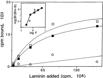
FIG. 2. Bindingof "25I-labeledmouse laminin topurified gp43 ofP.
brasiliensis. Binding was saturable (OI),total binding; 0,nonspecific
binding; *,specific binding), withaKdof3.7 nM and n = 1.09, as
measuredbyHillanalysis(insert).Results refer to the bphaseof the
curve.
Binding of laminin to purified gp43wasspecific and
satura-ble(Fig. 2).When the data obtainedwereplottedaccordingto
Scatchard (not shown), a bell-shaped curve was observed,
suggesting positive cooperativity (14). Hill analysis, which foresees thatpossibility (18)and inwhich resultsareplottedas
log [BI(Bm - B)] versus log F, where Bm is the maximal binding capacity and F is the amount of free ligand at
equilibrium,wasthen used. Anestimated Kd of 3.7 nM, withn
= 1.09,wasobtained. The aphase of the curveis
nonanalyz-ableand could representanartifact. The bphaseof thecurve
shows a correlation ofr = 0.98(Fig. 2, b, insert). Bindingof laminin towhole yeast forms ofP. brasiliensis showedspecific
binding in the same nanomolar range (not shown), but the number ofbinding sites found per cell(15 x
104)
maynotbe representative since large amounts of gp43 are secreted intoA B C kDa
U ~~43
FIG. 3. SDS-PAGE andimmunoblottingof cell-freeantigenfrom
P.brasiliensis. Wholecell-freeantigen (lane A;SDS-PAGE and silver staining) was transferred electrophoretically to nitrocellulose
mem-branes and sequentially treated with purified laminin, rabbit anti-lamininantibody,andperoxidase-conjugated anti-rabbit
immunoglob-ulin G(lane B). Reactionsweredevelopedwith4-chloro-1-naphthol.
Inthecontrolreaction(laneC),lamininwasomitted andnoprotein
bandwasrevealed.
INFEcr. IMMUN.
on January 22, 2014 by UNESP - Universidade Estadual Paulista
http://iai.asm.org/
NOTES 1467
VllP E S.. t ~ r-- .*
FIG. 4. Histology of hamster testicles after injection of P. brasiliensis.Hamstertesticleswereinjected with 10" viable yeast forms ofP.brasiliensis inthe presenceorabsence of 20 ,ug ofmouselaminin. Animalsweresacrificed after4weeks ofinfection, and testicleswereremoved, fixed, cut, andstained withhematoxylin-eosin.Thenumber and extension ofgranulomasweregreatly increased by the addition of laminin(b) comparedwith theinfection seenin the absence of laminin (a). Magnification, x40.
theculture medium. Indirect immunoblot analysisof P.
brasil-iensistotal cell-free antigen showed that lamininstrongly and
specifically bound togp43, asrevealed withantilaminin rabbit serum (Fig. 3).
Histological analysis of infected hamster testicles showed
that tissue destruction and replacement by P. brasiliensis granulomas,after 2 weeksof injection,wasmuch more intense
in thegroupinfectedwith yeast cells coated with laminin (Fig. 4b) than in the group injected with the fungusonly (Fig.4a).
Similar results were observed after4 weeks of injection (not
shown). Evaluation ofthe testicular area occupied by granu-lomas in both animal groups (four hamsters per group) for infections of 2 and 4 weeks demonstrated that infection with
fungipretreated with lamininwassignificantlymore extensive
than thatwith untreatedfungus (P < 0.05; not shown).
Themechanismsinvolvingthepathogenesis ofP. brasiliensis
in humans are not well understood. Since the infection may
disseminate to different organs and tissues, the ability of the
fungusto adhere toand escape the compartmentlinedby the basal membrane is admitted.Experimentsweredoneto
deter-mine whether interaction of the fungus with laminin could influencethe pathogenesis of P. brasiliensis.
Affinity ofthe fungusfor laminin could be demonstratedby
theenhancement ofyeastadhesionto amonolayerof MDCK
cells, asobserved byquantitative bindingassays and scanning
EM.These resultsaresimilartothosepreviouslydescribed for Trichomonas vaginalis and Trichomonas foetus (25). Their
adhesiontoand subsequentdisruptionoftheepithelial mono-layerwere greatly enhanced by the addition of laminin tothe
system. Since previous findings suggested that the increased VOL.62, 1994
on January 22, 2014 by UNESP - Universidade Estadual Paulista
http://iai.asm.org/
1468 NOTES
adhesion produced by laminin was not dependent on electro-static interactions (25), the present results pointed to the existence of laminin-binding proteins on the fungal surface.
To investigate this possibility, cell-free antigen of P.
brasil-iensiswas analyzed by immunoblotting. A single 43-kDa
gly-coprotein could be detected after incubationwithlamininand
antilaminin serum. This receptor protein, although biologically
similar to others already described, differs in size, since the laminin receptors are generally in the 50- to 70-kDa range, as
shown in Staphylococcus aureus, fibrosarcomas, macrophages,
polymorphonuclearcells(17),and epithelial cancers(31). Our data on the binding of labeled laminin topurifiedgp43 showed that the affinityof the protein for lamininappears tobe inthe
same nanomolar range. It should be noted that gp43 is a major antigenic component of P. brasiliensis. This molecule has
already been purified and characterized in our laboratory (21-23, 27) in its native and deglycosylated forms. It is found on the surface of the fungusand isalsosecreted in measurable
amounts into the culture medium (2, 27). Its recognition by sera of most patients with paracoccidioidomycosis has led
some groups to develop diagnostic assays for the disease that use the gp43 as the sole antigen, with detectable increase in
specificity and sensitivity (2, 6, 23, 30). The spontaneous
secretion of gp43 alsooccurs in vivo andcould act as a fungal
evasion mechanism againstthe host'simmune response. Thus,
the secreted antigen could bind to the host's antibodies,
leaving thecell-bound gp43 free tobind toextracellular matrix
laminin,thereforeincreasingthe ability of the fungus to invade
and disseminate to distant organs and tissues.
Laminin is known to change the metastatic behavior of
cancer cells, increasing their invasivenessafterbinding to their
specific surfacereceptors (15,28). The same seems to happen
with some parasites like T. vaginalis and T foetus, whose
adherence and virulence to the subjacent epithelial cells is greatly increased in thepresence oflaminin(25). To determine
whether laminin could influence P. brasiliensis behavior in a
similar way, weinjectedthefungusintohamster testicles in the
presence and intheabsence of laminin. In this in vivo model
for the study offungalpathogenicity,ourresultsconfirmed the
assumption that addition oflaminin to the system causes the
infection to be much more intense and severe, with much
largerareas ofthe testicles being occupied by fungal
granulo-mas. To explain the failure of macrophages in arresting the
infection (4, 8), one can assume that because these cells
express laminin receptors (11), as well ascomplement recep-tors,which are themselves activated by P. brasiliensis, ingestion oflaminin-coated fungi could be enhanced in such a way as to
override their ability of fungal intracellular destruction. Since
gp43was theonly detectable P. brasiliensis protein specifically
binding laminin, it probably can act as a receptor for the
extracellular matrix protein, thus inducing intracellular changesafterthe binding. The nature of these changes are not
yetdefined, but they seem sufficient to greatly increase fungal pathogenicity in the hamster model and could be one of the
factorsresponsible for the ability of the fungus to disseminate
in the humanhost.
We are indebted toCelia R. W. Carneiro and Roger Chammas for helpful discussions, Creuza Rosa de Oliveira for technical assistance, Marcia L. Sturaro for typing the manuscript, M. Cristina F. I. de Mattos for photographic documentation, and Denise Fecchio for statistical analysis.
Thiswork was supported by Fundacao de Amparo a Pesquisa do Estado deSaoPaulo (FAPESP) and Programa de Apoio a Ciencia e Tecnologia (PADCT-CNPq).
REFERENCES
1. Barsky, S. R., C. N. Rao, D. Hyams, and L. A. Liotta. 1984. Characterization of a laminin receptorfromhuman breast
carci-nomatissue. Breast Cancer Res. Treat. 4:181-188.
2. Blotta, M. M. S. L., and Z. P. Camargo. 1993. Immunological
response to cell-free antigens of Paracoccidioides brasiliensis:
relationship with clinical forms ofparacoccidiodomycosis. J. Clin. Microbiol. 31:671-676.
3. Bradford, M. M. 1976. A rapid and sensitive method for the quantification of microgram quantities of protein utilizing the principle of protein-dye binding. Anal. Biochem.72:248-254. 4. Brummer, E., E. Castaneda, and A. Restrepo. 1993.
Paracoccid-ioidomycosis: anupdate. Clin. Microbiol.Rev. 6:89-117. 5. Bryant,G.,C. N. Rao, M. Brentani, W. Martins, J. D. Lopes, S. E.
Martin, L. A. Liotta, and E. Schiffman. 1987. A role for the laminin receptor in leukocyte chemotaxis. J. Leukocyte Biol.
41:220-227.
6. Camargo, Z. P., C. P. Taborda, E. G. Rodrigues, and L. R.
Travassos. 1991. The use of cell-free antigenofParacoccidioides brasiliensis in serological tests.J. Med. Vet.Mycol.29:31-38. 7. Fraker, J. P., and J. C. Speclk 1978. Protein andcellmembrane
iodinations with a sparingly soluble chloroamide
1,3,4,6-tetra-chloro 3a,6a-diphenylglycoluril. Biochem. Biophys. Res. Com-mun. 80:849-857.
8. Franco, M. 1986. Host-parasite relationships in
paracoccidiodo-mycosis. J. Med. Vet. Mycol. 25:5-18.
9. Gold, L. I., and E.Pearistein. 1980. Fibronectin collagenbinding
and requirementduring cellularadhesion. Biochem. J. 186:551-559.
10. Grover, A., G. Andrews, and E. D. Adamson.1983.Role oflaminin
inepitheliumformationby F9 aggregates. J. Cell Biol. 97:137-144. 11. Huard, T. K., H. L. Malinoff, and M. S. Wicha. 1986.Macrophages
express a plasma membrane receptor for basement membrane
laminin. Am. J. Pathol. 123:365-370.
12. Kleinman, H. K., F. B. Cannon, G. W. Laurie, J. R. Hassel, M. Aumailley, V. P. Terranova, G. R. Martin, andM.Dubois-Daleq.
1985. Biological activities oflaminin.J.Cell.Biochem. 27:317-325. 13. Kleinman, H. K., M. L. McGarvey, L. A.Liotta, P. G.Robey,K.
Tryggvason, and G. R. Martin. 1982. Isolation and
characteriza-tion oftype IV procollagen, laminin andheparan sulfate proteo-glycan from the EHS sarcoma. Biochemistry21:6188-6193. 14. Levitzki, A. 1984. Receptors-aquantitative approach. The
Ben-jamin/Cummings Publishing Co. Inc.,MenloPark, Calif.
15. Liotta, L. A. 1986. Tumor invasion and metastases-role ofthe extracellular matrix: Rhoads Memorial Award Lecture. Cancer Res.46:1-7.
16. Liotta, L. A., C. N. Rao, and S. H. Barski. 1983. Tumor invasion
and theextracellular matrix. Lab. Invest.49:636-649.
17. Lopes, J. D., M. dos Reis, and R. R. Brentani. 1985. Presence of laminin receptors inStaphylococcus aureus. Science 229:275-277. 18. Marquezini, M. W., R. Chammas, S. Sonohara, and M. M. Brentani. 1991.Multiple laminin receptors-aradioligandbinding
approach. Mem. Inst. Oswaldo Cruz86(Suppl. III):119-120.
19. McCarthy, J. B., S. L. Palm, and L. T. Furcht. 1983. Migration of
heptotaxis of a Schwann cell tumor line to the basement
mem-brane glycoproteinlaminin. J. Cell Biol. 97:772-777.
20. McEween, J. G., V. Bedoya,M.M.Patino, M. E. Salazar,and A. Restrepo. 1987. Experimental murine paracoccidiodomycosis in-duced by the inhalation ofconidia. J. Med. Vet. Mycol. 25:165-175.
21. Puccia, R., S. Schenkman, P. A. J. Gorin, and L. R. Travassos.
1986. Exocellular components of Paracoccidioides brasiliensis: identification of a specificantigen. Infect. Immun. 53:199-206. 22. Puccia, R., and L. R.Travassos. 1991. The 43-kDa glycoprotein
from the human pathogen Paracoccidioides brasiliensis and its deglycosylated form: excretion and susceptibility to proteolysis.
Arch. Biochem. Biophys. 289:298-302.
23. Puccia, R., andL. R. Travassos. 1994.43-kilodalton glycoprotein
fromParacoccidioides brasiliensis: immunochemical reactions with serafrom patients withparacoccidiodomycosis, histoplasmosisor
Jorge Lobo's disease. J. Clin. Microbiol. 29:1610-1615.
24. San-Bias,G., A.Restrepo, K.Clemons,D. A.Stevens,F.San-Blas,
R. Puccia, L. R. Travassos, J.I.Figueroa, A.J.Hamilton, M. A. INFECT.IMMUN.
on January 22, 2014 by UNESP - Universidade Estadual Paulista
http://iai.asm.org/
NOTES 1469
Bartholomew, T.Harada, L. Fenelon,etal.1992.
Paracoccidiodo-mycosis. J. Med. Vet. Mycol. 30(Suppl. 1):59-71.
25. Silva Filho, F., W. Souza, and J. D. Lopes. 1988. Presence of laminin-binding proteins in trichomonads and their role in adhe-sion. Proc. Natl. Acad. Sci. USA 85:8042-8046.
26. Speziale, P., M. Hook, T. Wadstrom, and R.Timpl. 1982. Binding of the basement membrane protein laminin toEscherichia coli. FEBS Lett. 146:55-58.
27. Stambuck, B. V., R. Puccia, M. L. C. de Almeida, L. R. Travassos,
and S. Schenkman. 1988. Secretion of the 43-kDa glycoprotein antigen by Paracoccidioides brasiliensis. J. Med. Vet. Mycol. 26:
367-373.
28. Switalski,L. M., H. Murchison, R.Timpl,R. Curtiss III, and M. Hook.1987.Binding of laminintooral and endocarditis strains of viridans streptococci. J. Bacteriol. 169:1095-1101.
29. Switalski, L. M., P. Speziale, M. Hook, T. Waldstrom, and R.
Timpl.1984. Binding of Streptococcuspyogenestolaminin. J. Biol. Chem. 259:3734-3738.
30. Taborda, C. P., andZ. P. Camargo. 1993. Diagnosisof
paracoc-cidioidomycosis by passive haemagglutination assay ofantibody
using a purified and specificantigen-gp43. J. Med. Vet. Mycol.
31:155-160.
31. Terranova, J. P., C. N. Rao,T.Kalebic,I.M.Margulies,and L.A.
Liotta. 1983. Lamininreceptoronhuman breastcarcinomacells.
Proc.Natl.Acad. Sci. USA 80:444-448.
32. Towbin, H., T. Staehelin, and J.Gordon. 1979. Electrophoretic transfer of proteins from polyacrylamide gels to nitrocellulose
sheets: procedure and some applications. Proc. Natl. Acad. Sci.
USA 76:4350-4354. VOL.62, 1994
on January 22, 2014 by UNESP - Universidade Estadual Paulista
http://iai.asm.org/