doi: 10.1590/0037-8682-0230-2017
Consensus
Brazilian guidelines for the clinical management of
paracoccidioidomycosis
Maria Aparecida Shikanai-Yasuda
[1], Rinaldo Pôncio Mendes
[2], Arnaldo Lopes Colombo
[3],
Flávio de Queiroz-Telles
[4], Adriana Satie Gonçalves Kono
[5], Anamaria M. M. Paniago
[6],
André Nathan
[7], Antonio Carlos Francisconi do Valle
[8], Eduardo Bagagli
[9],
Gil Benard
[10],Marcelo Simão Ferreira
[11], Marcus de Melo Teixeira
[12],
Mario León Silva-Vergara
[13], Ricardo Mendes Pereira
[14], Ricardo de Souza Cavalcante
[2],
Rosane Hahn
[15], Rui Rafael Durlacher
[16], Zarifa Khoury
[17], Zoilo Pires de Camargo
[18],
Maria Luiza Moretti
[19], and Roberto Martinez
[20]Corresponding author: Profa. Maria Aparecida Shikanai-Yasuda.
email: [email protected]
Received 01 June 2017
Accepted 30 June 2017
Abstract
Paracoccidioidomycosis is a systemic fungal disease occurring in Latin America that is associated with rural environments and agricultural activities. However, the incidence and prevalence of paracoccidiodomycosis is underestimated because of the lack of compulsory notiication. If paracoccidiodomycosis is not diagnosed and treated early and adequately, the endemic fungal infection could result in serious sequelae. While the Paracoccidioides brasiliensis (P. brasiliensis) complex has been known to be the causal agent of paracoccidiodomycosis, a new species, Paracoccidioides lutzii (P. lutzii), has been reported in Rondônia, where the disease has reached epidemic levels, and in the Central West and Pará. Accurate diagnoses and availability of antigens that are reactive with the patients’ sera remain signiicant challenges. Therefore, the present guidelines aims to update the irst Brazilian consensus on paracoccidioidomycosis by providing evidence-based recommendations for bedside patient management. This consensus summarizes etiological, ecoepidemiological, molecular epidemiological, and immunopathological data, with emphasis on clinical, microbiological, and serological diagnosis and management of clinical forms and sequelae, as well as in patients with comorbidities and immunosuppression. The consensus also includes discussion of outpatient treatments, severe disease forms, disease prevalence among special populations and resource-poor settings, a brief review of prevention and control measures, current challenges and recommendations.
Keywords: Paracoccidioidomycosis. Guidelines. Clinical management. Diagnosis. Treatment follow-up. [1]. Departamento de Moléstias Infecciosas e Parasitárias, Faculdade de Medicina, Universidade de São Paulo, São Paulo, SP, Brasil.
[2]. Departamento de Doenças Tropicais e Diagnóstico por Imagem, Faculdade de Medicina Botucatu, Universidade Estadual Paulista, Botucatu, SP, Brasil. [3]. Departamento de Medicina, Escola Paulista de Medicina, Universidade Federal de São Paulo, São Paulo, SP, Brasil. [4]. Departamento de Saúde Comunitária,
Universidade Federal do Paraná, Curitiba, PR, Brasil. [5]. Divisão de Moléstias Infecciosas, Hospital das Clínicas, Faculdade de Medicina, Universidade de São Paulo, São Paulo, SP, Brasil. [6]. Faculdade de Medicina, Universidade Federal de Mato Grosso do Sul, Campo Grande, MS, Brasil. [7]. Divisão de
Pneumologia, Hospital das Clínicas, Faculdade de Medicina, Universidade de São Paulo, São Paulo, SP, Brasil.
[8]. Instituto de Pesquisa Clínica Evandro Chagas, Fundação Oswaldo Cruz, Rio de Janeiro, RJ, Brasil. [9]. Departamento de Microbiologia e Imunologia, Instituto de Biociências, Universidade Estadual Paulista, Botucatu, SP, Brasil. [10]. Departamento de Dermatologia, Faculdade de Medicina, Universidade de São Paulo, São Paulo, SP, Brasil. [11]. Serviço de Infectologia, Faculdade de Medicina, Universidade Federal de Uberlândia, Uberlândia, MG, Brasil. [12]. Translational Genomics Research Institute, Northern Arizona University, AZ, USA. [13]. Departamento de Clínica Médica, Faculdade de Medicina, Universidade Federal do Triângulo Mineiro, Uberaba, MG. Brasil. [14]. Departamento de Pediatria, Faculdade de Ciências Médicas, Universidade Estadual de Campinas, Campinas, SP, Brasil. [15]. Núcleo de Doenças Infecciosas e Tropicais, Faculdade de Ciências Médicas, Universidade Federal de Mato Grosso, Cuiabá, MT, Brasil. [16]. Centro de Medicina Tropical de Rondônia, Porto Velho, RO, Brasil. [17]. Instituto de Infectologia Emílio Ribas, Secretaria de Estado
da Saúde de São Paulo, São Paulo, SP, Brasil. [18]. Departamento de Microbiologia e Imunologia, Universidade Federal de São Paulo, São Paulo, SP, Brasil. [19]. Departamento de Clínica Médica, Faculdade Ciências Médicas, Universidade Estadual de Campinas, Campinas, SP, Brasil.
PS2
PS3 Pl
S1a S1b
PS4
FIGURE 1 - Geographic distribution of Paracoccidioides lutzii and Paracoccidioides brasiliensis cryptic species. Modiied (update on P. lutzii distribution) from: Muñoz JF, Farrer RA, Desjardins CA, Gallo JE, Sykes S, Sakthikumar S, et al. Genome diversity, recombination, and virulence across the major lineages of Paracoccidioides. mSphere 2016; 1(5): e00213-163. PS2; PS3; PS4; S1a; S1b: phylogenetic species of Paracoccidioides brasiliensis; Pl: Paracoccidioides lutzii.
ETIOLOGY
Paracoccidioidomycosis (PCM) is caused by thermo-dimorphic fungi that currently encompasses two species:
Paracoccidoides brasiliensis (P. brasiliensis) and
Paracoccidioides lutzii (P. lutzii)1. P. brasiliensis contains a
complex of at least ive phylogenetic clusters ranked as the following phylogenetic species: S1a, S1b, PS2, PS3, and PS42,3,4. The phylogenetic species S1a and S1b are predominantly found in lower South America, especially in southeastern and southern Brazil, Argentina, and Paraguay. The PS2 species has a sporadic distribution and is less frequently reported, with human cases only being reported thus far in Venezuela and southeast Brazil (Figure 1). The PS3 and PS4 species are exclusively endemic
to Colombia and Venezuela, respectively. P. lutzii encompasses a single species and is predominantly distributed in the Central West and Amazon regions of Brazil and Ecuador1,5,6,7,8. However,
the real incidence of each phylogenetic species and its implication on clinical practice is dificult to establish because of the lack of guided studies comparing PCM forms and manifestations
with their genetic background9. Radial immunodiffusion
against the commonly used exoantigens containing a 43-kDa glycoprotein (gp43) suggests that Paracoccidiodes spp. exhibit major antigenic variability. According to phylogenetic studies, different Paracoccidioides spp. isolates are distributed in different genotypes across multiple PCM endemic areas of Latin America. In particular, Paracoccidioides spp. in central Brazil (i.e. Mato Grosso and Rondônia) exhibit a lower rate of genetic similarity. Yet, P. lutzii isolates exhibit high species-speciic antigen variability9, which has already been assessed
in proteomic studies.
ECOEPIDEMIOLOGY
In nature, P. brasiliensis and P. lutzii develop as ilamentous
structures and produce infective propagules called conidia10,11
(Figure 2). If inhaled, the propagules give rise to yeast forms of
the fungus that become parasitic to the host. Paracoccidioides
spp. can cause infection and disease in humans and domestic and wild animals, although only a few active disease cases have been observed in animals, such as dogs10,12. The armadillo is
known to be a reservoir of P. brasiliensis, and the fungus can be easily cultured from the animal’s internal organs (spleen, liver, and lymph nodes), indicating a systemic process. P. lutzii
has not yet been isolated from armadillos.
Epidemic outbreaks of PCM have never been observed. Further, fungal recovery (culture) directly from the fungi’s environmental saprophytic form has been shown to be particularly dificult to obtain with reproducibility. Thus, the region where the disease is acquired is referred to as reservaria. Yet, sensitive molecular screening techniques have detected the fungus in soils and aerosols, especially among samples taken from animal burrows or sites with medium to high moisture content protected by vegetation cover13.
In recent decades, changes in the demographic characteristics and geographical distribution of PCM incidence have been observed. These shifts could be attributed to the rise of
urbanization, application of diagnostic methods, and the presence of comorbidities and immunosuppression. In addition, environmental factors, such as the expansion of settlements, clearing of forests, and increased coffee production, could contribute to the current high levels of PCM incidence in some regions of Rondônia14.
In addition, between 1982 and 1983, a region of Southeast Brazil experienced climatic changes related to El Niño, resulting in elevated soil moisture levels and temperatures between 18-28ºC, which are favorable for fungal sporulation and aerial dispersion. During this same time period, the region experienced an outbreak of acute cases of PCM15.
How is Paracoccidioides infection acquired?
En
vir
onme
nt
Hos
ts
Coffee and sugar cane plantation / rural workers
Inhalation of arthroconidia
Armadillo (Pb) (Pl?) immunotolerant / signalling
Agriculture activities / Climate conditions (soil humidity)
Human beings
predominance of infection predisposing factors for disease
genetic gender (male) great inoculum
Yeast phase (body temperature)
Animals – infection; rare cases of disease
P. lutzii P. brasiliensis
Mendes & Bagagli
FIGURE 2 - Spread of Paracoccidioides brasiliensis and Paracoccidioides lutzii. Pb: Paracoccidioides brasiliensis; Pl: Paracoccidioides lutzii.
if clinical manifestations appeared many years later11. Most of
these patients seeked medical attention many years after they left the endemic area and resided in urban centers where were engaged in other activities unrelated to soil management. For example, smoking (> 20 cigarettes/day for > 20 years) and alcoholism (> 50g/day)16 are frequently associated with mycosis. Unlike other mycoses, such as cryptococcosis, disseminated histoplasmosis, and candidiasis, PCM is not usually related to immunosuppressive diseases. However, cases of PCM associated with HIV infection, neoplasia and, more rarely, organ transplants and use of immunobiologicals have been reported17-20.
Incidence, prevalence and mortality
Since PCM is not a compulsory notification disease, we do not have precise data on its incidence in Brazil. Mycosis prevalence, incidence, and morbidity estimates are based on reports from epidemiological surveys, case series, hospitalization records, and mortality data21. Based on the
experiences of reference services caring for patients with PCM, the disease’s incidence in endemic areas ranges from three to four new cases per one million inhabitants and one to three new cases per 100,000 inhabitants per year. About 80% of PCM cases are registered in Brazil, particularly in the States of São Paulo, Paraná, Rio Grande do Sul, Goiás, and Rondônia (Figure 3)21. In
Latin America, cases are most frequently reported in Argentina, Colombia, Venezuela, Ecuador, and Paraguay. Estimates of annual incidence in Brazil range from 0.71 to 3.7 cases per
100,000 inhabitants21. However, recent records of incidence in
Rondônia report 9.4 cases per 100,000 inhabitants, with two municipalities reporting incidences close to 40 cases per 100,000 inhabitants14. Between 1980 and 1995, the Ministry of Health documented 3,181 cases of PCM-related deaths, resulting in a PCM mortality rate of 1.45 cases per one million inhabitants (2.59 for the South, 2.35 for the Midwest, 1.81 for the Southeast, 1.08 for the North, and 0.2 for the northeast regions)22. In this
study, among all chronic infectious and parasitic disease, PCM was listed as the eighth highest cause of mortality and had the highest mortality rate among the systemic mycoses, even having a higher mortality rate than leishmaniasis. Recent data collected from 13,683 patients who were hospitalized with systemic mycoses between January 1998 and December 2006 showed that PCM accounts for the largest number of hospitalizations (49%) among all mycoses23, with emphasis on hospitalization
rates in the north and Midwest regions, without major difference in the mortality of hospitalized patients (Figure 3).
Age group and distribution between genders
PCM infection is primarily acquired in the irst two decades of life, with a peak incidence between 10 and 20 years of age. However, the presentation of clinical manifestations or evolution to disease is uncommon in this age group. Instead, PCM occurs more frequently in adults between the ages of 30- and 50-years-old as a result of endogenous latent foci reactivation10.
CW
SE
S N
NE
Acre
Rondônia
Mato Grosso
Goiás
Para
Maranhão
Tocantins
Rio de Janeiro São Paulo Paraná
Rio Grande do Sul
BRAZIL (states)
FIGURE 3 - Geographical areas of paracoccidioidomycosis endemicity in Latin America. Reproduction from: Martinez R. New trends in Epidemiology. J Fungi.
2017;3(1):121.
(■) irst recognized areas of high endemicity
(■) high endemicity observed since the last decades of the 20th century (■) areas with some recent evidence of increasing endemicity (■) areas of moderate endemicity
(■) low endemicity
( ) no cases or rare cases of paracoccidioidomycosis report in these countries or regions
age of 20-years-old, while the remaining 90% occurring later in life. Further, in childhood, the frequency of PCM cases is evenly distributed between both genders, with a slight predominance in young male adults; however, in adulthood, the frequency ranges from ten to 15 men for one woman.
IMMUNOPATHOGENESIS
Control of Paracoccidioides spp infection depends on the host´s cellular immune response, with T cells playing a prominent role. PCM has a range of clinical presentations along a spectrum,
disease severity25,26. In fact, patients with infections that evolve to the more severe forms, such as the acute/subacute form disease (A) or eventually the severe disseminated chronic form (CF), develop Th-2 and Th-9 immune response patterns that do not form compact granulomas, but instead activate B lymphocytes, high levels of speciic antibodies, including the the IgE subclass, hypergammaglobulinemia, and eosinophilia27.
Patients with the severe/disseminated unifocal or multifocal CF who bear lower fungal burdens, also exhibit deicient Th-1 responses, often at a lesser degree that that of patients with the AF or severe disseminated CF. In addition, these patients can still experience the formation of compact granulomas that can suppress, at least partially, fungal replication25,26. In these patients, the loss of Th-1 function would be partially compensated by the development of Th-17 and Th-22 responses, both of which drive intense mucosal inlammatory responses rich in neutrophils27. In fact, a characteristic feature of the CF
is the involvement of the mucosa, especially in the respiratory tract. In addition, it has been demonstrated that regulatory T cells (Tregs) suppress T cell immunity and contribute to the T cell anergy observed in the more severe forms of the disease28,29.
The factors that determine the different outcomes of the PCM host-parasite interaction remain unknown. Preliminary data suggest that the host’s immunogenetic background may play a role30. Regardless, clinical experiences have indicated
that treatment of PCM should persist for long periods until effective cellular immune responses are elicited. However, for unknown reasons, yeast cells may remain in quiescent foci that can reactivate the disease and cause relapses. Usually, immune alterations subside with treatment and the protective Th-1 responses appear/reappear. This observation is corroborated by in vitro experiments that have demonstrated deicient Th-1
responses can be reverted31, although this response reconstitution
has not been shown to reach the magnitude of that observed in healthy individuals without disease32. The role of the observed
high serum levels of speciic antibodies in any mechanism of protection could not yet be determined.
CLASSIFICATION OF CLINICAL FORMS AND ASSESSMENT OF SEVERITY
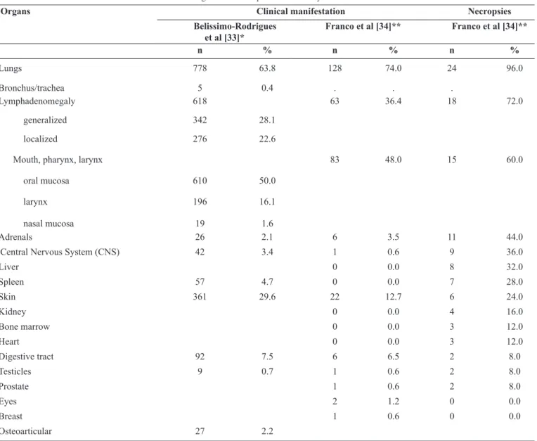
PCM can compromise any organ, apparatus or system, as revealed in Table 133,34, which presents clinical and autopsy indings. Further, PCM’s diversiication tends to hinder its classiication.
Several classiications of PCM clinical forms have been published based on different criteria, such as lesion topography, disease natural history, severity of clinical presentation, and serological reaction results. This consensus adopted the classiication presented in the
International Colloquium on Paracoccidioidomycosis held in February 1986 in Medellin, Colombia35.
I.Paracoccidioidomycosis infection II.Paracoccidioidomycosis (disease) A. Acute/subacute form (juvenile)
• - Moderate • - Severe
B. Chronic form (adult) • - Mild
• - Moderate • - Severe
III. Residual form or sequelae
Paracoccidioidomycosis infection
Paracoccidioidomycosis infection is contracted when a healthy individual comes into contact with a Paracoccidioides
spp. The infection is diagnosed by a positive intradermal reaction to speciic antigens and necropsy indings of latent fungi36.
Clinical forms of Paracoccidioidomycosis
Acute/subacute form (juvenile)
The acute/subacute form of PCM is responsible for 5-25% of cases and may be more frequent in certain endemic regions while almost never observed in others. In Brazil, this form is more commonly observed in the following States: Maranhão, Minas Gerais, Pará, Goiás, and São Paulo.
The incidence of PCM appears to be declining in some
endemic areas37. Acute/subacute PCM predominantly affects
children, adolescents, and young adults, but can occur in adults between the ages of 30- and 40-years-old. The incidence of PCM tends to be evenly distributed between genders, especially among the adolescent population33,37-39.
This clinical form of PCM rapidly evolves and disseminates the infection to multiple organs and systems. In general, patients are diagnosed within a few weeks of symptoms onset. Most symptoms involve the phagocytic-mononuclear system, including the presence of localized or generalized lymphadenomegaly, which may present suppuration, istulization, and hepatosplenomegaly. Symptoms may also include digestive manifestations, cutaneous (or mucosal) lesions, osteoarticular involvement, and rarely, pulmonary involvement. Fever, weight loss, and anorexia often accompany the clinical presentation. Intra-abdominal lymphadenomegaly may coalesce, producing tumor masses that exert compression on various organs, such as the bile duct and intestinal loops38 (Figure 4 and Figure 5).
A prominent inding of laboratory alterations in this form is peripheral eosinophilia, which occurs in 30% to 50% of cases38-40. Under certain conditions, eosinophilia may be signiicant (up to 70% of peripheral blood leukocytes).
Chronic form (adult)
Organs Clinical manifestation Necropsies
Belissimo-Rodrigues et al [33]*
Franco et al [34]** Franco et al [34]**
n % n % n %
Lungs 778 63.8 128 74.0 24 96.0
Bronchus/trachea 5 0.4 . . .
Lymphadenomegaly
generalized
localized
618 342 276
28.1 22.6
63 36.4 18 72.0
Mouth, pharynx, larynx
oral mucosa
larynx
610
196
50.0
16.1
83 48.0 15 60.0
nasal mucosa 19 1.6
Adrenals 26 2.1 6 3.5 11 44.0
Central Nervous System (CNS) 42 3.4 1 0.6 9 36.0
Liver 0 0.0 8 32.0
Spleen 57 4.7 0 0.0 7 28.0
Skin 361 29.6 22 12.7 6 24.0
Kidney 0 0.0 4 16.0
Bone marrow 0 0.0 3 12.0
Heart 0 0.0 3 12.0
Digestive tract 92 7.5 6 6.5 2 8.0
Testicles 9 0.7 1 0.6 2 8.0
Prostate 1 0.6 2 8.0
Eyes 2 1.2 0 0.0
Breast 1 0.6 0 0.0
Osteoarticular 27 2.2
TABLE 1
Organs affected in paracoccidioidomycosis.
A B C D
FIGURE 4 - Acute form of PCM in children. A. Abscesses in frontal and clavicular regions resulting from osteo-articular involvement. B. Female child presenting
with abscessed lymphatic involvement. C. Inguinal lymphoadenomegaly. D. Lymphatic-abdominal involvement with ascites and hepatosplenomegaly. PCM: paracoccidioidomycosis. Reproduction with modiication: Shikanai-Yasuda MA, Telles Filho F de Q, Mendes RP, Colombo AL, Moretti ML, Grupo de Consultores do Consenso em Paracoccidioidomicose. Consenso Brasileiro em Paracoccidioidomicose. Rev Soc Bras Med Trop. 2006;39(3):297-31039.
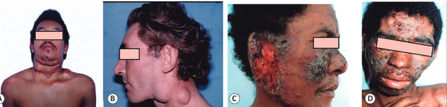
A B C D
FIGURE 5 - Patients with the acute/subacute (juvenile) form of PCM. A. Ganglionic mass in supraclavicular, cervical, and submandibular region. B.
Lymphadenomegaly of PCM, which must be differentiated from hematological diseases, such as lymphoma. C. Verrucous ulcerative lesions on the face and pavilion caused by hematogenous dissemination. D. Papulonodular ulcerative lesions caused by hematogenous dissemination. PCM: paracoccidioidomycosis. Reproduction with modiication: Shikanai-Yasuda MA, Telles Filho F de Q, Mendes RP, Colombo AL, Moretti ML, Grupo de Consultores do Consenso em Paracoccidioidomicose. Consenso Brasileiro em Paracoccidioidomicose. Rev Soc Bras Med Trop. 2006;39(3):297-31039.
A B
FIGURE 6 - Oral involvement in chronic PCM 47,48. A. Gingivostomatitis.
B. Moriform stomatitis of Aguiar-Pupo. The lesions may present as extensive, ulcers, or ulcer-vegetative, with characteristic hemorrhagic dots (moriform lesion). Although infrequent, the lesions may extend into the nasal vestibule, causing perforation of the palate and nasal septum, with inaesthetic and functional sequelae. In acute PCM, the oral mucosa is not frequently involved. PCM: paracoccidioidomycosis. Reproduction with modiication: Shikanai-Yasuda MA, Telles Filho F de Q, Mendes RP, Colombo AL, Moretti ML, Grupo de Consultores do Consenso em Paracoccidioidomicose. Consenso Brasileiro em Paracoccidioidomicose. Rev Soc Bras Med Trop. 2006;39(3):297-31039. Chronic PCM can be classiied as either mild, moderate, or
severe35. Severe cases are deined by meeting three or more of the following criteria: a) weight loss greater than 10% of the normal body weight; b) intense pulmonary involvement; c) involvement of other organs, such as adrenal glands, central nervous system, and bones; d) the presence of lymph nodes affected in multiple chains in supericial or deep, pseudotumoral form (>2.0cm in diameter, without suppuration) or suppurative form; e) high antibody titers. In fact, severe cases are represented by patients presenting clinical instability due to respiratory insuficiency, adrenal dysfunction, neurological syndrome or acute abdomen.
Mild cases, which constitute a small portion of patients, are those with weight loss below 5% of normal body weight and involvement of unique or a few organs or tissues without disfunction.
In some cases, patients present with clinical manifestations of both acute/subacute and chronic forms, making it dificult to properly classify the disease. Most of these patients present with intense suppression of cellular immunity and are labelled as having mixed form PCM43.
Residual forms (sequelae)
Residual forms, also referred to as sequelae, are clinical manifestations of anatomical and functional changes observed
FIGURE 7 - Clinical aspects of chronic PCM. A. Papulonodular ulcerative lesions on the face. B. Perioral and mentonian involvement. C. Fistulated cervical
and submandibular lymph nodes. D. Vegetative lesion with irregular borders in the perianal region. Through hematogenous dissemination, especially traumatic fungal implantation, the cutaneous lesions contiguously develop from compromised mucosa, istulated lymph nodes, or bone involvement47. The lesions are
characterized by ulcero-crusted polymorphisms, molluscoids, papules, or acneiforms, and are located primarily in the cephalic pole and perioriicial regions48.
Of note, patients with Addison's syndrome typically present with cutaneous and mucosal hyperpigmentation. PCM: paracoccidioidomycosis. Reproduction with modiication: Shikanai-Yasuda MA, Telles Filho FQ, Mendes RP, Colombo AL, Moretti ML, Grupo de Consultores do Consenso em Paracoccidioidomicose. Consenso Brasileiro em Paracoccidioidomicose. Rev Soc Bras Med Trop. 2006;39(3):297-31039.
after PCM treatment. Sequelae are observed in multiple organs, but have a higher rate of incidence in the lungs, skin, larynx, trachea, adrenals, mucosa of the upper aerodigestive tract, central nervous system, and lymphatic system, thus explaining the diversity of clinical presentation44-46.
INITIAL TREATMENT, DIAGNOSTIC APPROACH, AND OUTPATIENT FOLLOW-UP ROUTINE OF PATIENTS
WITH PCM
Since PCM is systemic, any organ can be affected. The attention of the observer should initially be directed to the general condition of the patient and then the organs and systems that are most frequently committed according to the forms of the disease presentation: acute/subacute PCM and chronic PCM. According to routine medical care, all patients should have a detailed physical examination, reporting weight and height evaluation, to allow the characterization of nutritional status.
General evaluation of a patient with acute/subacute form
In acute/subacute PCM, anamnesis and physical examination play an important role in the determination of disease severity and systemic involvement. For example, the presence of lymphadenomegaly in various lymphatic chains, hepatosplenomegaly, cutaneous lesions, or abdominal masses can be conirmed during a patient’s physical examination. In addition, clinical examination can also detect the presence of jaundice, ascites, and peripheral edema, which prompt investigation of hypoalbuminemia. In acute/subacute PCM, signs of adrenal and neurological involvement are rare. Fever, weight loss, and digestive complaints, such as abdominal pain, chronic malabsorptive diarrhea, and vomiting, are also quite frequent40. The presence of tumefaction or pain in the bone region requires the identiication of bone lesions.
Laboratory tests and imaging: Chest X-ray (posterior, anterior, proile)
Complete blood count and erythrocyte sedimentation rate (ESR) Liver biochemical tests (alanine aminotransferase (ALT), alkaline phosphatase)
Total proteins and fractions
Evaluation of renal and metabolic function (serum creatinine, Na, K)
Imaging tests, such as ultrasound, CT, magnetic resonance imaging (MRI), and scintigraphic mapping should only be performed when there is clinical suspicion or laboratory results suggestive of organ involvement that cannot be solely assessed by physical examination.
General evaluation of a patient with chronic form
In chronic PCM, anamnesis and physical examination must include the evaluation of signs and symptoms related
to pulmonary, tegumentary, and laryngeal involvement41,46
(cough, dyspnea, mucus/purulent expectoration, ulcerated
lesions of the skin and naso-oropharyngeal mucosa47,48,
odynophagia, dysphagia, and dysphonia). Both diagnostic methods must also evaluate signs and symptoms of lymphatic
adenomegaly, adrenal involvement49,50 (asthenia, weight
loss, hypotension, skin darkening, abdominal pain), central nervous system involvement51,52 (headache, motor deicit, convulsive syndrome, and alteration of behavior and/or level of consciousness), and digestive impairment (diarrhea and malabsorption syndrome)53,54 (Figure 8).
A B
C D
E F
FIGURE 8 - Images of PCM. A. Conventional butterfly wing radiological image showing bilateral, parahilar, and symmetrical pulmonary involvement with a predominance of alveolar lesions, sparing apices and lower thirds. These manifestations are very suggestive of PCM but occur infrequently. B. Diffuse and symmetric nodular and micronodular opacities. C. Computed tomography (CT) of lungs with multiple sub-pleural cavities. Pulmonary lesions may be interstitial of the thin or coarse reticular-nodular type, or, less frequently of the tumor type. D. Bilateral adrenal increase. E and F. CNS: hypodense and ring-shaped contrast enhancement images with small mass effect. PCM: paracoccidioidomycosis. Reproduction with modiication: Shikanai-Yasuda MA, Telles Filho F de Q, Mendes RP, Colombo AL, Moretti ML, Grupo de Consultores do Consenso em Paracoccidioidomicose. Consenso Brasileiro em Paracoccidioidomicose. Rev Soc Bras Med Trop. 2006; 39(3): 297-31039.
Laboratory tests and imaging
Chest X-rays (posterior, anterior, proile)
Complete blood count and erythrocyte sedimentation rate (ESR) Liver biochemical tests (ALT, alkaline phosphatase) Evaluation of renal and metabolic function (serum creatinine, Na, K)
imagining and functional tests should be performed under the guidance of medical experts. Given the high frequency of adrenal involvement and its clinical impact, patients suspected of having chronic PCM should undergo an assessment of the functional reserve when available.
Differential diagnosis
Other conditions to consider in the differential diagnosis for PCM include the following: acute lymphoma, leukemia, histoplasmosis, tuberculosis, toxoplasmosis, visceral leishmaniasis, and infectious mononucleosis. For chronic cutaneous-mucosal PCM, the conditions to consider in the differential diagnosis are cutaneous or mucosal leishmaniasis, tuberculosis, chromoblastomycosis, leprosy, sarcoidosis, lues, neoplasia and in the chronic pulmonary form, tuberculosis (Table 2)55, coccidioidomycosis, histoplasmosis, sarcoidosis, pneumoconiosis, and interstitial pneumonitis. For digestive
Epidemiology Tuberculosis Paracoccidioidomycosis
Age Wide range Restricted (30- to 60-years-old)
Gender Indistinct Prevalent in males (15:1)**
Incidence 45/100,000 1-3/100,000
Mortality 2.3/100,000 1.65/1,000,000
Geographic distribution Worldwide, more urban Latin America, more rural
Microbiology
etiologic agent Mycobacterium tuberculosis Paracoccidioides spp. source of infections Man/animal Soil
infection Contagious Non-contagious
cultivation Fastidious Fastidious
Clinical aspects
signs and symptoms Well deined Non-speciic
weight loss ++/++++ ++/++++
fever ++++ +/-***
cough ++++
+/-hemoptoic sputum +++/++++
+/-pleural involvement Yes No
association PCM (10-15%) TB (10-15%)
Radiology
image localization Predominance in upper zone Predominance in medium thirds, bilateral, and diffuse zones
cavities +++/++++ ++/++++
pleural images Yes No
dissemination Uni/multifocal Uni/multifocal
Laboratorial changes
red cells Normocytic normochromic anemia Normocytic normochromic anemia white cells Leukocytosis/leukopenia Leukocytosis/leukopenia
ESR +++/++++ +/++
serum proteins Normal/low Normal/low
Natural evolution +++ +++
consumption Yes Yes
anergy Yes Yes
death Yes Yes
* Modiied from: Queiroz-Telles F and Escuissato D. Pulmonary paracoccidioidomycosis. Semin Respir Crit Care Med. 2011;32:764-7455.**Pneumonia may occur
rarely in acute/subacute PCM. In these cases, both sexes can be affected; ***Fever may occur in patients with associated infections. PCM: paracoccidioidomycosis; TB: tuberculosis; ESR: erythrocyte sedimentation rate.
TABLE 2
Pulmonary involvement: differences between tuberculosis and paracoccidioidomycosis*.
PCM, conditions to consider in the differential diagnosis are tuberculosis and Chron’s disease, while for forms of PCM that affect the central nervous system, the conditions are tuberculosis, cryptococcosis, cysticercosis, and neoplasias.
Laboratory examinations for speciic diagnosis
The identiication of Paracoccidioides spp. through the examination of fresh sputum or other clinical specimens, such as lesion sample, lymph node aspiration, or biopsy fragment, is the gold standard for PCM diagnosis.
Aspects of PCM laboratory diagnosis are presented in
Figure 9.
In an attempt to standardized PCM diagnosis, the following deinitions are offered:
diseases that occur with a similar condition, for at least four weeks:
• Cough with or without sputum and dyspnea • Sialorrhea, odynophagia, or hoarseness • Lesion (ulcerated) in the nasal or oral mucosa
• Skin lesions (ulcers, vegetation, nodules, plaques, etc.) • Cervical or generalized adenomegaly, with or without
suppuration and istulization.
• Child or young adult with hepatosplenomegaly and/or abdominal tumefaction
Probable case: a patient with clinical manifestations compatible with PCM and anti-P. brasiliensis/P. lutzii serum antibody titers detected preferably by quantitative double immunodiffusion test or counterimmunoelectrophoresis.
Conirmed case: Patient with clinical manifestations compatible with PCM with secretions, bodily luids, or lesion material presenting with fungal elements suggestive of P. brasiliensis/P. lutzii infection (Figure 9). Note, the
micromorphology of P. brasiliensis/P. lutzii parasitic forms in the biological material of infected patients cannot distinguish between the two species. Therefore, the identiication of the involved species requires culture isolation and application of molecular techniques56,57.
A B
C D
E F
FIGURE 9 - Laboratory diagnosis of paracoccidioidomycosis. A and B. Cultivation of P. brasiliensis. A. mycelial phase. B. yeast phase. C. Fresh examination in KOH. D. Lactophenol staining showing yeast cells with multiple buds. E. Histological section stained by the Grocott method. F. Histological section stained by PAS. KOH: Potassium hydroxide; PAS: Periodic acid - Schiff stain. Reproduction with modiication: Shikanai-Yasuda, MA, Telles Filho F de Q, Mendes RP, Colombo AL, Moretti ML, Grupo de Consultores do Consenso em Paracoccidioidomicose. Consenso Brasileiro em Paracoccidioidomicose. Rev Soc Bras Med Trop. 2006;39(3):297-31039.
Critical evaluation of serological tests: serologic diagnosis and follow-up
Speciic serological tests are important in not only the diagnosis of PCM, but also in the assessment of host response to speciic treatments. Currently, double immunodiffusion (DID), counterimmunoelectrophoresis (CIE), immunoenzymatic assays (ELISA), and immunoblots (IB) are the serological tests available in different reference services58-60.
These tests use standardized techniques and adequate
antigens61, and display a sensitivity between 80% and 95%.
The titer of speciic anti-P. brasiliensis antibodies58,59,60 correlates with the severity of the clinical forms, with higher levels detected in the acute/subacute and disseminated forms. In cases of PCM caused by P. lutzii, such information remains unknown, which has motivated multicenter studies in endemic areas. PCM cases with false negative results from any of the previously mentioned tests are most often associated with very localized lesions and hosts with AIDS or immunodepressive conditions. Antigens prepared from P. brasiliensis that are rich in gp43KDA62 have excellent accuracy in the diagnosis
of P. brasiliensis infections58-60 , but have a low sensitivity in the diagnosis of P. lutzii infections63,64. These serological tests display a speciicity between 85% and 100%, with immunodiffusion having the highest rate of speciicity. False-positive reactions may occur in sera from patients with histoplasmosis, and eventually aspergillosis and leishmaniasis; however, gel immunodiffusion provides the highest rate of speciicity for these conditions.
Currently, the main method of PCM serological diagnosis is double agar gel immunodiffusion (DID) because of its simplicity, cost-effectiveness, sensitivity (>80%), speciicity (>90%), and extensive application over the last decades. For DID, or any other test used in the diagnosis of PCM, serums should be titrated to increase the accuracy of therapeutic response interpretation because antibody titers progressively decrease with successful clinical control. To meet serological cure criteria, negative or stabilization results at a dilution of 1:2 or less should be achieved. In certain cases, patients may already have titers below 1:4 upon diagnosis; therefore, the serological criteria will have limited value during treatment follow-up. Additional resources and techniques for PCM diagnosis have been developed, but they are not available for PCM routine assessment. These examinations include the immunoblot technique, the triage ELISA test, the detection of speciic antigens and PCR65,66.
To date, there are no validated serological techniques for the accurate diagnosis of infection by P. lutzii. Unfortunately,
there is no commercial system available for PCM diagnosis,
and all available tests are based on systems developed in
Frequency of outpatient visits and completion of examinations
During the irst 3 months of infection, monthly consultations are recommended to optimize patient adherence to the established regimen, to assess drug tolerability, and to ensure good clinical response (Table 3). If a satisfactory
clinical response is observed, the consultations should become quarterly until the end of the irst year. After 90 days of follow-up if a satisfactory clinical response is observed, patients should undergo complete blood count and biochemical tests every 3 months during the irst year. Radiological and serological examinations should be requested every 6 months, or at shorter intervals if a satisfactory clinical response is not observed or if laboratory results indicate no change in activity. The reduction of speciic antibody titers should occur approximately 6 months after treatment and should be either negative or stabilized at low titers for approximately 10 to 24 months after treatment. These estimations depend on the
Examinations 1st medical
appointment
1st, 2nd,
3rd month
6th, 9th,
12th monh
18th,
24th
month
≥ 2 years
6/6 months
>1 year after treatment interruption: 6/6
months*
Medical
appointment
X X X X X X
Hemogram, ALT, Alkaline phosphatase
Na, K creatinine
X 3rd month X X X X
Erythrocyte
sedimentation rate X X
Serology X 6th,
12th
18th,
24th
X X
Thorax X-ray X X 6th,
12th
X* X* If necessary**
TABLE 3
Guidance for clinical-laboratory follow-up of patients with paracoccidioidomycosis undergoing therapy.
ALT: alanine aminotransferase; Na: sodium; K: potassium. * After 1 year of treatment discontinuation, the patient should be released from follow-up if cure criteria has been met. **Thorax X-ray: according to the presence of changes.
clinical form, severity, and antifungal treatment administered, as itraconazole promotes a faster response than cotrimoxazole in the acute form rather than the chronic form. In the second year of follow-up, the consultations should become semi-annual. Once the cure criteria have been met, treatment should be discontinued and patients should be followed-up on an outpatient basis for up to two years. After this period, if the patient continues to meet cure criteria, the patient can be released from outpatient follow-up and instructed to return if necessary.
According to the clinical presentation of PCM, speciic follow-up examinations, such as ultrasound (e.g. to evaluate the evolution of nodal masses or nodular images in abdominal organs), and CT, or MRI to evaluate cephalic lesions should be requested.
Clinical specialty outpatient support
for evaluation and specialized examinations, such as nasoibroscopy, for early diagnosis and treatment of complications.
Despite adequate treatment and satisfactory therapeutic response, patients who develop persistent dyspnea associated with cutaneous and mucosal lesion scarring and body weight recovery should be referred to a pulmonologist to treat cicatricial lung disease or associated respiratory pathologies.
Similarly, patients with Addison’s syndrome caused by adrenal impairment as a result of active or residual PCM should be followed up by an endocrinologist, especially those patients who were unresponsive to usual treatments. Patients with PCM with neurological, intestinal, or abdominal lymphatic involvement, accompanied by malabsorption syndromes, reproductive organ lesions, or other clinical situations, should be followed-up by a specialized physician because management of disease in these patients often presents additional dificulties.
INDICATIONS FOR HOSPITALIZATION
The following types of patients must be hospitalized:
1. Patients with disseminated forms presenting one of the following complications: neurological alterations, respiratory insuficiency, nutritional status deiciency, gastrointestinal involvement, jaundice, ascites, or hemodynamic changes.
• Patients presenting comorbidities, such as AIDS, tuberculosis, and/or neoplasia, and if there is a need for better diagnostic investigation or observed clinical deterioration.
• Patients with sequelae and clinical instability, such as decompensated COPD, cor pulmonale, Addison’s disease, laryngeal, or tracheal stenosis (Figure 10).
ASSESSMENT OF COMORBIDITIES AND IMMUNOSUPPRESSION
Many patients with chronic PCM are smokers and present with chronic obstructive pulmonary disease (COPD) prior to the diagnosis of mycosis. Other diseases are relatively common in patients with PCM, particularly chronic infectious parasitic diseases and neoplasias. Predisposing factors of PCM are believed to promote the occurrence of certain diseases, including tuberculosis, leishmaniasis, Chagas disease, leprosy, and strongyloidiasis33. Further, Paracoccidioides spp. may be
opportunistic in patients with reduced cellular immunity as a result of underlying disease or immunosuppressive treatments.
Tuberculosis
Tuberculosis has been reported in approximately 2% to 20% of PCM cases, and the disease can be diagnosed before, after, or concurrently with PCM. When the two infections occur simultaneously, dificulties may arise in the diagnosis, selection of antifungal therapy, and recognition of therapeutic response68. In patients with lung injuries related to Paracoccidioides spp.
A B
C
D
E F
FIGURE 10 - Sequelae of PCM in the respiratory system. A and B. Microstomia
resulting from perioral lesions. C. Tracheostomy as a result of tracheal stenosis. D. Chest X-ray showing residual bilateral reticular iniltrate. E and F. CT showing post-treatment septal thickening, peripheral reticulation, and peribroncovascular thickening. PCM: paracoccidioidomycosis. Reproduction with modification: Shikanai-Yasuda, MA, Telles Filho F de Q, Mendes RP, Colombo AL, Moretti ML, Grupo de Consultores do Consenso em Paracoccidioidomicose. Consenso Brasileiro em Paracoccidioidomicose. Rev Soc Bras Med Trop. 2006;39(3):297-31039.
infection, sputum smear microscopy is recommended to evaluate the occurrence of tuberculosis, especially when there is pulmonary iniltrate affecting the upper lobes.
Cancer
Several studies have reported that between 0.16% and 14.1% of patients with PCM also present with a neoplasm at
some point in their lives19. Carcinomas were observed more
frequently in the lungs, oropharynx, and larynx. Patients with
Paracoccidioides spp.-related airway lesions may develop carcinomas at or near the fungal lesion several years later19. Whether PCM represents a risk factor for cancer or if both diseases have similar predisposing factors remains controversial. In patients with PCM, the frequency of cancer is higher
in smokers than in nonsmokers69. Diagnostic suspicion is
These patients are generally treated with antifungal therapy and any deaths are attributed to neoplasia. In areas endemic to PCM, this mycosis should be considered in cases of cancer patients experiencing clinical worsening.
Organ transplantation and immunosuppressive therapy
Opportunistic PCM has been observed in patients with reduced cellular immunity as a result of renal or hepatic transplantation, use of immunosuppressants, such as corticosteroids and cytotoxic and immunobiological drugs for the treatment of several diseases, or, in rare cases, primary immunodeiciency. This is exempliied by a case report of rheumatoid arthritis and bone sarcoma in which the patient was medicated with adalimumab, methotrexate, and lelunomide, and presented with lung and bone disease related to
Paracoccidioides spp. infection20. In a small number of cases, PCM
manifested a few days to 14 years after kidney transplantation, which enabled the evaluation of PCM’s clinical and laboratory characteristics under these conditions. Chest X-rays revealed that these patients had bilateral nodules18, pulmonary cavitation71,72,
or bronchopneumonic iniltrate73. Lymphatic impairment was
uncommon74. In these cases, microbiological diagnosis should be preferential because serological tests generally show low titers of anti-Paracoccidioides spp. antibodies73,75. As in other immunosuppressed patients, unusual clinical and serological expressions may confuse and delay the diagnosis of PCM. In addition, the response to therapy in renal transplant patients may be slow, especially with the administration of oral antifungal agents72-74. In half of these cases, death was reported. The use of effective and intravenous antifungal agents is thus recommended, as well as rigorous monitoring of immunosuppressed patients who have been exposed to areas endemic to PCM.
HIV infection and AIDS
Opportunistic PCM has been observed in HIV-infected patients, with up to 1.5% of Brazilian AIDS cases reporting simultaneously PCM infection17,76. In these co-infected patients, PCM progresses more rapidly and lesions are more widespread, involving lymphadenomegaly, umbilicated cutaneous lesions, hepatosplenomegaly, pulmonary iniltrates, and lesions of the central nervous system and other tissues. Most cases present mixed clinical manifestations with lesions predominantly of acute/subacute PCM, but with frequent pulmonary lesions,
which may be atypical43. Many patients have a low CD4 +
lymphocyte count and may present PCM as the irst manifestation of AIDS. Although about 30% of the cases do not present
anti-Paracoccidioides spp. antibodies, the fungus is easily recognized or isolated in mycological or histopathological examinations. HIV/Paracoccidioides spp. co-infection can lead to death, but most patients can be cured with intensive antifungal treatment combined with antiretrovirals and secondary prophylaxis17.
Recommendations for patients suspected of comorbidities and immunosuppression
1. Request an acid-fast bacilli smear and culture in three samples of sputum from patients with pulmonary PCM, particularly those patients who present with fever, night
sweats, and iniltrate and/or cavitation in the upper lobes of the lungs.
2. Perform an otorhinolaryngological follow-up in patients with laryngeal lesions that persist with dysphonia to perform a differential diagnosis with tuberculosis or neoplasia.
3. In patients with pulmonary involvement and declining re -spiratory function despite appropriate treatment, the fol-lowing conditions should be considered: bacterial infec-tion, smoking-related lung disease (COPD), or associated neoplasia or sequelae with functional consequences. 4. Investigate a possible HIV infection in patients with
suggestive epidemiology and in patients with acute/ subacute or mixed form PCM.
5. In immunocompromised patients, the absence of antibodies, even in disseminated disease, does not rule out the diagnosis of PCM, which should be investigated with microbiological testing, and, if possible, with tissue biopsy and histopathological examination.
SEQUELAE
PCM is a systemic disease whose host response to the infecting agent consists of chronic granulomatous inlammation associated with an overlying ibrosing process. Thus, in addition to granuloma formation, there is an increased production of cytokines, including TNF-α and TGF-β, which can induce collagen and reticulin accumulation in the infected tissue. This ibrosing response may then lead to anatomical and functional changes in the organs affected during infection, especially the lungs.
Pulmonary
Despite treatment, chronic PCM may continue to present symptoms, such as varying degrees of cough, hyaline expectoration, and dyspnea41,42. In addition, changes in spirometry, including an obstructive pattern, may also be observed in most cases, which is justiied by the association of peribronchial ibrotic sequelae with the history of smoking in most patients42. A complete lung function test may also show air trapping and diffusion reduction, which in more severe cases can lead to chronic hypoxemia42. Most patients, even after completing treatment, show scarring alterations upon imaging tests, especially CT scans44 (Figure 10), which can reveal architectural distortion (90%), septal and reticulated thickening (88%), centrolobular or parasseptal emphysema (82%), bronchial thickening (82%), parenchymal bands (74%), scarring in emphysema areas (66%), nodules <3 cm (62%), and pulmonary cysts (10%)42. In gasometry, an increased alveolar-arterial O2 gradient, hypoxemia, and hypercapnia may be present,
with the latter signaling greater severity. Pulmonary hypertension is rare, and secondary to parenchymal changes.
Adrenal glands
clinical manifestations, and 3.5% of these patients present with Addison’s disease, which requires frequent hormonal replacement therapy throughout one’s life49,50.
Larynx
Laryngeal sequelae are characterized by dysphonia, as hoarseness and alterations in airlow and indicate poor closure of the ibrotic vocal cords. These complications are associated with an increased risk of pulmonary infections by aspiration and even dificulty of socializing because the voice is so altered77.
Sequelae of the trachea can lead to obstruction of the airway and subsequent respiratory insuficiencies that can require tracheostomy or even surgical correction of tracheal stenosis.
Central nervous system
Patients frequently develop motor deficits, convulsive syndromes (epilepsy), and/or hydrocephalus. Cerebellar impairment occurs in about 20% to 30% of PCM cases with neurological involvement, and these cases often rapidly evolve to intracranial hypertension, which requires ventricular shunting51,52. Neurological forms of PCM have substantial risks for sequelae.
Skin
Cutaneous sequelae can lead to esthetic and oral mucosa alterations and even microstomia, which require special care for patient feeding and surgical correction after antifungal treatment.
Main differential diagnoses Sputum microbiology Sputum cytology Chest X-ray and tomography Serum (antibody/antigen)
PCM –
relapse
+ or –Paracoccidoides
-Recurrence of previous lesions, radiological indings of
activity*
+ For PCM*
+ para PCM
Infectious diseases
Fungi + or -
-Variable: Chronic histoplasmosis, pulmonary cryptococcosis, and chronic
cavitary aspergillosis
+ histoplasmosis,/Aspergillosis antibodies,Antigenemia
+:criptococcosis
Bacteria
Gram
+/-+ or - Culture
-Alveolar consolidation, air bronchogram, sometimes
interstitial iniltrate
-Tuberculosis AFB +
+ culture
-Centerlobular nodules, distr. segmentar or conluent micronodules, budding tree pattern, cavities: thick walls, upper thirds; pleural effusion
-Neoplasia - + neoplastic cells
Variable, neoplastic cells, with higher frequency of nodule or solitary mass excavated or not;
localized atelectasis
-PCM - sequela -
-Scarring emphysema, bronchial thickening, signs
of interstitial ibrosis, and peribroncovascular.
Low titers possible in PCM sequelae
TABLE 4
Diagnostic approach to respiratory complaints in treated patients.
PCM: paracoccidioidomycosis;: AFB: acid-fast bacilli; +: positive ; -: negative ; ±: positive or negative. *Consolidation, frosted glass lesions, thick-walled cavities, thickening of alveolar septa, nodules, and conluent masses.
Lymphatic
The sequelae of the abdominal lymphatic system can lead to obstructive jaundice, as a result of lymph node involvement in the hepatic hilum78 and malabsorption syndrome of proteins and
fats54,79. The social and economical cost of these complications has not been adequately evaluated by the scientiic community or health authorities.
Evaluation and follow-up of pulmonary sequelae
Patients with PCM that involves the lungs may present with persistent respiratory symptoms despite undergoing adequate treatment. Thus, the presence of respiratory sequelae in the lung that are associated with the pathologies or recurrence of PCM should be actively investigated in symptomatic patients80.
Spirometry should be performed and, in the presence of an obstructive disorder, the introduction of bronchodilator medication is suggested, following guidelines for the treatment of COPD81. Chest X-rays and CTs not only aid in the investigation
analysis may indicate the need for supplemental oxygen in cases of severe hypoxemia (PaO2 <55mmHg), and should be requested in patients with hypoxemia measured via pulse oximetry81. In
more advanced cases from a clinical, spirometric, or laboratory point of view, a pulmonologist should perform an evaluation to characterize the severity and orientation of the therapy.
TREATMENT
Unlike other pathogenic fungi, P. brasiliensis and P. lutzii
are susceptible to most systemic antifungal agents – even sulfonamide derivatives can inhibit their growth. There is no solid evidence to support primary or secondary resistance to the drugs used in PCM treatment. Therefore, several antifungal drugs have been shown to be effective in treating different clinical forms of the disease, including azole derivatives (ketoconazole, fluconazole, itraconazole, voriconazole, posaconazole, and isavuconazole82-87), sulfonamide derivatives
(cotrimoxazole, sulfadiazine, etc.)88, amphotericin B
(formulations in deoxycholate, lipid and liposomal complex) for severe forms89-91, and even terbinaine92.
Despite the vast therapeutic arsenal available for disease management, itraconazole, cotrimoxazole (sulfamethoxazole/ trimethoprim combination), and amphotericin B are more commonly used in clinical practice. There is currently no in vitro or in vivo evidence showing that PCM by P. brasiliensis
and P. lutzii respond differently to the antifungal agents used in the treatment of the disease. Therefore, the therapeutic recommendations are valid for all patients with PCM.
Although Latin America has high PCM incidence, morbidity, and mortality rates, only two randomized clinical trials involving patients with PCM have been published. However, neither study had statistical power capable of identifying crucial deinitions, such as eficacy, safety, and treatment duration83,86. Thus, the suggested guidelines for PCM treatment are based on only two prospective, open randomized studies, and multiple
Drugs Dose Average duration
Itraconazole*
200mg daily
**Children < 30kg e > 5 years, 5 to 10mg/kg/day, adjust dose without opening the capsule
9-18 months
Cotrimoxazole*
Trimethoprim, 160mg + Sulfamethoxazole, 800mg (VO 8/8h or 12/12h) Children –Trimetoprim, 8 to 10mg/kg + Sulfamethoxazole, 40 to
50mg/kg, VO 12/12h
18-24 months***
Amphotericin B
Deoxycholate 0.5-0.7mg/kg/day (IV)
Lipid formulation 3-5mg/kg/day (IV) 2-4 weeks**** (until improvement)
TABLE 5
Most commonly used drugs in patients with paracoccidioidomycosis.
CNS: central nervous system. *Do not use concomitantly with astemizole, antacids and H2 blockers, barbiturates, cisapride, cyclosporine, didanosine, digoxin, fentanyl, phenytoin, rifampicin, cisapride, and terfenadine. **Increased experience in children with sulfamethoxazole/trimethoprim treatment. ***Extend the duration of treatment when there is CNS involvement. ****Requires maintenance treatment with itraconazole or cotrimoxazole.
retrospective or prospective, comparative or non-comparative studies82,86,90,93,94. Although tested in a small number of patients, voriconazole, posaconazole, and isavuconazole have been shown to have inhibitory action in vitro against Paracoccidioides spp. isolates, and therefore, these agents are potentially useful in the treatment of PCM. However, drug interactions and adverse events of prolonged therapy should be taken into account.
Treatment of patients with mild and moderate forms
Itraconazole at a dose of 200mg daily has been widely used in the treatment of mild and moderate forms of PCM with high rates of eficacy and safety. Therefore, at present, this triazole is the treatment of choice for patients with mild to moderate forms of PCM. The duration of treatment may vary from 9 to 18 months, with an average duraction of 12 months, and the patient should always be evaluated by clinical, immunological, and radiological criteria. (Table 5). In general, the tegumentary lesions heal 30
days after the start of treatment and the lymphadenopathies regress between 45 and 90 days. Stabilization of radiological images is usually observed after 6 months of itraconazole use.
As with many triazole fungicides, the absorption of itraconazole may be impaired by a number of factors, such as drug interactions, achlorhydria, previous gastrectomy,
alkaline food intake, or fasted state (Table 5, Table 6,
Table 7, and Table 8). For increased serum levels in adult
Drugs Sulfamethoxazole + Trimethoprim (SXT) Amphotericin B
Azathioprine Risk of leukopenia by medullary suppression Monitor No interactions found
Aminoglycosides
(amikacin, gentamicin, tobramycin, netilmicin, and streptomycin)
No interactions found ↑ nephrotoxicity
Combination contraindicated
Cyclosporine ↓ cyclosporine
↑ risk of nephrotoxicity
Avoid combination
↑ nephrotoxicity
↑ ototoxicity
Combination contraindicated
Digoxin ↑ digoxin
Use with caution
↑ risk of toxicity of digoxin if ↓K
Monitor carefully
Phenytoin ↑ Phenytoin
Use with caution and monitor
No interactions found
Isoniazid No interactions found No interactions found
Loperamide No interactions found No interactions found
Methotrexate ↑ toxicity methotrexate
Avoid combination
No interactions found
Metformin ↑Metformin
Minor interaction
No interactions found
Rifampicin ↓SXT – Monitor No interactions found
Pyrimethamine ↑ risk of megaloblastic anemia Monitor
Warfarin ↑ Warfarin effect
Avoid combination
No interactions found
Thiazides ↑ risk of thrombocytopenic purpura Monitor No interactions found
TABLE 6
Drug interactions of sulfamethoxazole + trimethoprim and amphotericin B.
K: potassium; SXT: sulfametoxazole-thrimethoprim; References: http://reference.medscape.com/drug-interactioncheck95; https://online.epocrates.com/interaction-check96.
New azole derivatives
Although only a small number of patients have been treated with other drugs on the expanded triazole spectrum triazoles, including voriconazole, posaconazole (with expected future use with extended release capsules), and isavuconazol, these drugs can be considered as potential substitutes for itraconazole as their costs become more affordable and new evidence is published86,87; however, drug interactions should be taken into account95,96 (Table 6, Table 7 and Table 8).
Cotrimoxazole (sulfamethoxazole/trimethoprim)
Although cotrimoxazole is fungistatic and requires a longer treatment duration than that of itraconazole, cotrimoxazole is the second treatment option for patients with mild to moderate forms
Drugs Fluconazole Itraconazole Ketoconazole Voriconazole Posaconazole
Amitriptyline ↑ QT *Avoid combination
↑ QT* Avoid combination ↑ QT*Avoid combination ↑ QT*Avoid combination ↑ QT*Avoid combination Warfarin ↑ Warfarin
Avoid combination ↑ Warfarin Avoid combination ↑ Warfarin Avoid combination ↑ Warfarin Avoid combination ↑ Warfarin Avoid combination Calcium channel blockers ↑blocker effect Combination not contraindicated
↑ blocker effect Combination not contraindicated
↑ blocker effect Combination not contraindicated
↑ blocker effect Combination not contraindicated
↑ blocker effect Combination not contraindicated H2 blockers, antacids,
sucralfate
No interactions found ↓ Itraconazole Avoid combination
↓ Ketoconazole Avoid combination
No interactions found ↓ Posaconazole Avoid combination
Carbamazepine ↑Carbamazepine Monitor serum carbamazepine level
Use with caution
↑Carbamazepine ↓Itraconazole Monitor serum carbamazepine level
Use with caution
↑Carbamazepine ↓Ketoconazole Monitor serum carbamazepine level
Use with caution
↑Carbamazepine ↓Voriconazole Combination contraindicated ↑Carbamazepine ↓Posaconazole Monitor serum carbamazepine level
Use with caution Cyclosporine ↑Cyclosporine
Use with caution
↑Posaconazole ↑Cyclosporine Monitor clinical treatment ↑Cyclosporine Monitor serum level ↑Cyclosporine Monitor serum level
↑Posaconazole ↑Cyclosporine Monitor clinical treatment Phenytoin ↑Phenytoin
Use with caution ↓Itraconazole Avoid
combination ↑Phenytoin ↓Ketoconazole Avoid combination ↑Phenytoin ↓Voriconazole Avoid combination or monitor ↑Phenytoin ↓Posaconazole Avoid combination
Isoniazid No interactions found ↓Itraconazol e Monitor serum level
↓Ketoconazole Combination contraindicated
↑Voriconazole Monitor serum level
No interactions found
Proton pump inhibitors (omeprazole)
↑effect of proton pump inhibitors Use with caution
↓Itraconazole Avoid combination
↓Ketoconazole Combination contraindicated
↑ effect of proton pump inhibitors Use with caution
↓Posaconazole Avoid combination
Lovastatin /
Simvastatin
↑ Lovastatin /
Simvastatin Combination contraindicated
↑ Lovastatin /
Simvastatin Combination contraindicated Risk of myopathy and rhabdomyolysis
↑ Lovastatin /
Simvastatin Combination contraindicated Risk of myopathy and rhabdomyolysis
↑ Lovastatin /
Simvastatin Combination contraindicated Risk of myopathy and rhabdomyolysis
↑ Lovastatin /
Simvastatin Combination contraindicated Risk of myopathy and rhabdomyolysis Midazolam/ triazolam oral ↑ Midazolam Risk of respiratory depression Avoid combination
↑ Midazolam Risk of respiratory depression Combination contraindicated
↑ Midazolam Risk of respiratory depression Combination contraindicated
↑ Midazolam Risk of respiratory depression
Avoid combination
↑ Midazolam Risk of respiratory depression Avoid combination Rifampicin/ rifabutin ↓Fluconazole ↓Rifampicin Consider increasing the dose of luconazole ↓Itraconazole ↓Rifampicin Monitor treatment eficacy ↓Ketoconazole ↓Rifampicin Monitor treatment eficacy ↓Voriconazole ↓Rifampicin Combination contraindicated ↓Posaconazole ↓Rifampicin Monitor treatment eficacy Sirolimus ↑Sirolimus Combination contraindicated ↑Sirolimus
Use with caution ↑Sirolimus Combination contraindicated ↑Sirolimus Combination contraindicated ↑Sirolimus Combination contraindicated Tacrolimus ↑ QT
↑ Tacrolimus Avoid combination ↑ Tacrolimus Avoid combination ↑ Tacrolimus Avoid combination ↑ Tacrolimus
Use with caution ↑ Tacrolimus Use with caution
TABLE 7
Main drug interactions for azole antifungals.
Antiretroviral Fluconazole Itraconazole Voriconazole Posaconazole
Zidovudine ↓Zidovudine
Minor interaction
No interactions found No interactions found No interactions found
Tenofovir No interactions found No interactions found No interactions found No interactions found
Lamivudine No interactions found No interactions found No interactions found No interactions found
Efavirenz
(avoid combination with azoles)
Minor interaction ↑Efavirenz
↓Itraconazole
↑Efavirenz
↓Voriconazole
↑Efavirenz
↓Posaconazole
Dolutegravir No interactions found No interactions found No interactions found No interactions found
Darunavir ↑Darunavir ↑Darunavir
Avoid combination
↑Darunavir ↑Darunavir
Ritonavir ↑Ritonavir
Use with caution
↑Ritonavir; ↑Itraconazol
Avoid combination
↑Ritonavir; ↓Voriconazole
Combination contraindicated
↑Ritonavir; ↑Posaconazole
Lopinavir/
ritonavir
↑Lopinavir
↑Ritonavir
Avoid combination
↑Lopinavir
↑Ritonavir
↑Itraconazol
Avoid combination
↑Ritonavir
↓Voriconazole
Combination contraindicated
↑Lopinavir
↑Ritonavir
↓Posaconazole
Use with caution
Atazanavir ↑Atazanavir
Minor interaction
↑Atazanavir
↑Itraconazol
Use with caution
↑Atazanavir
Combination contraindicated
↑Atazanavir
Minor interaction
Abacavir No interactions found No interactions found No interactions found No interactions found TABLE 8
Drug interaction between antiretrovirals and azole antifungals.
References: http://reference.medscape.com/drug-interactioncheck95; https://online.epocrates.com/interaction-check96.
Treatment of patients with severe and disseminated forms
For severe and disseminated forms of PCM, amphotericin B in deoxycholate or in lipid formulation (liposomal or lipid complex) is indicated for use. The recommended induction dose of conventional amphotericin B is 0.5-0.7mg/kg/day, with a maximum of 50mg/day. Lipid formulations should be prescribed at doses of 3 to 5mg/kg/day. The duration of treatment depends on the clinical stability of the patient and amphotericin B should be administered for the shortest possible time (on average 2 to 4 weeks). Transition to oral medication during the consolidation phase should occur after clinical stabilization once the drug’s oral absorption has been conirmed.
If the lipid formulations of amphotericin B cannot be used, intravenous cotrimoxazole formulation is recommended at a dose of 800mg/160mg every 8 hours. Despite having little clinical experience, intravenous luconazole (600 to 800mg/ day) can also be a therapeutic option.
The main therapeutic challenges of PCM management are the continuous use of systemic antifungal agents for extended periods, the possibility of relapse, and the appearance of sequelae, mainly in the respiratory tract. In addition to the use of antifungal drugs, PCM therapeutic management should include measures that improve the patient’s general condition, treatment of infectious or non-infectious comorbidities, application of cure criteria, and post-therapeutic follow-up.
Corticosteroids