Submitted30 December 2015 Accepted 11 April 2016 Published17 May 2016
Corresponding authors Virginia I. Rich,
[email protected], [email protected] Matthew B. Sullivan, [email protected]
Academic editor Budiman Minasny
Additional Information and Declarations can be found on page 15
DOI10.7717/peerj.1999
Copyright 2016 Trubl et al.
Distributed under
Creative Commons CC-BY 4.0
OPEN ACCESS
Optimization of viral resuspension
methods for carbon-rich soils along a
permafrost thaw gradient
Gareth Trubl1,2, Natalie Solonenko2,3, Lauren Chittick2,3, Sergei A. Solonenko3,4, Virginia I. Rich1,2and Matthew B. Sullivan2,3,4,5
1Department of Soil, Water and Environmental Science, University of Arizona, Tucson, AZ, United States 2Current affiliation: Department of Microbiology, Ohio State University, Columbus, OH, United States 3Department of Ecology and Evolutionary Biology, University of Arizona, Tucson, AZ, United States 4Current affiliation: Department of Ecology, Evolution and Organismal Biology, Ohio State University,
Columbus, OH, United States
5Current affiliation: Department of Civil, Environmental and Geodetic Engineering, Ohio State University,
Columbus, OH, United States
ABSTRACT
Permafrost stores approximately 50% of global soil carbon (C) in a frozen form; it is thawing rapidly under climate change, and little is known about viral communities in these soils or their roles in C cycling. In permafrost soils, microorganisms contribute significantly to C cycling, and characterizing them has recently been shown to improve prediction of ecosystem function. In other ecosystems, viruses have broad ecosystem and community impacts ranging from host cell mortality and organic matter cycling to horizontal gene transfer and reprogramming of core microbial metabolisms. Here we developed an optimized protocol to extract viruses from three types of high organic-matter peatland soils across a permafrost thaw gradient (palsa, moss-dominated bog, and sedge-dominated fen). Three separate experiments were used to evaluate the impact of chemical buffers, physical dispersion, storage conditions, and concentration and purification methods on viral yields. The most successful protocol, amended potassium citrate buffer with bead-beating or vortexing and BSA, yielded on average as much as 2-fold more virus-like particles (VLPs) g−1of soil than other methods tested. All method combinations yielded VLPs g−1of soil on the 108order of magnitude across all three soil types. The different storage and concentration methods did not yield significantly more VLPs g−1of soil among the soil types. This research provides much-needed guidelines for resuspending viruses from soils, specifically carbon-rich soils, paving the way for incorporating viruses into soil ecology studies.
SubjectsEcology, Environmental Sciences, Microbiology, Soil Science, Virology
Keywords Viral diversity, Viral ecology, Soil viruses, Phages, Microbial ecology, Humic-laden, Permafrost, Viral methods, Active layer, Peatland
INTRODUCTION
Global carbon (C) cycling is intensively studied and modeled partly due to its impact on climate change (IPCC, 2014). About 50% of global soil C (∼1,300 Pg;Hugelius et al., 2014;
>1 cm yr−1(Elberling et al., 2013) and predicted to have∼85% loss (according to RCP 8.5 fromIPCC, 2014) by the end of the 21st century (Slater & Lawrence, 2013). Further, the rate of permafrost thaw is highly dependent on the location’s environmental parameters, with some habitats experiencing abrupt thaw and others a slower thaw progression (Jansson & Taş, 2014;Abbott & Jones, 2015;Schuur et al., 2015). The fate of the thawing organic matter is a key unknown for improving predictive models of climate change (Tarnocai et al., 2009;
Schuur et al., 2015).
Permafrost and active layer (the seasonally-thawed soils overlying permafrost) C cycling is mediated by diverse microorganisms including many novel phylotypes (Prater, Chanton & Whiting, 2007;Schimel, Balser & Wallenstein, 2007;Mackelprang et al., 2011;Jansson & Taş, 2014;Frank-Fahle et al., 2014). Active layer microbiota are likely most relevant to understanding C cycling during ongoing thaw, since the deeper habitat’s characteristics are becoming more like those of the active layer under climate change (i.e., seasonally thawed, more hydrologically connected, and more plant-impacted). Microorganisms in the active layer of permafrost soils reach biomasses comparable to those in temperate soils (Hansen et al., 2007;Frank-Fahle et al., 2014) with microbial diversity highest in the surface and decreasing with depth (Frank-Fahle et al., 2014). Resident microbiota have numerous roles in soil carbon processing including degradation of complex molecules, fermentation, and methane cycling (e.g.,Hultman et al., 2015), and have been quantitatively linked to aspects of ecosystems’ carbon gas emissions (McCalley et al., 2014).
While the key roles that microorganisms play in biogeochemical cycling in soils are now widely recognized (e.g.,Paul, 2014), virtually nothing is known about the viruses that infect these microorganisms. In the oceans, where viruses have been intensively studied, they are known to be major modulators of microbial metabolic outputs and ecosystem function. Marine viruses are abundant and dynamic, with viral-caused host mortality lysing ∼1/3 of host cells per day, and horizontal gene transfer moving ∼1029 genes per day (reviewed inFuhrman, 1999;Paul, 1999;Wommack & Colwell, 2000;Weinbauer, 2004;Suttle, 2007). These viral caused events liberate C and nutrients, and impact global ocean C cycling (Suttle, 2005). Marine viruses also alter C cycling by manipulating core microbial metabolisms of their hosts via metabolic reprogramming during infection, which now includes viral-encoded genes for photosynthesis, central C metabolism, and sulfur cycling (Sullivan et al., 2006;Roux et al., 2014;Anantharaman et al., 2014;Hurwitz, Brum & Sullivan, 2015). Recent discoveries have been aided by the development of a quantitative sample-to-sequence pipeline for studying double-stranded DNA viral communities via metagenomics (Duhaime et al., 2012;Solonenko & Sullivan, 2013). Such quantitative viral metagenomes (viromes) have enabled the formation of systematic ocean virome datasets (Hurwitz & Sullivan, 2013;Brum et al., 2015), revealing extensive new biology ranging from estimating the size of the global virosphere to documenting ‘‘core’’ and ‘‘flexible’’ genes of varied ocean viral communities (reviewed inBrum & Sullivan, 2015).
of viruses tend to adsorb to soil particles (Goyal & Gerba, 1979;Moore et al., 1981;Moore et al., 1982). Resuspension of viruses from soils has thus been attempted by varying chemical resuspension and physical dispersion methods. On the chemical side, many resuspension buffers have been utilized, including 10% beef extract, 250 mM glycine buffer, 10 mM sodium pyrophosphate (all used in e.g.,Williamson, Wommack & Radosevich, 2003), 1% potassium citrate (Paul et al., 1993;Williamson, Wommack & Radosevich, 2003), and sodium deoxycholate (Williamson et al., 2013). To aid in resuspension, physical dispersion methods have been implemented, including vortexing (Ashelford, Day & Fry, 2003), sonication (Duhamel & Jacquet, 2006;Danovaro & Middelboe, 2010), bead-beating (Ashelford, Day & Fry, 2003;Williamson et al., 2013), mechanical homogenization (Willner et al., 2009), and blending (Williamson et al., 2007;Swanson et al., 2009). These methods have been explored across diverse soil and sediment environments including freshwater sediment (Danovaro & Middelboe, 2010) and different types of soil (Williamson, Radosevich & Wommack, 2005;Williamson et al., 2013). Viruses from a peatland have been obtained once, but without methodological optimization (Quaiser et al., 2015). No universal solution has been identified, and the comparisons of methods across different soils have indicated that one is unlikely to be found (unsurprisingly given the physicochemical diversity of soils). Therefore, this first, crucial step of extracting viruses from soils remains a major bottleneck for studying soil viral ecology.
In spite of these challenges, there is some evidence that viruses impact soil microorganisms and their ecosystem outputs. Metagenomic studies in rainforest soils indicate that viral species richness is greater than bacterial species richness by an order of magnitude where moisture content and OM are high (Fierer et al., 2007; reviewed inKimura et al., 2008). Additionally, viral diversity may be greater in soils than in marine water column environments, because soil microorganisms are thought to be more diverse than marine microorganisms (Torsvik, Øvreås & Thingstad, 2002;Weinbauer & Rassoulzadegan, 2004;
Smalla & Van Elsas, 2010;Tamames et al., 2010;Crump, Amaral-Zettler & Kling, 2012), and viral and microbial diversity are correlated (Maranger & Bird, 1995;Anesio et al., 2004;
Ballaud et al., 2015). In addition, soil viruses have clear seasonal population dynamics with different temporal and spatial distributions (Ashelford et al., 1999;Ashelford et al., 2000;
Ballaud et al., 2015), which could act as mechanisms for maintaining the coexistence of a diverse community (Chesson, 2000). Still, little is known about how soil viruses contribute to natural ecosystem function because prior work was motivated by agronomical and epidemiological research (Kimura et al., 2008).
matter), a particularly climatically important soil type due to ongoing thaw from climate change, as described above. The active layer soils span three stages of permafrost thaw in Stordalen Mire, northern Sweden, and vary physicochemically (Hodgkins et al., 2014) and biologically (Mondav et al., 2014;McCalley et al., 2014). Water content and active layer depth increase along the permafrost thaw gradient, and OM becomes more labile, while pH ranges from 4.0 (bog) to 6.0 (fen) (Hodgkins et al., 2014). The dominant microbiota include Acidobacteria, Proteobacteria and Euryarchaeota, the latter including both hy-drogenotrophic and acetoclastic methanogens (Mondav et al., 2014;McCalley et al., 2014).
METHODS
Sample collection
Peatland soil cores were collected in July 2013 and July 2014 from palsa (raised hummock underlain by intact permafrost), moss-dominated (Sphagnum spp.) bog, and sedge-dominated (Eriophorum) fen habitats in Stordalen Mire, in Abisko, northern Sweden. Further description of the study site and habitat classification is reported inHodgkins et al. (2014). Soil was gathered with an 11 cm-diameter custom circular push corer at palsa sites, and with a 10 cm×10 cm square Wardenaar corer (Eijkelkamp, The Netherlands) at the bog and fen sites. Each core was carefully removed from the coring device, while maintaining the integrity of the core and its layers, and placed on a sterile surface. A measuring tape and camera were used to measure and document the core. Using a sterile knife, the outer 1 cm of the core was removed and then the knife was re-sterilized (cleaned with 70% ethanol after each use) and used to cut the core in five centimeter increments from 1 to 40 cms (if applicable). Intervals were placed in 50 ml conical tubes and treated in one of two ways, per ‘‘storage’’ methods below. Samples were transported to nearby Abisko Naturvetenskapliga Station, and then to the University of Arizona for processing.
Experimental overview
Three sets of experiments evaluated the impact of buffers, physical dispersion, storage conditions, and concentration and purification methods on viral yields. Experiment 1A/B used 2013 samples, pooled from 1–40 cm (evenly) of each soil core that was refrigerated at 4◦C during transportation (∼1–2 week storage while at the field site and then∼1 week for transportation to Arizona) and then either stored at−80◦C or 4◦C. The 2013 samples were used to test chemical and physical dispersion methods. Experiment 2 used 2014 samples from two depths and tested frozen versus chilled storage methods. Experiment 3 was an extension of Experiment 2, with the same samples, and tested the efficacy of a common concentration and purification method.
in Cunningham et al., 2015). Counts of virus-like particles and beads were compared to enumerate the viruses (Cunningham et al., 2015). All treatment comparisons of viral yields were tested for significance using two-tailed pairedt-tests. Additional viral characterization was obtained by analysis of VLPs via TEM from 10 fields of view for each sample using previously describe preparation techniques (Fig. S1;Brum, Schenck & Sullivan, 2013). Experiment 1A: optimizing resuspension buffers
The purpose of this experiment was to determine the best buffer for resuspending viruses from peatland soils. Three chemical desorption buffers were used: 1% potassium citrate (‘‘KC’’;Paul et al., 1993;Williamson, Wommack & Radosevich, 2003), amended 1% potas-sium citrate (‘‘AKC’’; derived fromHewson & Fuhrman, 2003), and amended 5 mM sodium pyrophosphate (‘‘PP’’; Middelboe, Glud & Finster, 2003). AKC was 1% potassium citrate amended with 10% phosphate buffered-saline (PBS), 5 mM ethylenediaminetetraacetic acid (EDTA), and 150 mM magnesium sulfate (MgSO4). The PP was 5 mM sodium pyrophosphate, 10% Phosphate buffered saline, 5 mM EDTA, and 150 mM MgSO4.
Each sample was thawed at room temperature (∼23◦C), and 5±1 g were weighed out into 50 mL super-speed centrifuge tubes (Thermo Fisher Scientific Inc. Waltham, MA, USA), and 10 mL of a buffer was added. The tubes were placed horizontally on a platform shaker and shaken at 400 rpm for 15 min at 4◦C.
Sonication was then used as a physical dispersion method for all of Experiment 1A samples, following the protocol ofWilliamson, Wommack & Radosevich (2003). Tubes were inserted into a styrofoam 50 mL tube rack with the bottoms pushed out, such that the styrofoam holder acted as a floatation device, and added to a sonicator (Branson 2510, Branson Ultrasonics Corp, Danbury, CT, USA) containing deionized water at 4◦C in a walk-in refrigerator. The samples were sonicated at 42 kHz for one minute, followed by 30 s of manual shaking; these two steps were then repeated twice more (perWilliamson, Wommack & Radosevich, 2003).
The samples were then centrifuged for 20 min at 15,000 g at 4◦C to pellet debris, and the supernatant was transferred into a new 50 ml tube. Then, following previous evidence that sequential re-extraction of the initial soil maximizes virus recovery (Williamson, Radosevich & Wommack, 2005), the pelleted debris was re-resuspended, beginning again at the initial addition of 10 ml of buffer and continuing through shaking and sonication. The pellet was then re-resuspended a third and final time. The final filtrates for each sample were combined into one 50 mL tube. If there was visible debris in the supernatant, we then filtered it through a 0.45 µm vacuum filter (Corning, Corning, NY, USA) into 50 mL tubes. The final filtrates for each sample were combined into one 50 mL tube. Samples were stored at 4◦C until enumerated (within a week).
Experiment 1B: physical dispersion methods
and stored the same as experiment 1A, and processed using AKC buffer. We followed the same protocol as before, except rather than sonicating, both vortexing and bead-beating were tested. Vortexing was on high for one minute, followed by 30 s of manual shaking using vortex adapters. Bead-beating involved the addition of 1.65 g of 1.4 mm ceramic beads (33% bead weight to sample weight; bead size was chosen to shear plant material) to each tube, then vortexing on high for one minute, followed by 30 s of manual shaking using vortex adapters. The samples were centrifuged, supernatants were collected, and pellets were re-resuspended two additional times as in Experiment 1A. Supernatants were pooled among sequential resuspensions and filtered, and particles were counted by EFM as previously described.
Experiment 2: optimization of storage conditions
The first group of experiments was performed in duplicate to assess which buffer and physical dispersion method worked best before the 2014 field season. With the new methods in mind, we collected more soil samples from all three habitats and tested two distinct storage methods, while also comparing protocol success on peats from two depths (1–5 cm, known as ‘‘shallow’’, and 36–40 cm, known as ‘‘deep’’). In 2014, two sets of samples were collected in triplicate and one set was immediately flash frozen in liquid nitrogen and stored at−80◦C, while the other was chilled at 4◦C upon collection and throughout storage (∼1–2 week storage while at field site,∼1 week for transportation to Arizona, and then samples were processed and enumerated within a week).
All samples were weighed and moved into 50 mL super-speed centrifuge tubes as previously described, now in triplicate. Based on results from the initial experiments, we utilized AKC buffer and vortexing for virus resuspension. The samples were centrifuged, filtered, and a total of three resuspensions were performed before particles were counted under EFM as previously described.
Experiment 3: optimization of concentration and purification methods We used deep samples from experiment 2 testing the effect bovine serum albumin (BSA) had on viral adsorption to 100 kDa Amicon filters (EMD Millipore, Darmstadt, Germany) and then CsCl purification. Amicon filter membranes were tested with and without coating with 2 mL of 1% BSA (w/v) in PBS to reduce adhesion of viruses to the membrane (Deng et al., 2012). The resulting viral concentrates were layered each onto a CsCl step gradient (1.65, 1.5, 1.4, 1.2 g ml−1in sterile water,sensuThurber et al., 2009), then centrifuged in an SW41 rotor (Beckman) at 24,000 rpm (102,000 g) for 2 h at 4◦C. Fractions were collected by piercing the bottom of the desired density layer with a needle. Droplets were collected into in 0.5 ml volumes for a total of 7 fractions. Viruses were harvested from fractions with densities of 1.4–1.52 g ml−1.
RESULTS AND DISCUSSION
Experiment 2: Storage
Deep Only
Amicon,
+ BSA/ - BSA Storage Tested:
1. Flash Frozen (−80°C)
2. Chilled (4°C)
B. Physical Dispersion Tested: 1. Bead-Beating
2. Sonication 3. Vortexing
Sonication A. Chemical Buffers Tested:
1. Amended Pyrophosphate 2. Potassium Citrate 3. Amended Potassium Citrate
EFM Soil Cores
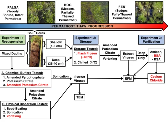
Shallow (1-5 cm) Cesium Chloride Extract Viruses Amended Potassium Citrate Vortexing TEM Amended Potassium Citrate Experiment 1: Resuspension Experiment 3: Purification PALSA (Woody Shrubs, Intact Permafrost BOG (Mosses, Partially-Thawed Permafrost) FEN (Sedges, Fully-Thawed Permafrost)
PERMAFROST THAW PROGRESSION
Mixed Depths
Deep (36-40 cm)
Figure 1 Optimizing viral recovery.Three experiments tested resuspension, storage, and purification
conditions for viral recovery on a range of peatland soils from this thawing permafrost site. Red font color indicates the best-performing option within each set. EFM, epifluorescence microscopy; TEM, transmis-sion electron microscopy; BSA, bovine serum albumin. Experiment 1 used peatland samples from 2013 and Experiments’ 2 & 3 used peatland samples from 2014. Palsa and bog pictures contributed by Anthony Garnello.
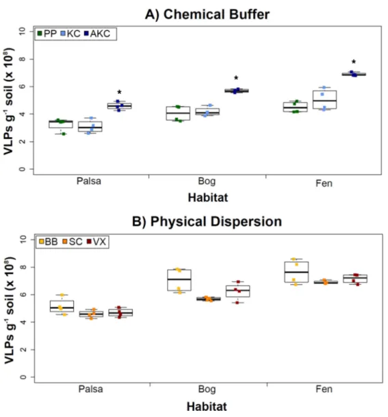
Experiment 1A: optimizing resuspension buffers
Figure 2 The impact of buffers and physical dispersion methods on viral yields.(A) Viral yields from different buffers (Experiment 1A). PP, sodium pyrophosphate; KC, potassium citrate; AKC, amended potassium citrate. Each treatment was followed by sonication for physical dispersion. (B) Viral yields from different physical dispersion methods (Experiment 1B). VX, vortexing; BB, bead-beating; SC, sonication. Each replicate was in AKC buffer. An∗denotes statistically significant difference (p<0.05) within the soil type.
of magnitude as or higher than previously reported using KC across wetland, clay, dune, forest, and park soils enumerated with the same epifluorescence microscopy technique (Williamson et al., 2013). (In that study, KC gave higher viral recoveries across soil types than water or sodium deoxycholate as a buffer;Williamson et al., 2013).
Experiment 1B: Physical dispersion methods
With an empirically optimized resuspension buffer, AKC, we next explored how physical dispersion methods (all with AKC) impacted viral yields across our three soil types. These three methods were chosen as follows: the sonication method has been most heavily tested in the literature, the beads in bead-beating might help with desorption of viruses (both reviewed inKimura et al., 2008), and vortexing might lead to the least tail breakage (Williamson, Helton & Wommack, 2012).
However, in our samples, Experiment 1B revealed no statistically significant difference in VLP yields across the physical dispersion methods tested (Fig. 2B). VLP counts ranged from 4.59×10⁸(±2.75×10⁷) VLPs g−1soil for sonication in palsa to 7.65×10⁸(±8.83
×107) VLPs g−1 soil for bead-beating in fen. As in Experiment 1A’s results, VLP yields were again lowest in palsa and highest in fen. Although bead-beating was not significantly better, it did have on average higher VLPs than the other physical dispersion methods. We hypothesize shearing of the humic material helped viruses desorb from the peatland soil.
While no significant differences across the methods were detected, we note that there are many variations of each method reported in the literature, which were not tested here. Variants of the sonication method include storing the samples on ice during sonication (Duhamel & Jacquet, 2006;Danovaro & Middelboe, 2010) and varying the sonication times (Danovaro & Middelboe, 2010). Sonication has been the most widely used method across sediment and soil environments and has performed better when combined with a well-paired resuspension buffer such as the KC buffer examined in Experiment 1A (Williamson, Wommack & Radosevich, 2003;Duhamel & Jacquet, 2006;Danovaro & Middelboe, 2010). Bead-beating methods have also been used frequently (Ashelford, Day & Fry, 2003;Williamson et al., 2013), but there is no evidence for its increased efficacy over other methods. However, not all types and sizes of beads have been tested. In our case, vortexing was performed using the same equipment and method as bead-beating, but without beads. Vortexers come with many different speed settings and attachments, and each could potentially influence viral yields. Finally, while vortexing is thought to reduce (by 20%) tail breakage as compared to sonication methods (Williamson, Helton & Wommack, 2012), we did not investigate this further since our goals were largely to obtain VLPs for metagenomic sequencing.
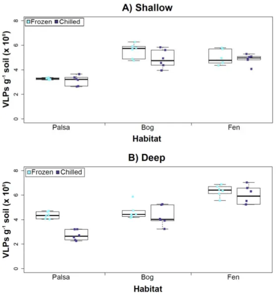
Experiment 2: optimization of sampling conditions
Figure 3 The impact of storage conditions on viral yields for different depth soils.Samples from shal-low (1–5 cm; A) or deep (36–40 cm; B) depths, and stored frozen (flash frozen and kept at−80 ◦C) or
chilled (4 ◦C), were counted using epifluorescence microscopy.
temperature had no significant influence on VLP yields either from shallow or deep samples (Fig. 3), and this finding is consistent with results in aquatic systems and sediments (Brum, Schenck & Sullivan, 2013). Soil virus literature to date, suggests viral community structure is not different with depth (based on genetic material; Adriaenssens & Cowan, 2014), nor does abundance change (comparing 5–10 cm and 10–15 cm from soil pore water;
Ballaud et al., 2015), but viral abundance was reported to vary seasonally (Ballaud et al., 2015). To this end, we tested storing both shallow and deep soil samples at chilled (4◦C) and frozen temperatures (−80◦C). In this experiment, the same protocol was followed from Experiment 1, but with AKC buffer and vortexing. Since there was not a significant difference in physical dispersion method from Experiment 1B, vortexing was chosen because bead-beating has been shown to release more humics from soil (i.e., ghost particles;
concentration (humics interact with minicolumns inHarry et al., 1999) and downstream processing of the samples (i.e., DNA extraction and sequencing;Saano et al., 1995;Zhou, Bruns & Tiedje, 1996).
Our results revealed that frozen samples had higher VLPs g−1soil than chilled samples for shallow (Fig. 3A) and deep samples (Fig. 3B), but this difference was not significant (i.e., the lowestpvalue was 0.067 in the deep palsa sample). This general finding of no significant difference in VLP recoveries by storage temperate is consistent with results in aquatic systems and sediments (Brum, Schenck & Sullivan, 2013). One caveat is that these samples are Arctic samples, whose in situhabitat temperatures at the time of collection spanned ‘‘chilled’’ temperatures (∼0–15◦C at 36–40 cm depth, although∼14–20◦C at 1–5 cm; the coldest conditions were in the deep palsa samples). Therefore, storage at 4◦C may allow microbial—and associated viral—communities (especially those at depth) to remain active, and thus drift fromin situcomposition, even though total viral recovery numbers did not change. In addition, while freeze-thaw cycles are known to broadly decrease infectivity for viruses (Ward et al., 2004), here the primary goal of recovery is enumeration and genomic sequencing, not culturing, and these viruses are also likely less sensitive to freeze-thaw given that it is a characteristic of their habitat. For these reasons, we conclude that frozen storage is the best choice for obtaining an unaltered viral community for these sample types.
The second part of this experiment tested an ecological difference rather than a methodological one: whether virus abundance changed with depth. Our frozen-storage data show significantly higher VLPs in deep than shallow palsa and fen (28% and 27% higher, respectively,p<0.05;Fig. 3). Conversely, bog had higher virus abundance in the shallow samples (18%), although it was not significant. In the chilled samples, there was no significant difference between shallow and deep (Fig. 3); the trends were the same as for frozen samples for bog and fen, but opposite in palsa (shallow was 14% higher than deep).
In some aquatic systems virus abundance decreases with depth (Cochlan et al., 1993;
Noble & Fuhrman, 1998;Parada et al., 2007), correlating with the decrease in microbial abundance with depth (Buitenhuis et al., 2012; Mediterranean Sea data inMapelli et al., 2013). This is not surprising, as viral and bacterial abundances have been shown to be moderately correlated in at least some systems (Maranger & Bird, 1995;Anesio et al., 2004). If this trend also occurs in terrestrial environments, then we would expect viral abundance to decrease with depth, as it has been shown that microbial abundance decreases with depth (Mackelprang et al., 2011). In this study, this trend (viral abundance decreasing with depth) is supported only at the bog site (for both storage conditions) and at the palsa site with chilled storage. The palsa and bog habitats both have permafrost, with palsa’s being intact and the bog’s being partially thawed. The presence of permafrost would decrease microbial activity and thus viral activity (Frank-Fahle et al., 2014). However, it is contradicted at the palsa site with frozen storage and the fen site (for both storage conditions). At the palsa site, the depth increases in VLPs may be due to a concentration of virus particles and microbial cells due to the permafrost/active layer boundary for palsa samples, as has been observed for microbial cells at the permafrost boundary in other studies (permafrost in palsa starts
permafrost, the depth increases in VLPs could be due to a temperature-stratification-based water density gradient in the inundated soil column (average fen temperature at 1–5 cm was 15.8◦C, and 13.5◦C at 30–36 cm). The difference in frozen versus chilled results for the palsa site may be due to the similarity of chilled storage to thein situpalsa deep conditions, due to its proximity to the permafrost table. Therefore, the stored chilled samples could be active and therefore likely to give artificial results. The frozen palsa results of higher VLP abundance at depth may representin situbiological patterns whereas the chilled samples may be artificial and not represent thein situviral communities. The chilled samples may instead reflect the viral communities in autumn or spring months when soil temperatures average∼4◦C because that was the temperature at which these viruses were stored. The palsa habitat is not waterlogged (like a majority of bog habitats and all fen habitat at our site) and therefore the viruses may be more resistant to temperature change because soil has a lower heat capacity than water (Bristow, 1998).
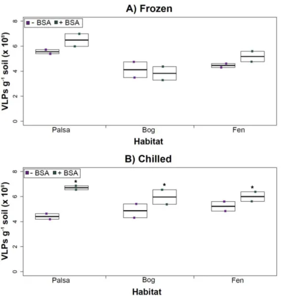
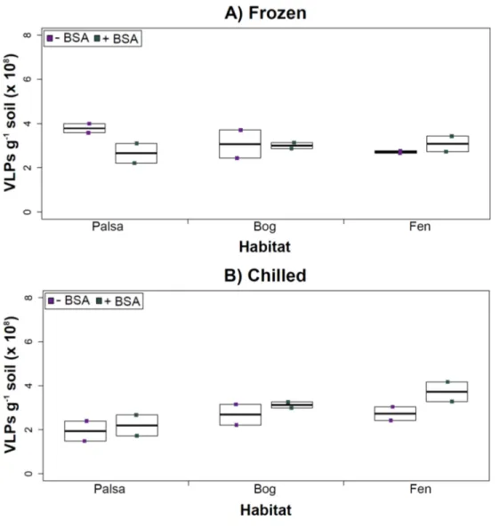
Experiment 3: optimization of concentration and purification methods Having explored how buffer, dispersion, and sampling conditions impact viral yields (and used improvements therein to assess depth differences in viral abundances at the site), we next tested the efficacy of several concentration and purification methods. Specifically, we tested concentration on Amicon filters with and without BSA addition (for ‘‘blocking’’ of the membranes to decrease viral adhesion), followed by purification on a CsCl gradient.
The addition of BSA to Amicon filters quantitatively, though not significantly, increased viral yields (except bog stored frozen), but only significantly in chilled samples (Fig. 4). This is curious, as BSA has previously been shown to increase viral recovery yield by 2-fold with seawater viruses (Deng et al., 2012). We hypothesize that soil viruses across the three habitats in Stordalen mire may be less ‘‘sticky’’ than ocean viruses so that the blocking agent had little effect on yields off the Amicon filters (lack of ‘‘stickiness’’ of soil viruses characterized inKlitzke et al., 2015). This might reflect the highly charged soil environment, relative to seawater, selecting for reduced charges in soil virus tail fibers and structural proteins as compared to those in ocean viruses (Sobsey et al., 1980;Dowd et al., 1998). Additionally, soil viruses may be less ‘‘sticky’’ because they need to be able to attach to their host for infection and viral transport in soils is slow compared to marine environments because in marine environments viruses are transported by currents (Brum et al., 2015).
Comparing samples with or without BSA in the Amicon step showed no significant effect after CsCl (although half hadpvalues around 0.058;Fig. 5). Purification by CsCl gradient caused significant loss (p<0.05, two-tailed pairedt-test;Fig. S2) of VLPs in half of the samples, with 22–67% loss observed (Fig. S2).This loss is not surprising given that non-virus ‘‘ghost’’ particles such as humics and cell debris, which can be mistakenly counted as VLPs by EFM, are typically present in environments with high organic matter (e.g., as discussed inSuttle & Fuhrman, 2010;Forterre et al., 2013) and the CsCl densities (rho=
Figure 4 The impact of BSA on virus concentration by Amicon filter.Deep samples, stored frozen (A) or chilled (B), were used for this comparison and post concentration yields were counted by epifluores-cence microscopy. An∗denotes statistically significant (p<0.05) within the soil type.
techniques have been carefully examined and in each, the use of EFM was suggested (Hermes & Suttle, 1995;Weinbauer & Suttle, 1997;Noble & Fuhrman, 1998;Brum, Schenck & Sullivan, 2013;Williamson et al., 2013). The lowered VLP counts could be due to the CsCl damaging viral particles during purification, which is known to be osmotically challenging for many viral isolates. In seawater samples, where there is less concern for organic matter and ghost particle background, viral loss was observed from CsCl purification (Hurwitz et al., 2013). Also, others have observed viral recovery for isolates after CsCl purification to average between 53–66% with a range of 13–100% recovery (Chen, Estes & Ramig, 1992;
Figure 5 Comparison of samples with or without BSA after CsCl gradient purification.Counts were taken of deep samples that were CsCl gradient purified after Amicon filter concentration (with and with-out BSA) for samples from each habitat that were (A) frozen or (B) chilled.
represent ‘‘a loss step’’ they are important for purifying viral particles from contaminating nucleic acids or inhibitory substances for downstream processing (Saano et al., 1995;
Zhou, Bruns & Tiedje, 1996), and outperform other purification options such as column chromatography or commercial purification kits (Duffy, O’Brien & Strappe, 2005).
CONCLUSIONS
via CsCl density gradient centrifugation. This optimized protocol can now be used to help further viral research in soil, particularly in challenging, humic-laden highly organic soils. At the same time, a revolution in viral ecology is being born out of experimental and informatic advances for studying viral signals at the single cell level (reviewed inDang & Sullivan, 2014), in microbial datasets (Labonté et al., 2015;Roux et al., 2015a;Roux et al., 2015b) and through the development of quantitative sample-to-sequence pipelines for surveying viral communities (reviewed inWommack et al., 2012;Solonenko & Sullivan, 2013;DeLong, 2013;Brum et al., 2016). The findings presented here, in parallel with these technical advances in viral ecology, should soon enable a more thorough understanding of viral diversity and viral impacts on permafrost-associated soil microorganisms, specifically impacts of viral infection upon host community structure, viral–host gene transfer, and viral influence on host cell physiology. These key findings can then feed into modeling efforts that should help elucidate the role(s) of viruses in biogeochemical cycles and, ultimately, improve our ability to incorporate them into climate change models.
ACKNOWLEDGEMENTS
We thank Bonnie Poulos, Christine Schirmer, and Mario Moreno for their advice, encouragement, and assistance. Thanks also to the Rich and Sullivan Labs and the Abisko Naturvetenskapliga Station for added guidance and logistical support. Special thanks to Moira Hough, Robert Jones, and Dr. Rachel Wilson for helping collect soil cores from Stordalen mire in Sweden. We thank Anthony Garnello for contributing the palsa and bog pictures in Fig. 1. We thank everyone from the IsoGenie consortium, in particular, Suzanne Hodgkins, Dr. Rachel Wilson, and Dr. Jeff Chanton for their guidance and added expertise.
ADDITIONAL INFORMATION AND DECLARATIONS
Funding
This work was supported by the US Department of Energy Office of Biological and Environmental Research under the Genomic Science program (Award DESC0010580 to VR and MBS), and by a Gordon and Betty Moore Foundation Investigator Award (GBMF# 3790 to MBS). VR & GT received support through the Ecosystem Genomics Initiative, by the University of Arizona Technology and Research Initiative Fund, through the Water, Environmental and Energy Solutions Initiative. We thank the Abisko Scientific Research Station for sampling infrastructure. The funders had no role in study design, data collection and analysis, decision to publish, or preparation of the manuscript.
Grant Disclosures
The following grant information was disclosed by the authors:
US Department of Energy Office of Biological and Environmental Research: DESC0010580. Gordon and Betty Moore Foundation Investigator Award: GBMF#3790.
Competing Interests
The authors declare there are no competing interests.
Author Contributions
• Gareth Trubl conceived and designed the experiments, performed the experiments, analyzed the data, wrote the paper, prepared figures and/or tables, reviewed drafts of the paper.
• Natalie Solonenko conceived and designed the experiments, performed the experiments, wrote the paper, reviewed drafts of the paper.
• Lauren Chittick performed the experiments.
• Sergei A. Solonenko performed the experiments, wrote the paper, reviewed drafts of the paper.
• Virginia I. Rich conceived and designed the experiments, contributed reagents/materi-als/analysis tools, wrote the paper, prepared figures and/or tables, reviewed drafts of the paper.
• Matthew B. Sullivan conceived and designed the experiments, contributed reagents/materials/analysis tools, wrote the paper, reviewed drafts of the paper.
Data Availability
The following information was supplied regarding data availability: The raw data has been supplied as aData S1.
Supplemental Information
Supplemental information for this article can be found online athttp://dx.doi.org/10.7717/ peerj.1999#supplemental-information.
REFERENCES
Abbott BW, Jones JB. 2015.Permafrost collapse alters soil carbon stocks, respiration, CH4, and N2O in upland tundra.Global Change Biology21(12):4570–4587 DOI 10.1111/gcb.13069.
Adriaenssens EM, Cowan DA. 2014.Using signature genes as tools to assess envi-ronmental viral ecology and diversity.Applied and Environmental Microbiology
80(15):4470–4480DOI 10.1128/AEM.00878-14.
Anantharaman K, Duhaime MB, Breier JA, Wendt KA, Toner BM, Dick GJ. 2014. Sulfur oxidation genes in diverse deep-sea viruses.Science344(6185):757–760 DOI 10.1126/science.1252229.
Anesio AM, Hollas C, Granéli W, Laybourn-Parry J. 2004.Influence of humic sub-stances on bacterial and viral dynamics in freshwaters.Applied and Environmental
Microbiology70:4848–4854DOI 10.1128/AEM.70.8.4848-4854.2004.
Ashelford KE, Day MJ, Fry JC. 2003.Elevated abundance of bacteriophage infect-ing bacteria in soil.Applied and Environmental Microbiology69(1):285–289 DOI 10.1128/AEM.69.1.285-289.2003.
Ashelford KE, Norris SJ, Fry JC, Bailey MJ, Day MJ. 2000.Seasonal population dynamics and interactions of competing bacteriophages and their host in the rhizosphere.Applied and Environmental Microbiology 66(10):4193–4199 DOI 10.1128/AEM.66.10.4193-4199.2000.
Ballaud F, Dufresne A, Francez AJ, Colombet J, Sime-Ngando T, Quaiser A. 2015. Dynamics of viral abundance and diversity in aSphagnum-dominated peatland: temporal fluctuations prevail over habitat.Frontiers in Microbiology6:1494 DOI 10.3389/fmicb.2015.01494.
Bristow KL. 1998.Measurement of thermal properties and water content of unsaturated sandy soil using dual-probe heat-pulse probes.Agricultural and Forest Meteorology
89(2):75–84DOI 10.1016/S0168-1923(97)00065-8.
Brum JR, Ignacio-Espinoza JC, Kim EH, Trubl G, Jones RM, Roux S, VerBerkmoes NC, Rich VI, Sullivan MB. 2016.Illuminating structural proteins in viral dark matter with metaproteomics.Proceedings of the National Academy of Sciences of the United States of America113(9):2436–2441DOI 10.1073/pnas.1525139113.
Brum JR, Ignacio-Espinoza JC, Roux S, Doulcier G, Acinas SG, Alberti A, Chaffron S, Cruaud C, De Vargas C, Gasol JM, Gorsky G. 2015.Ocean plankton. patterns and ecological drivers of ocean viral communities.Science348(6237):1261498 DOI 10.1126/science.1261498.
Brum JR, Schenck RO, Sullivan MB. 2013.Global morphological analysis of marine viruses shows minimal regional variation and dominance of non-tailed viruses.The ISME Journal7(9):1738–1751DOI 10.1038/ismej.2013.67.
Brum JR, Sullivan MB. 2015.Rising to the challenge: accelerated pace of discov-ery transforms marine virology.Nature Reviews Microbiology 13(3):147–159 DOI 10.1038/nrmicro3404.
Buitenhuis ET, Li WKW, Vaulot D, Lomas MW, Landry MR, Partensky F, Karl DM, Ulloa O, Campbell L, Jacquet S. 2012.Picophytoplankton biomass distribution in the global ocean.Earth System Science Data4(1):37–46DOI 10.5194/essd-4-37-2012. Chen DY, Estes MK, Ramig RF. 1992.Specific interactions between rotavirus outer
capsid proteins VP4 and VP7 determine expression of a cross-reactive, neutralizing VP4-specific epitope.Journal of Virology66(1):432–439.
Chesson P. 2000.Mechanisms of maintenance of species diversity.Annual Review of Ecology and Systematics31:343–366.
Cochlan WP, Wikner J, Steward GF, Smith DC, Azam F. 1993.Spatial distribution of viruses, bacteria and chlorophyll a in neritic, oceanic and estuarine environments.
Marine Ecology Progress Series92:77–87DOI 10.3354/meps092077.
Crump BC, Amaral-Zettler LA, Kling GW. 2012.Microbial diversity in arctic fresh-waters is structured by inoculation of microbes from soils.The ISME Journal
6(9):1629–1639.
Cunningham BR, Brum JR, Schwenck SM, Sullivan MB, John SG. 2015.An inexpensive, accurate, and precise wet-mount method for enumerating aquatic viruses.Applied and Environmental Microbiology 81(9):2995–3000DOI 10.1128/AEM.03642-14. Dang VT, Sullivan MB. 2014.Emerging methods to study bacteriophage infection at the
single-cell level.Frontiers in Microbiology5:Article 724DOI 10.3389/fmicb.2014.00724. Danovaro R, Middelboe M. 2010. Separation of free virus particles from sediments in
aquatic systems. In: Wilhelm SW, Weinbauer MG, Suttle CA, eds.Manual of aquatic viral ecology. Waco: ASLO, 74–81.
DeLong E. 2013.Microbial metagenomics, metatranscriptomics, and metaproteomics. Vol. 531. Cambridge: Academic Press.
Deng L, Gregory A, Yilmaz S, Poulos BT, Hugenholtz P, Sullivan MB. 2012.Contrasting life strategies of viruses that infect photo-and heterotrophic bacteria, as revealed by viral tagging.MBio3(6):e00373–12DOI 10.1128/mBio.00373-12.
Dowd SE, Pillai SD, Wang S, Corapcioglu MY. 1998.Delineating the specific influence of virus isoelectric point and size on virus adsorption and transport through sandy soils.Applied and Environmental Microbiology 64(2):405–410.
Duffy AMO, O’Brien T, Strappe PM. 2005.Purification of adenovirus and adeno-associated virus: comparison of novel membrane-based technology to conventional techniques.Gene Therapy12(Suppl 1):S62–S72DOI 10.1038/sj.gt.3302616.
Duhaime MB, Deng Li, Poulos BT, Sullivan MB. 2012.Towards quantitative metage-nomics of wild viruses and other ultra-low concentration dna samples: a rigorous assessment and optimization of the linker amplification method.Environmental Microbiology14(9):2526–2537DOI 10.1111/j.1462-2920.2012.02791.x.
Duhamel S, Jacquet S. 2006.Flow cytometric analysis of bacteria- and virus-like particles in lake sediments.Journal of Microbiological Methods64(3):316–332 DOI 10.1016/j.mimet.2005.05.008.
Elberling B, Michelsen A, Schädel C, Schuur EAG, Christiansen HH, Berg L, Tamstorf MP, Sigsgaard C. 2013.Long-term CO2production following permafrost thaw.
Nature Climate Change 3(10):890–894DOI 10.1038/nclimate1955.
Fierer N, Breitbart M, Nulton J, Salamon P, Lozupone C, Jones R, Robeson M, Edwards RA, Felts B, Rayhawk S, Knight R. 2007.F.Applied and Environmental Microbiology
73(21):7059–7066DOI 10.1128/AEM.00358-07.
Forterre P, Soler N, Krupovic M, Marguet E, Ackermann H-W. 2013.Fake virus particles generated by fluorescence microscopy.Trends in Microbiology21(1):1–5 DOI 10.1016/j.tim.2012.10.005.
Frank-Fahle BA, Yergeau É, Greer CW, Lantuit H, Wagner D. 2014.Microbial func-tional potential and community composition in permafrost-affected soils of the NW Canadian Arctic.PLoS ONE9(1):e84761DOI 10.1371/journal.pone.0084761. Fuhrman JA. 1999.Marine viruses and their biogeochemical and ecological effects.
Goyal SM, Gerba CP. 1979.Comparative adsorption of human enteroviruses, simian ro-tavirus, and selected bacteriophages to soils.Applied and Environmental Microbiology
38(2):241–247.
Graham DE, Wallenstein MD, Vishnivetskaya TA, Waldrop MP, Phelps TJ, Pfiffner SM, Onstott TC, Whyte LG, Rivkina EM, Gilichinsky DA, Elias DA. 2012.Microbes in thawing permafrost: the unknown variable in the climate change equation.The ISME Journal6(4):709–712DOI 10.1038/ismej.2011.163.
Hansen AA, Herbert RA, Mikkelsen K, Jensen LL, Kristoffersen T, Tiedje JM, Lom-stein BA, Finster KW. 2007.Viability, diversity and composition of the bacterial community in a high arctic permafrost soil from Spitsbergen, Northern Norway.
Environmental Microbiology9(11):2870–2884 DOI 10.1111/j.1462-2920.2007.01403.x.
Harry M, Gambier B, Bourezgui Y, Garnier-Sillam E. 1999.Evaluation of purification procedures for DNA extracted from rich organic samples: interference with humic substances.Analusis27(5):439–441.
Helton RR, Liu L, Wommack KE. 2006.Assessment of factors influencing direct enumeration of viruses within estuarine sediments.Applied and Environmental
Microbiology72(7):4767–4774DOI 10.1128/AEM.00297-06.
Hermes KP, Suttle CA. 1995.Direct counts of viruses in natural waters and labo-ratory cultures by epifluorescence microscopy.Limnology and Oceanography
40(6):1050–1055DOI 10.4319/lo.1995.40.6.1050.
Hewson I, Fuhrman JA. 2003.Viriobenthos production and virioplankton sorptive scavenging by suspended sediment particles in coastal and pelagic waters.Microbial Ecology 46(3):337–347DOI 10.1007/s00248-002-1041-0.
Hodgkins SB, Tfaily MM, McCalley CK, Logan TA, Crill PM, Saleska SR, Rich VI, Chanton JP. 2014.Changes in peat chemistry associated with permafrost thaw increase greenhouse gas production.Proceedings of the National Academy of Sciences of the United States of America111(16):5819–5824DOI 10.1073/pnas.1314641111. Hugelius G, Strauss J, Zubrzycki S, Harden JW, Schuur EAG, Ping CL, Schirrmeister L,
Grosse G, Michaelson GJ, Koven CD, O’Donnell JA, Elberling B, Mishra U, Camill P, Yu Z, Palmtag J, Kuhry P. 2014.Estimated stocks of circumpolar permafrost carbon with quantified uncertainty ranges and identified data gaps.Biogeosciences
11(23):6573–6593DOI 10.5194/bg-11-6573-2014.
Hultman J, Waldrop MP, Mackelprang R, David MM, McFarland J, Blazewicz SJ, Harden J, Turetsky MR, McGuire AD, Shah MB, Verberkmoes NC, Lee LH, Mavrommatis K, Jansson JK. 2015.Multi-omics of permafrost, active layer and thermokarst bog soil microbiomes.Nature521(7551):208–212 DOI 10.1038/nature14238.
Hurst CJ. 1988.Influence of aerobic microorganisms upon virus survival in soil.
Canadian Journal of Microbiology34(5):696–699DOI 10.1139/m88-117.
Hurwitz BL, Deng L, Poulos BT, Sullivan MB. 2013.Evaluation of methods to con-centrate and purify ocean virus communities through comparative, replicated metagenomics.Environmental Microbiology15(5):1428–1440.
Hurwitz BL, Sullivan MB. 2013.The Pacific Ocean Virome (POV): a marine viral metagenomic dataset and associated protein clusters for quantitative viral ecology.
PLoS ONE8(2):e57355DOI 10.1371/journal.pone.0057355.
IPCC. 2014. Climate change 2014: impacts, adaptation, and vulnerability. part a: global and sectoral aspects. In: Field CB, Barros VR, Dokken DJ, Mach KJ, Mastrandrea MD, Bilir TE, Chatterjee M, Ebi KL, Estrada YO, Genova RC, Girma B, Kissel ES, Levy AN, MacCracken S, Mastrandrea PR, White LL, (eds.)Contribution of working Group II to the fifth assessment report of the Intergovernmental Panel on Climate Change. Cambridge, New York: Cambridge University Press.
Jansson JK, Taş N. 2014.The microbial ecology of permafrost.Nature Reviews Microbiol-ogybiol12(6):414–425 DOI 10.1038/nrmicro3262.
Kimura M, Jia ZJJ, Nakayama N, Asakawa S. 2008.Ecology of viruses in soils: past, present and future perspectives.Soil Science & Plant Nutrition54(1): Taylor & Francis Group: 1–32DOI 10.1111/j.1747-0765.2007.00197.x..
Klitzke S, Schroeder J, Selinka H-C, Szewzyk R, Chorus I. 2015. Attenuation and colloidal mobilization of bacteriophages in natural sediments under anoxic as com-pared to oxic conditions. In:Science of the total environment. Vol. 518. Amsterdam: Elsevier, 130–138.
Labonté JM, Swan BK, Poulos B, Luo H, Koren S, Hallam SJ, Sullivan MB, Woyke T, Wommack KE, Stepanauskas R. 2015.Single-cell genomics-based analysis of virus–host interactions in marine surface bacterioplankton.The ISME Journal
9(11):2386–2399DOI 10.1038/ismej.2015.48.
Lakay FM, Botha A, Prior BA. 2007.Comparative analysis of environmental DNA extraction and purification methods from different humic acid-rich soils.Journal of Applied Microbiology102(1):265–273.
Mackelprang R, Waldrop MP, DeAngelis KM, David MM, Chavarria KL, Blazewicz SJ, Rubin EM, Jansson JK. 2011.Metagenomic analysis of a permafrost micro-bial community reveals a rapid response to thaw.Nature480(7377):368–371 DOI 10.1038/nature10576.
Mapelli F, Varela MM, Barbato M, Alvariño R, Fusi M, Álvarez M, Merlino G, Daffon-chio D, Borin S. 2013.Biogeography of planktonic bacterial communities across the whole Mediterranean Sea.Ocean Science9(4):585–595DOI 10.5194/os-9-585-2013. Maranger R, Bird DF. 1995.Viral abundance in aquatic systems: a comparison
be-tween marine and fresh waters.Marine Ecology Progress Series121(1):217–226 DOI 10.3354/meps121217.
Michen B, Graule T. 2010.Isoelectric points of viruses.Journal of Applied Microbiology
109(2):388–397DOI 10.1111/j.1365-2672.2010.04663.x.
Middelboe M, Glud RN, Finster K. 2003.Distribution of viruses and bacteria in relation to diagenetic activity in an estuarine sediment.Limnology and Oceanography
48(4):1447–1456DOI 10.2307/3597468.
Mondav R, Woodcroft BJ, Kim E-H, McCalley CK, Hodgkins SB, Crill PM, Chanton J, Hurst GB, VerBerkmoes NC, Saleska SR, Hugenholtz P. 2014.Discovery of a novel methanogen prevalent in thawing permafrost.Nature Communications5:Article 3212 DOI 10.1038/ncomms4212.
Moore RS, Taylor DH, Reddy MM, Sturman LS. 1982.Adsorption of reovirus by minerals and soils.Applied and Environmental Microbiology 44(4):852–859.
Moore RS, Taylor DH, Sturman LS, Reddy MM, Fuhs GW. 1981.Poliovirus adsorption by 34 minerals and soils.Applied and Environmental Microbiology 42(6):963–975. Muniesa M, Blanco JE, de Simon M, Serra-Moreno R, Blanch AR, Jofre J. 2004.
Diversity of stx2 converting bacteriophages induced from Shiga-toxin-producing
Escherichia colistrains isolated from cattle.Microbiology150(9):2959–2971. Noble RT, Fuhrman JA. 1998.Use of SYBR green i for rapid epifluorescence counts
of marine viruses and bacteria.Aquatic Microbial Ecology14(2):113–118 DOI 10.3354/ame014113.
Parada V, Sintes Eva, Van Aken HM, Weinbauer MG, Herndl GJ. 2007.Viral abundance, decay, and diversity in the meso-and bathypelagic waters of the North Atlantic.Applied and Environmental Microbiology73(14):4429–4438 DOI 10.1128/AEM.00029-07.
Paul JH. 1999.Microbial gene transfer: an ecological perspective.Journal of Molecular Microbiology and Biotechnology 1(1):45–50.
Paul EA. 2014.Soil microbiology, ecology and biochemistry. Cambridge: Academic Press. Paul JH, Rose JB, Jiang SC, Kellogg CA, Dickson L. 1993.Distribution of viral
abun-dance in the reef environment of key largo, Florida.Applied and Environmental Microbiology59(3):718–724.
Prater JL, Chanton JP, Whiting GJ. 2007.Variation in methane production pathways associated with permafrost decomposition in collapse scar bogs of Alberta, Canada.
Global Biogeochemical Cycles21(4):GB4004DOI 10.1029/2006GB002866. Quaiser A, Dufresne A, Ballaud F, Roux S, Zivanovic Y, Colombet J, Sime-Ngando
T, Francez A-J. 2015.Diversity and comparative genomics of microviri-dae insphagnum-dominated peatlands.Frontiers in Microbiology6:1–10 DOI 10.3389/fmicb.2015.00375.
Roux S, Enault F, Hurwitz BL, Sullivan MB. 2015a.VirSorter: mining viral signal from microbial genomic data.PeerJ3:e985 DOI 10.7717/peerj.985.
Roux S, Hallam SJ, Woyke T, Sullivan MB. 2015b.Viral dark matter and virus–host interactions resolved from publicly available microbial genomes.eLife4:e08490 DOI 10.7554/eLife.08490.
uncultivated SUP05 bacteria as revealed by single-cell-and meta-genomics.Elife
3:e03125DOI 10.7554/eLife.03125.
Saano A, Tas E, Pippola S, Lindström K, Van Elsas JD. 1995. Extraction and analysis of microbial DNA from soil. In:Nucleic acids in the environment. Berlin Heidelberg: Springer, 49–67.
Schimel J, Balser TC, Wallenstein M. 2007.Microbial stress-response physiology and its implications for ecosystem function.Ecology88(6):1386–1394DOI 10.1890/06-0219. Schuur EAG, McGuire AD, Schädel C, Grosse G, Harden JW, Hayes DJ, Hugelius G,
Koven CD, Kuhry P, Lawrence DM. 2015.Climate change and the permafrost carbon feedback.Nature520(7546):171–179DOI 10.1038/nature14338.
Slater AG, Lawrence DM. 2013.Diagnosing present and future permafrost from climate models.Journal of Climate26(15):5608–5623DOI 10.1175/JCLI-D-12-00341.1. Smalla K, Van Elsas JD. 2010. The soil environment. In:Environmental molecular
microbiology. Norfolk: Caister Academic Press, 111–129.
Sobsey MD, Dean CH, Knuckles ME, Wagner RA. 1980.Interactions and survival of en-teric viruses in soil materials.Applied and Environmental Microbiology 40(1):92–101. Solonenko SA, Sullivan MB. 2013. Preparation of metagenomic libraries from naturally occurring marine viruses. In:Methods in enzymology. 1st edition. Vol. 531. Amster-dam: Elsevier Inc.
Speir JA, Bothner B, Qu C, Willits DA, Young MJ, Johnson JE. 2006.Enhanced local symmetry interactions globally stabilize a mutant virus capsid that main-tains infectivity and capsid dynamics.Journal of Virology 80:3582–3591 DOI 10.1128/JVI.80.7.3582-3591.2006.
Sperelakis N. 2012.Cell physiology sourcebook: essentials of membrane biophysics. Amster-dam: Elsevier.
Srinivasiah S, Bhavsar J, Thapar K, Liles M, Schoenfeld T, Wommack KE. 2008.Phages across the biosphere: contrasts of viruses in soil and aquatic environments.Research in Microbiology 159(5):349–357DOI 10.1016/j.resmic.2008.04.010.
Stevenson FJ. 1994.Humus chemistry: genesis, composition, reactions. 2nd edition. New York: John Wiley & Sons.
Sullivan MB, Lindell D, Lee JA, Thompson LR, Bielawski JP, Chisholm SW. 2006. Prevalence and evolution of core photosystem II genes in marine cyanobacterial viruses and their hosts.PLoS Biology4(8):e234 DOI 10.1371/journal.pbio.0040234. Suttle CA. 2005.Viruses in the sea.Nature437(7057):356–361DOI 10.1038/nature04160. Suttle CA. 2007.Marine viruses–major players in the global ecosystem.Nature Reviews
Microbiology5(10):801–812 DOI 10.1038/nrmicro1750.
Suttle CA, Fuhrman JA. 2010. Enumeration of virus particles in aquatic or sediment samples by epifluorescence microscopy. In:Manual of aquatic viral ecology. ASLO, 145–153.
Tamames J, Abellán JJ, Pignatelli M, Camacho A, Moya A. 2010.Environmental distri-bution of prokaryotic taxa.BMC Microbiology10(1):1DOI 10.1186/1471-2180-10-1. Tarnocai C, Canadell JG, Schuur EAG, Kuhry P, Mazhitova G, Zimov S. 2009.Soil
organic carbon pools in the northern circumpolar permafrost region.Global Biogeochemical Cycles23(2):GB2023 DOI 10.1029/2008GB003327.
Thurber RV, Haynes M, Breitbart M, Wegley L, Rohwer F. 2009.Laboratory procedures to generate viral metagenomes.Nature Protocols4(4):470–483 DOI 10.1038/nprot.2009.10.
Torsvik V, Øvreås L, Thingstad TF. 2002.Prokaryotic diversity–magnitude, dynamics, and controlling factors.Science296(5570):1064–1066DOI 10.1126/science.1071698. Ward CL, Dempsey MH, Ring CJA, Kempson RE, Zhang L, Gor D, Snowden BW,
Tisdale M. 2004.Design and performance testing of quantitative real time PCR assays for influenza A and B viral load measurement.Journal of Clinical Virology
29(3):179–188 ElsevierDOI 10.1016/S1386-6532(03)00122-7.
Weinbauer MG. 2004.Ecology of prokaryotic viruses.FEMS Microbiology Reviews
28(2):127–181DOI 10.1016/j.femsre.2003.08.001.
Weinbauer MG, Rassoulzadegan F. 2004.Are viruses driving microbial diversification and diversity?Environmental Microbiology6(1):1–11.
Weinbauer MG, Suttle CA. 1997.Comparison of epifluorescence and transmission electron microscopy for counting viruses in natural marine waters.Aquatic Microbial Ecology 13(3):225–232DOI 10.3354/ame013225.
Williamson KE, Corzo KA, Drissi CL, Buckingham JM, Thompson CP, Helton RR. 2013.Estimates of viral abundance in soils are strongly influenced by ex-traction and enumeration methods.Biology and Fertility of Soils49(7):857–869 DOI 10.1007/s00374-013-0780-z.
Williamson KE, Helton RR, Wommack KE. 2012.Bias in bacteriophage mor-phological classification by transmission electron microscopy due to breakage or loss of tail structures.Microscopy Research and Technique75(4):452–457 DOI 10.1002/jemt.21077.
Williamson KE, Radosevich M, Smith DW, Wommack KE. 2007.Incidence of lysogeny within temperate and extreme soil environments.Environmental Microbiology
9(10):2563–2574DOI 10.1111/j.1462-2920.2007.01374.x.
Williamson KE, Radosevich M, Wommack KE. 2005.Abundance and diver-sity of viruses in six delaware soils.Applied and Environmental Microbiology
71(6):3119–3125DOI 10.1128/AEM.71.6.3119-3125.2005.
Williamson KE, Wommack KE, Radosevich Mark. 2003.Sampling natural viral
communities from soil for culture-independent analyses.Applied and Environmental
Microbiology69(11):6628–6633DOI 10.1128/AEM.69.11.6628-6633.2003.
Willner D, Furlan M, Haynes M, Schmieder R, Angly FE, Silva J, Tammadoni S, Nosrat B, Conrad D, Rohwer F. 2009.Metagenomic analysis of respiratory tract DNA viral communities in cystic fibrosis and non-cystic fibrosis individuals.PLoS ONE
Wommack KE, Bhavsar J, Polson SW, Chen J, Dumas M, Srinivasiah S, Furman M, Jamindar S, Nasko DJ. 2012.VIROME: a standard operating procedure for analysis of viral metagenome sequences.Standards in Genomic Sciences6(3):421–433 DOI 10.4056/sigs.2945050.
Wommack KE, Colwell RR. 2000.Virioplankton: viruses in aquatic ecosystems.
Microbiology and Molecular Biology Reviews64(1):69–114 DOI 10.1128/MMBR.64.1.69-114.2000.
Zhou J, Bruns MA, Tiedje JM. 1996.DNA recovery from soils of diverse composition.