the Suprachiasmatic Nucleus and Optic Nerve of
Cryptochrome
-Deficient Mice
Takahiro J. Nakamura1,2,3*, Shizufumi Ebihara2, Kazuyuki Shinohara1
1Division of Neurobiology and Behavior, Department of Translational Medical Sciences, Graduate School of Biomedical Sciences, Nagasaki University, Nagasaki, Japan, 2Division of Biomodeling, Graduate School of Bioagricultural Sciences, Nagoya University, Nagoya, Japan,3Faculty of Pharmaceutical Sciences, Teikyo Heisei University, Ichihara, Chiba, Japan
Abstract
To examine roles of theCryptochromes(Cry1andCry2) in mammalian circadian photoreception, we recorded single-unit neuronal firing activity in the suprachiasmatic nucleus (SCN), a primary circadian oscillator, and optic nerve fibersin vivo
after retinal illumination in anesthetizedCry1andCry2double-knockout (Cry-deficient) mice. In wild-type mice, most SCN neurons increased their firing frequency in response to retinal illumination at night, whereas only 17% of SCN neurons responded during the daytime. However, 40% of SCN neurons responded to light during the daytime, and 31% of SCN neurons responded at night inCry-deficient mice. The magnitude of the photic response in SCN neurons at night was significantly lower (1.3-fold of spontaneous firing) inCry-deficient mice than in wild-type mice (4.0-fold of spontaneous firing). In the optic nerve near the SCN, no difference in the proportion of light-responsive fibers was observed between daytime and nighttime in both genotypes. However, the response magnitude in the light-activated fibers (ON fibers) was high during the nighttime and low during the daytime in wild-type mice, whereas this day–night difference was not observed inCry-deficient mice. In addition, we observed day–night differences in the spontaneous firing rates in the SCN in both genotypes and in the fibers of wild-type, but notCry-deficient mice. We conclude that the low photo response in the SCN ofCry-deficient mice is caused by a circadian gating defect in the retina, suggesting thatCryptochromesare required for appropriate temporal photoreception in mammals.
Citation:Nakamura TJ, Ebihara S, Shinohara K (2011) Reduced Light Response of Neuronal Firing Activity in the Suprachiasmatic Nucleus and Optic Nerve of
Cryptochrome-Deficient Mice. PLoS ONE 6(12): e28726. doi:10.1371/journal.pone.0028726
Editor:Shree Ram Singh, National Cancer Institute, United States of America
ReceivedSeptember 9, 2011;AcceptedNovember 14, 2011;PublishedDecember 21, 2011
Copyright:ß2011 Nakamura et al. This is an open-access article distributed under the terms of the Creative Commons Attribution License, which permits unrestricted use, distribution, and reproduction in any medium, provided the original author and source are credited.
Funding:This work was supported by Grants-in-Aid for the Promotion of Science for Young Scientists from Japanese Ministry of Education, Science and Culture 14011687 (to TJN). The funders had no role in study design, data collection and analysis, decision to publish, or preparation of the manuscript.
Competing Interests:The authors have declared that no competing interests exist.
* E-mail: [email protected]
Introduction
Circadian rhythms are oscillations with daily periodicities in physiological and behavioral functions of organisms. In mammals, the central circadian oscillator is located in the suprachiasmatic nucleus (SCN) of the ventral hypothalamus [1]. The rhythms are generated by a cell-autonomous circadian oscillator that is synchronized with the environment by light through the retinohypothalamic tract; hence, light synchronizes the behavior of the organism with the daily 24-hr light–dark (LD) cycle [2].
The mammalian retina mediates several nonvisual light-responsive functions, including circadian photoreception [3], acute suppression of locomotor activity by light (masking) [4], photic suppression of pineal melatonin synthesis [5], and pupillary light responses [6]. Candidate photoreceptors for these functions include opsins, such as classical rod and cone opsins and the novel photopigment melanopsin [7], and the blue-light photore-ceptive pigmentsCryptochromes[8]. In therd(retinal degeneration) mouse, a mutation in a rod-specific gene [9] leads to the rapid degeneration of rod cells. Rod degeneration is complete after 2 months whereas the secondary degeneration of cones is much slower [10]. While these rd/rd mice lack the photopigments for vision, they retain robust nonvisual responses to light [3,4,5,6,11],
indicating the involvement of other nonclassical photoreceptors. Melanopsin knockout mice (Opn42/2) exhibit a somewhat reduced sensitivity to circadian phase shifting [12,13] and masking [12] but display normal entrainment [12,13] and nearly normal pupillary light reflex [14,15] and gene induction by light in the SCN [13]. Indeed,rd/rd Opn42/2mice displayed severe defects in all tested photoreceptive tasks [15,16].
Cryptochromes are folate- and flavin-based members of the photolyase family of photopigments that are necessary for normal circadian phase shifting inArabidopsisandDrosophila[17]. Mammals have twoCryptochromefamily members, which are expressed in the inner retina [17,18] as well as many other tissues. Mice lacking
Cryptochromes(mCry12/2 mCry22/2) display severe defects in gene induction in the SCN [19,20], but retain normal pupillary light reflex [21] and masking [22]. Photoresponsiveness is markedly depressed in Rd/rd mCry12/2 mCry22/2 mice, as measured by masking, pupillary light reflex, and light-induced immediate-early gene expression in the SCN [19,21,23]. These studies indicate that both melanopsin andCryptochromescontribute to nonvisual photo-responses and play important roles in these processes.
cannot be assayed for circadian phase shifting. Therefore, in the present study, we performed extracellular single unit recordings of the neuronal firing activity in the SCN and the optic nerve of anesthetized mCry12/2 mCry22/2 mice in response to retinal illumination. Several rodent studies have shown that the photic responses of the electrical activities in the SCN are closely correlated with the photic entrainment properties in the locomotor activity rhythms [25,26,27]. The magnitude of the photic responses of electrical activities in the SCN depends on both the circadian phase [27] and the light intensity [25,26,27] in rats and hamsters. Recently, we successfully recorded the photic response in the firing of mouse SCNin vivo, which corresponds to the properties of photic entrainment in the locomotor activity of mice [28]. In the present study, to assay circadian photoreceptions inmCry12/2 mCry22/2 mice, we compared the light response of neuronal firing activity in the SCN and the optic nerve during the daytime and the nighttime.
Results
For recordings in the SCN and optic fibers, we used 46 wild-type, 69mCry12/2mCry22/2mice. We carried out a single recording per animal in which 16 neurons in wild-type mice and 26 neurons in
mCry12/2mCry22/2mice were recorded in the SCN. The rest of the animals were used for optic nerve fiber recordings.
Temporal differences in spontaneous neuronal firing activity in the SCN and optic nerve fibers
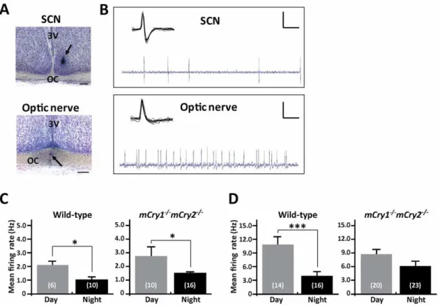
Representative photographs and oscilloscope traces for the single unit recordings of the neuronal firing activity in the SCN and the optic nerve fibers of mice are shown in Figure 1A and B. We clearly distinguished spikes between the SCN and the optic nerve by means of the spike form and additionally confirmed the recording site using the histological method after the recording. We first measured the baseline of spontaneous firing activity in the SCN and optic nerve fibers of mice during the daytime (Zeitgeber time [ZT] 4–8) and the nighttime (ZT 14–16). We collected the baseline firing frequency (Hz) for 60 sec without retinal illumina-tion. In the SCN, both wild-type andmCry12/2mCry22/2 mice displayed a day–night variation in spontaneous firing activity (2.0360.37 during the daytime vs. 1.0960.21 during the nighttime in wild-type mice and 2.8360.67 during the daytime vs. 1.5460.17 during the nighttime in mCry12/2 mCry22/2;
P,0.05for both genotypes, Student’st-test; Fig. 1C). In the optic nerve, wild-type mice showed a distinct day–night change (10.9961.53 during the daytime vs. 4.1360.72 during the nighttime;P,0.001, Student’st-test; Fig. 1D), whereasmCry12/2 mCry22/2 mice did not exhibit a day–night difference in spontaneous firing rate (8.7260.12 during the daytime vs. 6.2261.19 during the nighttime; Fig. 1D).
Figure 1. Temporal differences in spontaneous neuronal firing activity in the SCN and optic nerve of wild-type andmCry12/2 mCry22/2mice. A, Representative photographs show the recording sites of the SCN (top panel) or optic nerve fibers (bottom panel). Arrow-heads indicate the typical recording positions. 3V, 3rd ventricle; OC, optic chiasm. Scale bars indicate 200mm.B, Representative ‘‘light-activated’’ neuronal
firings recorded in the SCN neuron and the optic nerve fibers of mice are shown. Each inset shows the characteristic waveform of the spike. Vertical calibration bars, 0.5 mV; horizontal calibration, 0.1 sec (oscilloscope trace) and 10 msec (inset).CandD, Day–night differences in the frequencies of spontaneous neuronal firing activity in the SCN (C) and optic nerve (D) of wild-type andmCry12/2mCry22/2mice. Histograms depict data as mean
6 SEM; the number of animals is shown in parentheses. *P,0.05, ***P,0.001, Student’st-test.
SCN light responsiveness
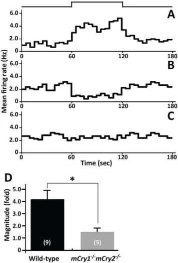
The spontaneously firing SCN neurons of mice showed three different types of responses to light [28]. The recordings consisted of before, during, and after retinal stimulation and each stage was applied for 60 sec. The first type of response waslight-activated, in which the firing frequency increased during light exposure (Fig. 2A), the second type of response waslight-suppressed, in which the firing rate decreased during light exposure (Fig. 2B), and the third type of response was unresponsive in which changes in the firing rate during light exposure were less than 10% of the basal firing rate (Fig. 2C).
We compared the differences in the populations of the three types of responses in the SCN during the daytime and nighttime in wild-type andmCry12/2 mCry22/2 mice (Table 1). In wild-type mice, the spontaneous activities of six SCN neurons were recorded in the daytime and one (17%) of the SCN neurons was light-activated whereas the remaining neurons (83%) were unrespon-sive. During the nighttime, 10 SCN neurons were recorded in
wild-type mice and nine (90%) of them were light-activated and one neuron (10%) was light-suppressed. Statistical analysis revealed that a day–night difference in the populations of response types in SCN neurons of wild-type mice (P,0.001, Dunn’s test). In contrast, among 26 SCN neurons that were recorded inmCry12/2
mCry22/2 mice, 16 were recorded during the nighttime. In the nighttime recording, five neurons (31%) were light-activated and 11 neurons (69%) were unresponsive. Among the 10 neurons recorded during the daytime, one neuron (10%) was light-activated, three neurons (30%) were light-suppressed, and the remaining neurons (60%) were unresponsive. A day–night variation was not observed in the populations of response types in the SCN neurons ofmCry12/2mCry22/2mice.
We next examined the magnitude of change in neuronal firing activities by retinal illumination recorded in the SCN of wild-type and mCry12/2 mCry22/2 mice (Fig. 2D). Because few light-responsive neurons were observed during the daytime, the recording was carried out only in light-activated neurons during the nighttime. We collected the mean firing rate at each stage of light stimulation: before (60 sec), during (60 sec), and after (60 sec) light stimulations. The magnitude of neuronal light response was calculated by using the following equation:
Magnitude foldð Þ
~
Mean firing rate during light stimulation Mean firing rate before and after light stimulation=2
In the SCN, wild-type mice showed a magnitude of 4.016 0.79-fold whereasmCry12/2 mCry22/2 mice showed 1.3560.15-fold. The magnitude of the neuronal light response in the SCN of
mCry12/2mCry22/2 mice during the nighttime was significantly lower than in the wild-type mice (P,0.05, Student’st-test). Optic nerve fiber light responsiveness
To determine whether the reduced light response in the SCN of
mCry12/2 mCry22/2 mice was caused by retinal defects, we examined the response to retinal illuminations in firing activity in the optic nerve, which the neural light information pathway to the SCN. The recordings and light stimulations were performed using the same procedure as for the SCN recording. We recorded three classes of light responses in optic nerve fibers, which is consistent with Hartline et al. [29] and other reports [30,31]. The first type was the ON fiber that discharges vigorously when the retina is illuminated (Fig. 3A), the second was the OFF fiber that discharges vigorously when the light is turned off (Fig. 3B), and the third was the ON/OFF fiber that responds to both the onset and the termination of light (Fig. 3C).
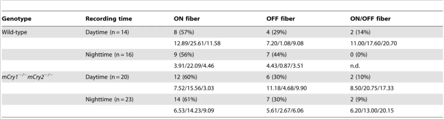
We compared the differences in the populations of the three fibers in the optic nerve during the daytime and the nighttime in wild-type andmCry12/2 mCry22/2 mice (Table 2). In wild-type mice, the spontaneous activities of 14 optic nerve fibers were recorded in the daytime and eight (57%) were ON fibers, four (29%) were OFF fibers, and two (14%) were ON/OFF fibers. During the nighttime, 16 fibers were recorded in wild-type mice and nine (56%) were ON fibers and seven (44%) were OFF fibers. InmCry12/2mCry22/2mice, 20 fibers were recorded in the optic nerve during the daytime and twelve (60%) were ON fibers, six (30%) were OFF fibers, and the remaining (10%) were ON/OFF fibers. During the nighttime, 23 fibers were recorded and 14 (61%) were ON fibers, seven (30%) were OFF fibers, and the remaining (9%) were ON/OFF fibers. No day–night variation was observed in the populations of the three classes of fibers in the optic nerve of wild-type ormCry12/2mCry22/2mice.
Figure 2. Reduced light response of neuronal firing activity in the SCN ofmCry12/2mCry22/2mice. A,B, andC, Peristimulus time histograms of three response patterns in firing activity are shown: (A) light-activated, (B) light-suppressed, and (C) unresponsive. The mean firing rate per second (Hz) is plotted every 5 min in the histogram. The light intensity was 1.061015photons?cm22
?s21
We also examined the magnitude of the neuronal light response recorded in the optic nerve fibers of wild-type and mCry12/2
mCry22/2mice (Fig. 3D). The recordings were carried out only in ON fibers during the daytime and the nighttime because OFF and ON/OFF fibers exhibit a specific response to retinal illuminations and thus these fibers could not be assayed in the present study. The magnitudes of the neuronal light response during the daytime and nighttime recordings were 2.6460.60-fold and 13.366 4.41-fold, respectively, in the ON fibers of wild-type mice. In contrast, the magnitude of the neuronal light response during the daytime and nighttime recordings were 3.2360.57-fold and 3.0260.82 fold, respectively, inmCry12/2mCry22/2mice. The magnitude of the neuronal light response in the ON fibers of wild-type mice during the nighttime was significantly higher than both the daytime value in wild-type mice and the day and night values in
mCry12/2 mCry22/2 mice (P,0.01; Tukey’s test). Thus, a day– night difference was observed in the magnitude of the neuronal light response in the ON fibers of wild-type mice but not in
mCry12/2mCry22/2mice. Discussion
In the present study, we investigated the effect of the loss of
Cryptochromeson the light response of neuronal firing activity in the SCN. We revealed thatmCry12/2mCry22/2mice have a reduced firing activity response to retinal illumination in nighttime recordings and did not exhibit a day–night variation in frequency of the response types in the SCN. We also determined that
mCry12/2 mCry22/2 mice have decreased optic nerve fiber photosensitivity of optic nerve fibers during the night. These results suggest thatCryptochromesplay a key role in the sensitivity of circadian photoreception in mammals.
Our previous study demonstrated that the photo response of firing activity in the mouse SCN had phase-dependent manner and showed a light-intensity relationship [28]. These data indicate that the electrophysiological properties of mice SCN are similar to those of rats and hamsters [25,26,27] and may correspond to properties of the light-induced phase shifting in locomotor activity of mice. In the present results, the SCN of wild-type mice showed a day–night difference in the populations of light responsive neurons. On the other hand, a day–night variation was not observed in the proportion of response types in the SCN of
mCry12/2mCry22/2mice. The magnitudes of the neuronal light responses in the SCN of mCry12/2 mCry22/2 mice were
significantly lower than those of wild-type mice during the night.
mCry12/2 mCry22/2 mice displayed a severe defect in c-fos
induction in the SCN [19,20] and no day–night difference inc-fos
induction in the SCN [32]. These data are consistent with the present results, suggesting that circadian photoreception sensitivity is reduced in the SCN ofmCry12/2mCry22/2mice.
BecauseCryptochromeshave a transcriptional regulatory function in the molecular clock mechanism,mCry12/2mCry22/2mice do not exhibit circadian rhythms of locomotor activity in DD [19,24], suggesting that the SCN lacks to the central clock function in
mCry12/2mCry22/2mice. Although it appears that the loss of the day–night difference in the photo response in the SCN of these animals causes the lack of SCN function, we consider this model unlikely because some evidence for partial clock function in the absence of Cryptochromes has been reported. For example, anticipatory wheel-running activity, where mice exhibit increased locomotor activity in the hours just prior to lights off, has been reported inmCry12/2mCry22/2mice [20,22]. Under normal LD conditions, a single peaks in circadian multiunit electrical activities were detected in thein vitro SCN slices from these animals [33]. Our present results also show a temporal difference in the spontaneous neuronal of the SCN inmCry12/2 mCry22/2mice. These data suggest that mCry12/2 mCry22/2 mice maintained under LD conditions have normal clock functions of the SCN through the duration of one circadian cycle. Because we recorded the neuronal firing activity of these animals under LD conditions, the SCN showed normal clock functions inCry-deficient mice in the present study.
Our data demonstrate that the light response of ON fibers also exhibited a day-night difference in wild-type mice, although the day–night variation was not observed in the populations of optic nerve fibers in wild-type ormCry12/2mCry22/2mice. Circadian rhythms have been detected in numerous invertebrate visual systems but are not unique to them [34]. A circadian clock in the brain ofLimulustransmits efferent optic nerve activity to the lateral eyes and increases their sensitivity at night [35] enabling them to see nearly as well at night as they do during the day. Circadian rhythms have been also detected in functions such as photorecep-tor disk shedding [36] and retina light sensitivity of the retina in rats [37] and humans [38]. Therefore, it is possible that the day– night difference in the sensitivity of ON fibers is relevant to circadian photoreceptions. The observation that the ON fibers of
mCry12/2 mCry22/2 mice lack these day–night variations may
Table 1.Temporal difference in frequency of the response types in the SCN of wild-type andmCry12/2mCry22/2mice.
Genotype Recording time Response Unresponsive
Activated Suppressed
Wild-type Daytime (n = 6) 1 (17%) 0 (0%) 5 (83%)
1.00/4.00/0.90 n.d. 2.24/2.22/2.20
Nighttime (n = 10)* 9 (90%) 1 (10%) 0 (0%)
0.98/3.00/0.93 2.10/1.30/2.10 n.d.
mCry12/2mCry22/2 Daytime (n = 10) 1 (10%) 3 (30%) 6 (60%)
1.45/1.50/1.25 1.60/1.17/1.57 3.67/3.63/3.67
Nighttime (n = 16) 5 (31%) 0 (0%) 11 (69%)
1.48/2.46/2.00 n.d. 1.56/1.63/1.65
The actual number and proportion (%) of responding cells in mouse SCN during daytime and nighttime are shown. The mean firing rates of each responding cell are exhibited in the bottom of each column described as before/during/after light stimulation.
*P,0.001, when compared with daytime group of wild-type mice andmCry12/2mCry22/2mice (Dunn’s test). n.d.; not determined.
lead to the finding thatCryptochromesplay a role in the retina for mammalian circadian photoreception.
The discovery of intrinsically photoresponsive retinal ganglion cells (ipRGCs) has given non-visual phototransduction an anatomical basis [39]. Berson and colleagues used retrograde dye tracing from the circadian pacemaking cells in the rat SCN to define direct retinohypothalamic-projecting ganglion cells, and patch-clamp recording showed that these cells are found to be intrinsically photosensitive whereas non-retinohypothalamic pro-jecting retinal ganglion cells had no intrinsic photosensitivity [40]. Although we could not determine whether all recorded optic nerve fibers were ipRGCs in the present study,CryptochromemRNAs are expressed in the retinal ganglion cells of mice [8]. Melanopsin is also expressed nearly exclusively in the ,1000 ipRGCs of the rodent retina [41]. The mammalian retina contains an intrinsic
circadian clock that controls melatonin synthesis and many other retinal functions [42]. In addition, retinal ganglion cells express
Period(Per)1and2,Clock, andBmal1, as well asCry1and2, which are core molecular components of circadian clocks and their expression is necessary for circadian rhythmicity [43]. Further-more, studies using real-time reporting of the PER2::LUC fusion protein revealed that clock gene rhythms persist for.25 days in cultured mouse retinas [43,44]. These data suggest that the ipRGCs of the mammalian retina contain functionally autono-mous circadian clocks. In the present study,mCry12/2mCry22/2 mice did not exhibit a day–night difference in spontaneous firing rate of the optic fibers and in the magnitude of neuronal light response in ON fibers. There results indicate that the low photosensitivity in the optic fibers is caused by the loss of
Cryptochromesin the retina.
In summary, this study provides several findings regarding the neuronal light response in the SCN and optic fibers ofCry-deficient mice: (1) A day–night variation in the populations of response types in SCN neurons was observed in wild-type mice but not in
mCry12/2 mCry22/2 mice. (2) The magnitude of the neuronal light response in the SCN ofmCry12/2 mCry22/2 mice during nighttime was significantly lower than in wild-type mice. (3) A day–night difference was observed in the magnitude of neuronal light response in the ON fibers of wild-type mice but not in
mCry12/2 mCry22/2 mice. These findings indicate that CRY deletion leads to the low photo response of the SCN and optic nerve fibers during the nighttime. In addition, we observed a day– night difference in the spontaneous firing rates in the optic fibers in wild-type mice but not inmCry12/2 mCry22/2 mice, suggesting that CRY deletion also disrupts the circadian rhythms of the neural system in the retina. We conclude that the low photo response in the SCN ofCry-deficient mice is caused by a circadian gating defect in the retina, which suggests thatCryptochromes are required for appropriate temporal photoreception in mammals.
Materials and Methods
Animals
The mCry12/2 mCry22/2 mice (originally from the colony of Dr. T. Todo [Kyoto University, Kyoto, Japan]) and wild-type mice of a similar mixed background were generated as described previously [19,20]. Genotyping was carried out by PCR using two sets of primers that amplified the wild-type or the disrupted gene for each of theCryptochromegenes. Animals were maintained under controlled air conditions (room temperature, 2461uC, and humidity, 50%65%) with food and water available ad libitum. Animals were housed under a LD cycle of 12 hr of light and 12 hr of darkness with a light intensity of 200–300 lux until the beginning of the experiment. All animal housing and experimental procedures were carried out in accordance with the guidelines of the Japanese Physiological Society and approved by the Institu-tional Animal Care and Use Committee of the Graduate School of Biomedical Sciences Nagasaki University (approval ID#: 0206090168).
Preparation
Male mCry12/2 mCry22/2 mice and wild-type mice ranging from 3 to 5 months of age were used in the experiments. The experiments were carried out during the daytime, ZT 4–8 (ZT12 is defined as the time of lights-off), and nighttime, ZT 14–16. The mice studied during the daytime were transferred to constant darkness from ZT 12 on the previous day in order to prevent light adaptation in animals and to create light conditions similar to the nighttime recording. On the day of the experiment, mice were Figure 3. Reduced light response of neuronal firing activity in
the optic nerve fibers ofmCry12/2mCry22/2mice. A,B, andC, Peristimulus time histograms of three distinct response patterns were recorded: ON fibers, which respond to light onset (A), OFF fibers, which respond to light offset (B), and ON/OFF fibers, which respond to both light onset and offset (C). The mean firing rate per second (Hz) is plotted every 5 min in the histogram. Light intensity was 1.061015
photons?cm22
?s21for 60 sec. The timing of the light pulse is indicated
in the step diagram above the records.D, The magnitude of change in the discharge rate for retinal illumination recorded in the ON fiber. Histograms depict data as mean 6 SEM; the number of animals is shown in parentheses. **P,0.01 vs. others, Tukey’s test.
transferred to the experimental room after their eyes were covered with blindfolds. The surgery described below, which occurred before the electrical recording, was performed under dim red light (,10 lux). The mice were anesthetized with 20% urethane solution (initial dose 2 g/kg, i.p.). Thereafter, the mice were placed in a stereotaxic instrument (Narishige, Tokyo, Japan) with the incisor bar set22 mm below the ear bar and cranial surgery was performed. The coordinates for the SCN in mice were 0.4 mm anterior to bregma, 0.1 mm lateral to the midline, and 5.0– 5.5 mm below the dural surface. The pupils of the animals were dilated 30 min before the recordings by application of 1% atropine sulfate to the cornea.
Electrophysiological recordings
Electrophysiological experiments were performed as previously described [28]. Extracellular single unit recordings were per-formed with a glass micropipette electrode (10–20 MV) filled with 2% Chicago Sky Blue (SIGMA, St. Louis, MO) in 0.5 M NaCl. The potentials were amplified, processed through a bandpass analog filter (100 Hz–3 KHz), and fed into a personal computer with an AD converter (PCI-6024E; National Instruments, Austin, TX). The frequency of neuronal single-unit firing was counted with customized software programmed by LabVIEW (National Instruments).
When spontaneous firings were recorded, we continued recording for 10 min without illumination to ensure stability of the firing activity. The stimulation was carried out after the variation of the mean spontaneous firing rate in 60 sec settled down to a rate that was within 10% of the value of the previous minutes. An increase or decrease in the firing activity was determined for the light stimulation of 60 sec in duration by a mean frequency change of more than 10% relative to the mean firing rate for the 60 sec prior to the stimulation.
Light stimulation
A photic stimulation pulse was applied with an assembled light source of 6 blue-green high-intensity light-emitting diodes (lmax:
500 nm, E1L51-KC0A2-02; Toyota Gosei, Kasugai, Japan) to the eye of the animal with pupils dilated. The intensity of the light stimulation at the plane of the eye was 1.061015 photons?cm22
?s21
. Light intensity was measured using a United Detector Technologies photometer (model S371; Hawthorne, CA).
Identification of the electrode position
At the end of the recording, a small negative current (3–5mA; 3–5 min) was passed through the microelectrode to mark the recording site. The brain was removed and fixed overnight with 4% paraformaldehyde in phosphate-buffered saline. The brain was sliced (100mm thick) with a micro slicer (Dosaka EM, Kyoto, Japan) and slices were counterstained with cresyl violet to verify the location of the SCN or optic nerve.
Data analysis and statistics
Differences between proportions of responsive neurons or fibers during the daytime and the nighttime in wild-type andmCry12/2
mCry22/2mice were analyzed using the non-parametric Kruskal– Wallis test followed by Dunn’s post-hoc analyses. In other cases, Student’st-tests were used to examine the difference between two groups and one-way ANOVA with post-hoc Tukey’s test was used to compare multiple groups. All results are presented as the mean
6SEM and were considered significant atP,0.05.
Acknowledgments
We thank Dr. Takeshi Todo for the generous gift ofmCry12/2mCry22/2 mice.
Author Contributions
Conceived and designed the experiments: TJN SE KS. Performed the experiments: TJN. Analyzed the data: TJN. Contributed reagents/ materials/analysis tools: TJN. Wrote the paper: TJN.
References
1. Inouye ST, Shibata S (1994) Neurochemical organization of circadian rhythm in the suprachiasmatic nucleus. Neurosci Res 20: 109–130.
2. Lowrey PL, Takahashi JS (2000) Genetics of the mammalian circadian system: Photic entrainment, circadian pacemaker mechanisms, and posttranslational regulation. Annu Rev Genet 34: 533–562.
3. Freedman MS, Lucas RJ, Soni B, von Schantz M, Munoz M, et al. (1999) Regulation of mammalian circadian behavior by non-rod, non-cone, ocular photoreceptors. Science 284: 502–504.
4. Mrosovsky N, Foster RG, Salmon PA (1999) Thresholds for masking responses to light in three strains of retinally degenerate mice. J Comp Physiol [A] 184: 423–428. Table 2.Temporal difference in frequency of the ON, OFF, and ON/OFF fibers of wild-type andmCry12/2mCry22/2mice.
Genotype Recording time ON fiber OFF fiber ON/OFF fiber
Wild-type Daytime (n = 14) 8 (57%) 4 (29%) 2 (14%)
12.89/25.61/11.58 7.20/1.08/9.08 11.00/17.60/20.70
Nighttime (n = 16) 9 (56%) 7 (44%) 0 (0%)
3.91/22.09/4.46 4.43/0.87/3.51 n.d.
mCry12/2mCry22/2 Daytime (n = 20) 12 (60%) 6 (30%) 2 (10%)
7.52/15.56/3.03 11.18/4.68/9.90 8.50/20.75/17.33
Nighttime (n = 23) 14 (61%) 7 (30%) 2 (9%)
6.53/14.23/9.09 5.61/2.67/6.06 6.20/13.00/20.15
The actual number and proportion (%) of types of the optic fiber during daytime and nighttime are shown. The mean firing rates of each responding cell are exhibited in the bottom of each column described as before/during/after light stimulation. n.d.; not determined.
5. Lucas RJ, Freedman MS, Munoz M, Garcia-Fernandez JM, Foster RG (1999) Regulation of the mammalian pineal by non-rod, non-cone, ocular photore-ceptors. Science 284: 505–507.
6. Lucas RJ, Douglas RH, Foster RG (2001) Characterization of an ocular photopigment capable of driving pupillary constriction in mice. Nat Neurosci 4: 621–626.
7. Provencio I, Rodriguez IR, Jiang G, Hayes WP, Moreira EF, et al. (2000) A novel human opsin in the inner retina. J Neurosci 20: 600–605.
8. Miyamoto Y, Sancar A (1998) Vitamin B2-based blue-light photoreceptors in the retinohypothalamic tract as the photoactive pigments for setting the circadian clock in mammals. Proc Natl Acad Sci U S A 95: 6097–6102. 9. Bowes C, Li T, Danciger M, Baxter LC, Applebury ML, et al. (1990) Retinal
degeneration in the rd mouse is caused by a defect in the beta subunit of rod cGMP-phosphodiesterase. Nature 347: 677–680.
10. Carter-Dawson LD, LaVail MM, Sidman RL (1978) Differential effect of the rd mutation on rods and cones in the mouse retina. Invest Ophthalmol Vis Sci 17: 489–498.
11. Yoshimura T, Ebihara S (1998) Decline of circadian photosensitivity associated with retinal degeneration in CBA/J-rd/rd mice. Brain Res 779: 188–193. 12. Panda S, Sato TK, Castrucci AM, Rollag MD, DeGrip WJ, et al. (2002)
Melanopsin (Opn4) requirement for normal light-induced circadian phase shifting. Science 298: 2213–2216.
13. Ruby NF, Brennan TJ, Xie X, Cao V, Franken P, et al. (2002) Role of melanopsin in circadian responses to light. Science 298: 2211–2213. 14. Lucas RJ, Hattar S, Takao M, Berson DM, Foster RG, et al. (2003) Diminished
pupillary light reflex at high irradiances in melanopsin-knockout mice. Science 299: 245–247.
15. Panda S, Provencio I, Tu DC, Pires SS, Rollag MD, et al. (2003) Melanopsin is required for non-image-forming photic responses in blind mice. Science 301: 525–527.
16. Hattar S, Lucas RJ, Mrosovsky N, Thompson S, Douglas RH, et al. (2003) Melanopsin and rod-cone photoreceptive systems account for all major accessory visual functions in mice. Nature 424: 76–81.
17. Sancar A (2000) Cryptochrome: the second photoactive pigment in the eye and its role in circadian photoreception. Annu Rev Biochem 69: 31–67. 18. Miyamoto Y, Sancar A (1999) Circadian regulation of cryptochrome genes in
the mouse. Brain Res Mol Brain Res 71: 238–243.
19. Selby CP, Thompson C, Schmitz TM, Van Gelder RN, Sancar A (2000) Functional redundancy of cryptochromes and classical photoreceptors for nonvisual ocular photoreception in mice. Proc Natl Acad Sci U S A 97: 14697–14702.
20. Vitaterna MH, Selby CP, Todo T, Niwa H, Thompson C, et al. (1999) Differential regulation of mammalian period genes and circadian rhythmicity by cryptochromes 1 and 2. Proc Natl Acad Sci U S A 96: 12114–12119. 21. Van Gelder RN, Wee R, Lee JA, Tu DC (2003) Reduced pupillary light
responses in mice lacking cryptochromes. Science 299: 222.
22. Mrosovsky N (2001) Further characterization of the phenotype of mCry1/ mCry2-deficient mice. Chronobiol Int 18: 613–625.
23. Van Gelder RN, Gibler TM, Tu D, Embry K, Selby CP, et al. (2002) Pleiotropic effects of cryptochromes 1 and 2 on free-running and light-entrained murine circadian rhythms. J Neurogenet 16: 181–203.
24. van der Horst GT, Muijtjens M, Kobayashi K, Takano R, Kanno S, et al. (1999) Mammalian Cry1 and Cry2 are essential for maintenance of circadian rhythms. Nature 398: 627–630.
25. Meijer JH, Groos GA, Rusak B (1986) Luminance coding in a circadian pacemaker: the suprachiasmatic nucleus of the rat and the hamster. Brain Res 382: 109–118.
26. Meijer JH, Rusak B, Ganshirt G (1992) The relation between light-induced discharge in the suprachiasmatic nucleus and phase shifts of hamster circadian rhythms. Brain Res 598: 257–263.
27. Meijer JH, Watanabe K, Schaap J, Albus H, Detari L (1998) Light responsiveness of the suprachiasmatic nucleus: long-term multiunit and single-unit recordings in freely moving rats. J Neurosci 18: 9078–9087.
28. Nakamura TJ, Fujimura K, Ebihara S, Shinohara K (2004) Light response of the neuronal firing activity in the suprachiasmatic nucleus of mice. Neurosci Lett 371: 244–248.
29. Hartline HK (1938) The response of single optic nerve fibers of the vertebrate eye to illumination of the retina. Am J Physiol 121: 400–415.
30. Bisti S, Gargini C, Chalupa LM (1998) Blockade of glutamate-mediated activity in the developing retina perturbs the functional segregation of ON and OFF pathways. J Neurosci 18: 5019–5025.
31. Wang GY, Liets LC, Chalupa LM (2001) Unique functional properties of on and off pathways in the developing mammalian retina. J Neurosci 21: 4310–4317.
32. Thompson CL, Selby CP, Partch CL, Plante DT, Thresher RJ, et al. (2004) Further evidence for the role of cryptochromes in retinohypothalamic photoreception/phototransduction. Brain Res Mol Brain Res 122: 158–166. 33. Albus H, Bonnefont X, Chaves I, Yasui A, Doczy J, et al. (2002)
Cryptochrome-deficient mice lack circadian electrical activity in the suprachiasmatic nuclei. Curr Biol 12: 1130–1133.
34. Block GD, Khalsa SB, McMahon DG, Michel S, Guesz M (1993) Biological clocks in the retina: cellular mechanisms of biological timekeeping. Int Rev Cytol 146: 83–144.
35. Barlow RB, Jr., Bolanowski SJ, Jr., Brachman ML (1977) Efferent optic nerve fibers mediate circadian rhythms in the Limulus eye. Science 197: 86–89. 36. LaVail MM, Ward PA (1978) Studies on the hormonal control of circadian outer
segment disc shedding in the rat retina. Invest Ophthalmol Vis Sci 17: 1189–1183.
37. Terman M, Terman J (1985) A circadian pacemaker for visual sensitivity? Ann N Y Acad Sci 453: 147–161.
38. Bassi CJ, Powers MK (1986) Daily fluctuations in the detectability of dim lights by humans. Physiol Behav 38: 871–877.
39. Berson DM, Dunn FA, Takao M (2002) Phototransduction by retinal ganglion cells that set the circadian clock. Science 295: 1070–1073.
40. Berson DM (2003) Strange vision: ganglion cells as circadian photoreceptors. Trends Neurosci 26: 314–320.
41. Hattar S, Liao HW, Takao M, Berson DM, Yau KW (2002) Melanopsin-containing retinal ganglion cells: architecture, projections, and intrinsic photosensitivity. Science 295: 1065–1070.
42. Green CB, Besharse JC (2004) Retinal circadian clocks and control of retinal physiology. J Biol Rhythms 19: 91–102.
43. Ruan GX, Zhang DQ, Zhou T, Yamazaki S, McMahon DG (2006) Circadian organization of the mammalian retina. Proc Natl Acad Sci U S A 103: 9703–9708.