Comparative cytogenetic studies of
Bufo ictericus
,
B. paracnemis
(Amphibia,
Anura) and an intermediate form in sympatry
MFC Azevedo
1, F. Foresti
2, PRR Ramos
3and J Jim
1 1Departamento de Zoologia, Instituto de Biociências, UNESP, Botucatu, SP, Brazil.
2Departamento de Morfologia, Instituto de Biociências, UNESP, Botucatu, SP, Brazil.
3Departamento de Física e Biofísica, Instituto de Biociências, UNESP, Botucatu, SP, Brazil.
Abstract
Specimens ofBufo ictericus, Bufo paracnemis and a third type, considered an intermediate subgroup between these species, were cytogenetically studied by conventional Giemsa staining, C-banding and staining of the nucleolus organizer region (NOR). The nuclear DNA content and seroproteins were also analyzed to characterize these species, and verify the possibility of hybridization between them. Karyotypes and cytogenetic markers were essentially equal on the basis of the methods used. The DNA nuclear content found was 6.25 ± 0.30 pg/DNA inBufo ictericus; 7.57 ± 0.40 pg/DNA in Bufo paracnemis and 7.04 ± 0.29 pg/DNA in the intermediate subgroup. Eletrophoresis of total blood serum inBufo ictericus, Bufo paracnemis and the intermediate specimens revealed a remarkable difference in the patterns of the protein bands whose molecular weight corresponded to that of albumin. While the parental species presented two different bands, the intermediate form presented 4. However, only three of these bands were seen in each specimen. The results obtained pointed to a high probability for natural hybridization betweenBufo ictericus and Bufo paracnemis in the site and specimens studied.
Key words: Bufo, hybridization, cytogenetics, nuclear DNA, seroproteins.
Received: July 26, 2002; Accepted: March 10, 2003.
Introduction
Anura have usually been studied from the morpho-logical and ecomorpho-logical point of view. TheBufogenus is of particular interest for studies regarding evolution and re-productive isolation mechanisms (Haddadet al., 1990), due to its cosmopolitan distribution and easy interbreeding in nature. Many authors have reported hybridization among the members of this genus in areas of sympatry (Blair, 1941; Thornton, 1955; Sullivan, 1986; Schlyter et al., 1991; Gerguset al., 1999 and Malmoset al., 2001). Ac-cording to Wells (1977), the high frequency of hybridiza-tion can be explained by the reproductive mechanisms, such as external fertilization or the non-territoriality of ob-served in the group.
“Pre-zygotic” mechanisms of reproductive isolation prevent inter-species breeding while “post-zygotic” mech-anisms do not always prevent the formation of descendents (Mayr, 1977). However, hybrids are not very successful due to sterility, or either partial or total lack of viability.
Blair (1955) believes that natural selection reinforces isola-tion mechanisms against hybridizaisola-tion in the form of a handicap against the hybrid in the competition with the pa-rental types. Hybridization has attracted the interest of tax-onomists and evolutionists because the study of this process in nature is of great value in obtaining answers to questions regarding the mechanisms involved in the pro-cess of evolution and species formation in the group. Even so, little effort has been made toward obtaining consistent data regarding hybridization success in nature. Therefore, the few isolated facts available in the literature are ex-tremely useful in the evaluation of this part of the evolu-tionary process, according to Blair (1974).
Kloss (1972) reported the existence of inter-gradient forms betweenBufo ictericusandB. paracnemisin transi-tion zones between brush, prairies, forest, and clearings. Jim (1980) also refers to the “topo da cuesta”, an area lo-cated in Botucatu, São Paulo, Brazil, as an intermediate area where these species occur, and reported the difficulties encountered in identifying the material collected in this re-gion due to the morphological variations presented by the samples collected. More recently, Kasaharaet al.(1998) have reported the occurrence of aBufospecimen with an in-termediate phenotype between B. paracnemis and B.
Send correspondence to Marisa Fagundes Carvalho de Azevedo. Departamento de Morfologia, Instituto de Biociências - UNESP, Campus de Botucatu, Distrito de Rubião Junior, C.P. 510, 18618-000 Botucatu, SP, Brazil. Phone/Fax: 0055-14-6802.6264. E-mail: [email protected].
ictericus. The occurrence of these intermediary types could have resulted from forest clearing and damming of water courses, which cause environmental alterations and affect the degree of isolation among the species (Blair, 1941). The increase in the degree of interaction among these species could be interfering in their reproductive strategies, affect-ing the hybridization frequency (Gerguset al., 1999).
Bufo ictericus and B. paracnemis are phylogeneti-cally closely related (Blair, 1972; Frost, 1985), and the in-termediate forms observed could be due to the anatomical similarities, or the possible occurrence of crossbreeding. The purpose of this study was to enable a better understand-ing of the relationships betweenBufo species and taxo-nomic studies of the group by characterizing the B. ictericusandB. paracnemissamples found in the region of Botucatu, São Paulo, Brazil, and also to test the hypothesis of hybridization between these species by comparing the results obtained from samples considered as parentals and supposedly hybrids (anatomically intermediate).
Material and Methods
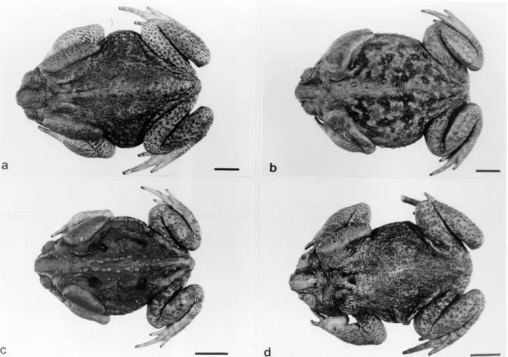
Seven of Bufo ictericus specimens, 4 of B. paracnemis, and 4 of an intermediate form (Figure 1), were
collected in a man-made lake with shoreline vegetation at Estância Funari, Botucatu, São Paulo, Brazil (S. 22°53’12,9” and W. 48°29’46,6”). The collection site was selected because it was an altered environment.
All the specimens, after sacrifice, were fixed in 10% formaldehyde, preserved in 70% alcohol and kept in the collection of the Department of Zoology - UNESP, Botucatu, São Paulo, Brazil.
Cytogenetic techniques
Mitotic cells were obtained by the injection of a bio-logical yeast solution, as described by Cole and Leavens (1971), followed by lymphocyte cellular culture as de-scribed by Fenocchio and Bertollo (1988). For staining, the slides were covered with Giemsa solution buffer. For the determination of the number and location of Nucleolus Or-ganizer Regions (NORs) in the karyotypes, silver-ion (AgNO3) impregnation was used according to a method
originally described by Howell and Black (1980). C-banding pattern was obtained as proposed by Sumner (1972). The karyotypes of the species under study were not different from those of other populations previously stud-ied. Therefore, the formats of distribution and chromosome
presentation were according to the system used by Blair (1972) and Kasaharaet al.(1996), among others.
Nuclear DNA quantification
Blood cell smears from each specimen were prepared as described by Gold and Price (1985), with 10-minute hy-drolysis. Quantitative analyses of the nuclei were per-formed with the software OPTIMAS, 4.1 version.
Seroprotein electrophoresis
Seroproteins were studied as described by Hames and Rickwood (1990). The method of alkaline electrophoresis (pH = 9.0) was used, in polyacrylamide gel, in separate containers, at 10% concentration in the separation gel, and at 4% in the topping gel.
Results
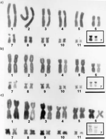
The diploid number was 2n = 22 inBufo ictericus,B. paracnemisand the individuals considered as intermediate forms. All of chromosomes were submetacentric and meta-centric in all samples analyzed. The presence of secondary constrictions was observed on the short arm of pair 7, coin-ciding with the NORs (Figure 2). This description is the same as that decrypted by Blair (1972) and Kasaharaet al.
(1996).
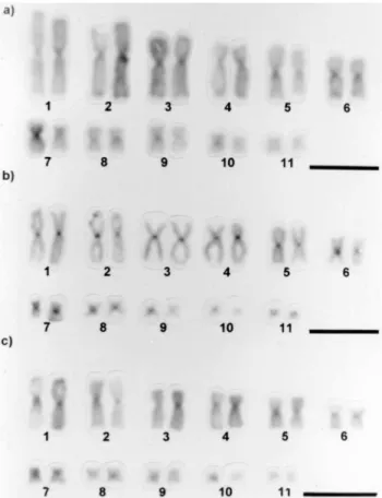
The detection of constitutive heterochromatin by the C-banding technique revealed that all preparations of B. ictericus,B. paracnemis, and the intermediate form studied showed positive marks in the centromeric regions of all chromosomes, which were more evident in the larger pairs. In the majority of metaphases, pair 7 presented a heterochromatic interstitial block, coincident with the sec-ondary constriction. Faint telomeric and interstitial marks could be observed in some chromosomes of some meta-phases (Figure 3). In one individual of theB. paracnemis
sample, 12 silver-stained metaphases were examined. Five of them presented centromeric marks in all chromosomes (Figure 4).
The average nuclear DNA content found in B. ictericus was 6.25 ± 0.30 pg DNA/nucleus, in B. paracnemis, it was 7.57 ± 0.40 pg and the intermediate form presented an average of 7.04 ± 0.29 pg DNA/nucleus (Table 1).
Eletrophoresis of total blood serum inBufo ictericus, Bufo paracnemisand the intermediate specimens revealed a remarkable difference in the patterns of the protein bands whose molecular weight corresponded to that of albumin (Figure 5). The analysis of these bands indicated that both species considered as parental presented at least one large band whereas the intermediate form presented at least three distinct bands.
Discussion
Cytogenetics
The karyotypes ofB. ictericus,B. paracnemisand the form with intermediate characteristics between the first two, shared the same karyotype pattern observed in practi-cally all the species of the genus, in line with the report by Bogart (1972). Henderson et al. (1972) and Hsu et al.
(1975) have already observed that NORs are usually found in zones of secondary constriction, as seen in the present work. In general, these marks exhibited equivalent sizes in the same metaphase.
Figure 2- Karyotypes with details of the NOR-bearing pair in the boxes. (a) Bufo ictericus; (b) Bufo paracnemis; (c) Intermediate Type. Bar = 10µm.
In amphibians, especially in Anura, NORs tend to be coincident with positive C-band regions, and may present large C-band positive blocks associated with them (King, 1988, 1990; King et al., 1990). With regard to constitu-tional heterochromatic detection, the speciesB. ictericus
andB. paracnemisand the intermediate form studied in this research were C-band positive in the centromeric region in all chromosomes. In the majority of the metaphases, pair 7 showed an interstitial C-band, coinciding with the region of secondary constriction and with NORs. This pattern did not differ from those found in the other species belonging to the
marinusgroup (Schmid, 1978, 1980; Schmid and Almeida, 1988; Kasaharaet al., 1996), based on which Kasaharaet al.(1996) suggested that the pattern observed could charac-terize this morphological group.
In one B. paracnemis specimen studied herein, centromeric marks were observed in metaphases submitted to silver-staining. According to Sumner (1990 although of wide spread use, this method is not specific for NORs, since heterochromatin, kinetocore or other acid proteins may also occasionally be marked. However, these structures, when marked, are easily distinguished from NORs by clear dif-ferences in shape and color, as observed here.
The results obtained in the application of these cytogenetic techniques did not permit the differentiation of the species. However, the karyotypic stability described for these species was also identified in the samples from the Botucatu region, São Paulo, Brazil.
Nuclear DNA content
Quantification of nuclear DNA has been conducted in various frog species before and the data obtained have dem-onstrated that the quantity of nuclear DNA is constant for each species. Therefore, as a particular characteristic, it might be used to distinguish each given species (Herreroet al., 1993; Cavalloet al., 2002). Mean nuclear DNA content was 6.25 ± 0.30pg DNA/nucleus inB. ictericusand 7.57 ± 0.40pg inB. paracnemis, while in the intermediary form it was 7.04 ± 0.29pg DNA/nucleus, characterizing the latter as an intermediate form between B. ictericus and B. paracnemis. Comparisons between parental and intermedi-ary types showed significantly different values only in pa-rental types (Table 1). Therefore, under the given conditions, the methodology utilized can be considered ef-fective at the level of species, in systematic studies of the group. Furthermore, the DNA-nuclear content found in the intermediate forms showed an intermediary value between the means obtained in the other two species, suggesting the occurrence of a hybridization process betweenB. ictericus
andB. paracnemis. The same analysis was done by Cavallo
et al.(2002), in a study of diploid and tetraploid popula-tions ofBufo viridis and they considered as hybrids the triploid individuals found in one population.
This approach was also used by Diaz (1986), who considered that the nuclear DNA content could be a useful
Table 1- Diploid number and DNA content values, in picograms (pg), observed in the Bufo species and in the Intermediate form analyzed.
Toads analyzed (M/F)
2n Nuclear DNA content (pg)
Bufo ictericus 5/2 22 6.25 + 0.30
Bufo paracnemis 3/1 22 7.57 + 0.40
Intermediate form 3/1 22 7.04 + 0.29
M: Male; F: Female.
Figure 5- Polyacrylamide gel electrophoresis showing the characteristic pattern of blood serum proteins ofBufo ictericus(Bi),Bufo paracnemis
(Bp) and the Intermediate Type (It). O: Origin; Alb: Bands corresponding to albumin.
Figure 4- Karyotypes stained according to the C-banding technique. (a)
parameter in the phylogenetic analysis of that group. This is based on the fact that many closely related species possess the same diploid number and very similar, or even identi-cal, chromosomal morphology, exhibiting, nonetheless, considerable difference in nuclear DNA content (revised by King, 1990). Northlandet al.(1990) also considered that comparative studies of nuclear DNA content were relevant, when comparisons were made among groups with rela-tively close phylogenic characteristics.
Seroprotein electrophoresis
With the results obtained with the electrophoresis technique for the protein bands whose mobility corre-sponded to that of albumin, it was possible to observe a clear and neat difference between the band patterns of the intermediate types and the species B. ictericus and B. paracnemis. The two latter species were identified by two distinct bands in the region corresponding to the albumin while four bands were observed in the intermediate forms, three in each animal.
This is probably not a populational polymorphism as described by Bertini and Cei (1960), because small popula-tions possess reduced heterozygosity levels (Futuyma, 1992). The polymorphism found in B. ictericus and B. paracnemisdiffers widely in the patterns of albumin bands from that found in the intermediate forms. Furthermore, there is also the possibility of hybridization between one of the two parental species studied in this work and another species found in the study site, such asBufo crucifer, for ex-ample. However, the species B. ictericus and B. paracnemiswere selected for this study because they are morphologically more similar to the intermediate form.
According to Sage and Selander (1979), protein elec-trophoresis can reveal hybridization zones because they sometimes contain rare alleles not found in any of the pa-rental semi-species. Such alleles may have resulted from a high mutation index in the hybrid genoma, or be formed by intra-genetic recombination between different alleles of the parental populations. The results, therefore, suggest that hybridization occurs between B. ictericus and B. paracnemis.
Acknowledgments
Our thanks to Shirlei M. Recco-Pimentel for the sup-port in methodology and to Celio F. B. Haddad for sugges-tions and to Claudio Oliveira for critical reviewing of the manuscript. This work was supported by CAPES, CNPq, and FAPESP.
References
Bertini F and Cei JM (1960) Observaciones electroforeticas en seroproteinas de poblaciones argentinas de Bufo arenarum. Rev Soc Argent Biol 36(7-8):356-362.
Blair AP (1941) Variation, isolating mechanismis and hybridiza-tion in certain toads. Genetics 26:398-417.
Blair WF (1955) Mating call and stage of speciation in the Microhyla olivacea – M. carolinensis complex. Evolution 9:469-480.
Blair WF (1972) Evidence from hibridization. Evolution in the genus Bufo. Austin, University of Texas Press, Texas, pp 196-232.
Blair WF (1974) Character displacement in frogs. Amer Zool 14:1119-1125.
Bogart JP (1972) Karyotypes. In: Blair WF (ed) Evolution in the genus Bufo. Austin, University of Texas Press, Texas, pp 171-195.
Cavallo D, De Vita R, Eleuteri P, Borkin L, Emechenko V, Odierna G and Balletto E (2002) Karyological and flow cytometric evidence of triploid specimens in Bufo viridis (Amphibia Anura). Eur J Histochem 16(2):159-164. Cole CJ and Leavens CR (1971) Chromosome preparations of
amphibians and reptiles: improved technique. Herpetol Rev 3(6):102.
Díaz NF (1986) Biosistemática de los Leptodactylidae chilenos. An Mus Hist Nat Valparaíso 17:65-85.
Fenocchio AS and Bertollo LAC (1988) A simple method for fresh-water fish lymphocyte culture. Rev Brasil Genet 11(4):847-852.
Frost DR (1985) Amphibian Species of the World. Allen Press and the Association of Systematics Collections. Lawrence, Kansas. pp 723.
Futuyma, DJ (1992) Biologia Evolutiva. Sociedade Brasileira de Genética/CNPq. Ribeirão Preto, São Paulo. pp 631. Gergus EWA, Malmos KB and Sullivan BK (1999) Natural
hy-bridization among distantly related toads (Bufo alvaris,Bufo cognatus, Bufo woodhousii) in Central Arizona. Copeia 2:281-286.
Gold JR and Price HJ (1985) Genome size variation among north american minnows (Cyprinidae). I. Distribution of the varia-tion in five species. Heredity 54:297-305.
Haddad CFB, Cardoso AJ and Castanho LM (1990) Hibridação natural entre Bufo ictericus e Bufo crucifer (Amphibia: Anura). Rev Brasil Biol 50(3):739-744.
Hames BD and Rickwood D (1990) Gel electrophoresis of pro-teins. Oxford University Press, New York, pp 383. Henderson AS, Warburton D and Atwood KC (1972) Location of
ribosomal DNA in the human chromosome complement. Proc Natl Sci 69:3394-3398.
Herrero P, Lopezjurado LF, Arano B and Garciaparis M (1993). Karyotype analyses and nuclear-DNA content of Bufo-brongersmai hoogmoed. J Herpetol 27(4):463-465. Howell WM and Black DA (1980) Controlled silver-staining of
nucleolus organizer regions with a protective colloidal de-veloper: a 1-step method. Experientia 36:1014-1015. Hsu TC, Spirito SE and Pardue ML (1975) Distribution of 18+28S
ribosomal genes in mammalian genomes. Chromosoma 53:25-36.
Jim J (1980) Aspectos Ecológicos dos Anfíbios Registrados na Região de Botucatu, Estado de São Paulo (Amphibia, Anura). Doctoral Thesis, Instituto de Biociências, Universidade de São Paulo, São Paulo, Brazil.
Kasahara S, Silva APZ and Gruber SL (1998) Use of lymphocyte cultures for BrdU replication banding patterns in anuran species (Amphibia). Genet Mol Biol 21(4):471-476.
King M (1988) The interrelationship of G-banding, C-banding pattern and nucleolus organizer structure in anuran amphibi-ans. In: Brandam PE (ed) Kew Chromosome Conference 111, pp 51-63.
King M (1990) Amphibia. In: John B (ed) Animal Cytogenetics. Gebrüder Borntraeger, Berlin. pp 2, 241.
King M, Contreras N and Honeucutt RL (1990) Variation within and between nucleolar regions in Australian hylid frogs (Anura) shown by 18S and 28S “in situ” hybridization. Genetica 80:17-29.
Kloss GR (1972) Os Rhabdias dosBufodo grupoMarinus. Um estudo de espécies crípticas (Nematoda: Rhabditoidea). Doctoral Thesis, Universidade de São Paulo, São Paulo, Brazil.
Malmos KB, Sullivan BK and Lamb T (2001) Calling behavior and directional hybridization between two toads (Bufo microscaphus x B-woodhousii) in Arizona. Evolution 55(3):626-630.
Mayr E (1977) Populações, Espécies e Evolução. EdUSP, São Pulo, Brazil. pp 485.
Northland I, Capetillo J, Iturra P and Veloso A (1990) Nuclear DNA content and karyosystematic relationships of species grouped in primitive tribes of Leptodactylidae (Amphibia – Anura). Rev Brasil Genet 13(2):247-254.
Sage RD and Selander RK (1979) Hybridization between species of the Rana pipienscomplex in central Texas. Evolution 33:1069-1088.
Schlyter F, Hoglund J and Stromberg G (1991) Hybridization and low numbers in isolated populations of the Natterjack,Bufo calamita, and the green toadBufo viridis, in Southern Swe-den: possible conservation problems. Amphibia-Reptilia 12:267-281.
Schmid M (1978) Chromosome banding in Amphibia. I. Constitu-tive heterochromatin and nucleolus organizer regions in
BufoandHyla. Chromosoma 66:361-388.
Schmid M (1980) Chromosome banding in Amphibia. IV. Differ-entiation of GC- and AT- rich chromosome regions in Anura. Chromosoma 77:83-103.
Schmid M and Almeida CG (1988) Chromosome banding in Amphibia. XII. Restriction endonucleases banding. Chromosoma 96:283-290.
Sullivan BK (1986) Hybridization between the toadsBufo micros-caphus and Bufo woodhousei in Arizona: Morphological variation. Journal of Herpetology 20(1):11-21.
Sumner AT (1972) A simple technique for demonstrating cen-tromeric heterochromatin. Exptl Cell Res 75:304-306. Sumner AT (1990) Chromosome banding. Unwin Human,
Lon-don. pp 433.
Thornton WA (1955) Interspecific hybridization in Bufo woodhouseiandBufo valliceps. Evolution 9:455-468. Wells KD (1977) The social behavior of anuran amphibians.
Anim Behav 25:666-693.