Journal of Feline Medicine and Surgery Open Reports
1 –6
© The Author(s) 2016 Reprints and permissions:
sagepub.co.uk/journalsPermissions.nav DOI: 10.1177/2055116916675801 jfmsopenreports.com
This paper was handled and processed by the European Editorial Office (ISFM) for publication in JFMS Open Reports
Creative Commons Non Commercial CC-BY-NC: This article is distributed under the terms of the Creative Commons Attribution-NonCommercial 3.0 License (http://www.creativecommons.org/licenses/by-nc/3.0/) which permits non-commercial use, reproduction and distribution of the work without further permission provided the original work is attributed as specified on the SAGE and Open Access pages (https://us.sagepub.com/en-us/nam/open-access-at-sage).
Case description
A 5-month-old female intact feral kitten presented to the local animal shelter unable to ambulate after being trapped underneath a garage door. A board-certified neurologist (KMV) localized the lesion to the C5 to brain-stem region. Cervical and thoracic radiographs showed no evidence of cervical fracture, but a mild bronchointer-stitial pulmonary pattern was noted. As per routine pro-tocol of the shelter, imidacloprid/moxidectin (Advantage Multi for Cats; Bayer) was applied to the kitten and a 5 day course of fenbendazole was initiated at 50 mg/kg PO q24h.
The kitten was placed in foster care once able to ambulate 5 days later, and although it continued to improve neurologically, respiratory signs of tachypnea, cough and wheezing suddenly worsened in frequency and severity 9 days later. On day 10, the cat presented to the University of California, Davis, William R Pritchard
Respiratory distress associated with
lungworm infection in a kitten
Melissa M Hawley
1, Lynelle R Johnson
2, Donato Traversa
3,
Dan Bucy
1, Karen M Vernau
4and William Vernau
5Abstract
Case summary A 5-month-old feral kitten developed worsening respiratory signs, including tachypnea, coughing and wheezing after standard anthelmintic treatment with fenbendazole at a local shelter. The kitten was referred to the University of California, Davis, William R Pritchard Veterinary Medicine Teaching Hospital for further evaluation. Thoracic radiographs revealed a severe diffuse bronchointerstitial pattern with bronchial cuffing, ill-defined nodules and lymphadenomegaly. Differentials included infectious etiologies such as toxoplasmosis, feline infectious peritonitis and cryptococcosis. Parasitic infection was considered less likely, owing to previous anthelmintic treatment. Bronchoalveolar lavage revealed marked neutrophilic and eosinophilic inflammation, and parasitic larvae were observed in a swab of trachea mucus. PCR confirmed the larvae as Aelurostrongylus abstrusus. The kitten recovered with two more rounds of anthelmintic treatment.
Relevance and novel information Parasitic pneumonia should be considered as a cause of respiratory distress in kittens and cats. Lungworm infections have been more commonly reported in free-roaming young and adult cats, but cannot be excluded as a differential diagnosis in cats from varied environments and in kittens. Kittens appear to be especially sensitive to lungworm infections, manifested by the development of more severe clinical signs; thus lungworm infection should always be considered when presented with a kitten in respiratory distress. In the absence of cytologic confirmation of infection via bronchoalveolar lavage or oropharyngeal swab, PCR provides a valuable means for identification of lungworms, such as A abstrusus and Troglostrongylus brevior.
Accepted: 16 September 2016
1 William R Pritchard Veterinary Medical Teaching Hospital,
University of California, Davis, Davis, CA, USA
2 Department of Medicine and Epidemiology, University of
California, Davis, Davis, CA, USA
3 University Veterinary Teaching Hospital, University of Teramo,
Teramo, Italy
4 Department of Surgical and Radiological Sciences University
of California, Davis, Davis, CA, USA
5 Department of Pathology, Microbiology and Immunology,
University of California, Davis, Davis, CA, USA
Corresponding author:
Lynelle Johnson DVM, MS, PhD, Diplomate ACVIM (Small Animal Internal Medicine), 2108 Tupper Hall, Department of Medicine and Epidemiology, University of California, Davis,
CA 95616, USA
Email: [email protected]
2 Journal of Feline Medicine and Surgery Open Reports
Veterinary Medicine Teaching Hospital for a further diagnostic work-up. On physical examination the kitten weighed 1.7 kg, heart rate was 180 beats per minute, res-piratory rate was 70 breaths per minute with wheezing and rectal temperature was 103.0ºF. Complete blood count results included a mild normocytic normochromic non-regenerative anaemia, likely age related (haemato-crit 27%; reference interval [RI] 30–50%), and a white blood cell count of 6098/μl with neutrophils (4025/μl; RI 2000–9000/μl), lymphocytes (1342/μl; RI 1000–7000/μl) and eosinophils (305/μl; RI 150–1100/μl) within RIs, and a slight left shift (band neutrophils 183/μl; RI 0/μl). Serum biochemistry results included hyperkalemia (5.3 mmol/l; RI 3.6–4.9 mmol/l), hypochloremia (115 mmol/l; RI 117–126 mmol/l), hyperphosphatemia (9.0 mg/dl; RI 3.2–6.3 mg/dl) and increased alanine ami-notransferase (103 IU/l; RI 27–101 IU/l) and creatine kinase (722 IU/l; RI 73–260 IU/l).
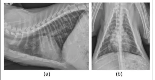
Thoracic radiographs revealed a severe diffuse bron-chointerstitial pulmonary pattern with peribronchial cuffing and ill-defined pulmonary nodules. Soft-tissue opacities dorsal to the carina and the second and third sternebrae were interpreted as lymphadenomegaly (Figure 1). Differentials included infectious etiologies such as toxoplasmosis, feline infectious peritonitis and cryptococcosis. Serology was considered but was thought less likely to be helpful in the diagnosis.1
Parasitic pulmonary disease was considered less likely due to previous treatment with fenbendazole and neoplasia (lymphoma) was also lower on the differen-tial list.
Given the severity of thoracic radiographic findings, the kitten was prepped for anesthesia to undergo a laryngoscopy/bronchoscopy and bronchoalveolar lavage (BAL). Premedication was provided with
butorphanol 0.6 mg/kg (1.03 mg) subcutaneously, flow-by 100% oxygen was provided and induction was performed with alfaxalone 1.0 mg/kg (1.7 mg) and midazolam 0.3 mg/kg (0.5 mg) intravenously. A jet ven-tilator was used to provide oxygenation at 180 pulses per minute, and anaesthesia maintained with an alfaxalone constant rate infusion to effect (4.12 mg/kg infused). A 2.8mm outer diameter video-endoscope (Flex XC; Karl Storz Veterinary Endoscopy) was utilized for bron-choscopy. Moderate laryngeal, tracheal and bronchial hyperemia was noted along with excessive mucus accu-mulation (Figure 2). BAL was performed in the left and right caudal lung lobes and was submitted for cytologic evaluation. Copious amounts of tracheal mucus were expectorated and a swab collected from the oropharynx was also submitted for cytologic evaluation.
BAL fluid was hypercellular (1010 and 1100 cells/µl, right and left, respectively; RI 300–400/μl) with 62% and 50% neutrophils (normal 5–8%) and 22% and 25% eosin-ophils (normal <20%) (Figure 3). Increased numbers of multinucleate giant cells, goblet cells and mast cells were also present, along with epithelial hyperplasia. Swabs of oropharyngeal mucus revealed a dense background of mucus with neutrophilic and eosinophilic inflammation, numerous free mucus granules and evidence of miner-alization. Five large parasitic (helminth) larvae were observed on one slide (Figure 4), including one that appeared to have a dorsal spine on the tail. The oro-pharyngeal sample was submitted to the University of Teramo for molecular identification of larvae. DNA was extracted using a commercially available kit (QIAgen Mini Kit; Qiagen), and nested PCR was performed tar-geting the ITS2 region of Aelurostrongylus abstrusus and Troglostrongylus brevior, as previously described.2,3 PCR
was positive for A abstrusus at the 233 base-pair region
and negative for T brevior at the 356 base-pair region (Figure 5).
The kitten was discharged with application of a sec-ond dose of imidiclopid/moxidectin, and fenbendazole was prescribed at 50 mg/kg PO q24h for 10 days. Three weeks later, the kitten was doing clinically well at home but still coughing intermittently. Radiographs revealed marked resolution in the bronchial pattern with partial resolution of peribronchial cuffing and ill-defined nod-ules (Figure 6). Fenbendazole was prescribed for an additional 5 days at 50 mg/kg/day. All respiratory signs had resolved at the time of ovariohysterectomy 3 weeks later, and radiographs 2 months after the final treatment were normal.
Discussion
Lungworm infection in domestic cats is most commonly caused by either A abstrusus or Capillaria aerophila, although infection of domestic cats with T brevior is increasingly reported in Europe.4,5 T brevior and A
abstrusus have a similar indirect biological life cycle with estimated prepatent period of 4–6 weeks, while C aerophila has a direct life cycle.4,6,7 It is possible that
T brevior, found primarily in the Old World, might also be passed vertically in a transmammary fashion from queen to offspring.8 Adults of A abstrusus reside within
the alveolar ducts and alveoli, while T brevior live within the bronchi and bronchioles; adults of C aerophila live within the submucosa of the trachea and bronchi.4,7
Figure 2 (a) Excessive pharyngeal mucus was observed on bronchoscopy and (b) the carina was diffusely reddened. *Jet ventilator catheter
Figure 3 Bronchoalveolar lavage cytology from a kitten with
Aelurostrongylus abstrusus lungworm infection. There are
increased numbers of neutrophils and eosinophils, along with scattered macrophages and mast cells (objective × 60, Wright-Giemsa stain; bar = 10 μm)
Figure 4 Oropharyngeal swab cytology from a kitten with
Aelurostrongylus abstrusus lungworm infection. A helminth
4 Journal of Feline Medicine and Surgery Open Reports
A abstrusus is considered the most common feline lungworm parasite and is found worldwide; it can infect all cats, regardless of their habitat, lifestyle, breed or sex. Indoor cats are at decreased risk, but possible infection should not be ruled out based upon lifestyle alone. Kittens, owing to their immature immune system, and adult cats that hunt are at an increased risk for lung-worm infection with A abstrusus.4 While lungworm
infection most commonly affects young adult cats, 16% of infected cats were <6 months of age in one study.9
Clinical signs of infection vary depending upon worm burden, age, immune response and overall health status of the feline host, and can range from subclinical to severe disease, occasionally including fatal pneumo-nia.4,10,11 Many cats are subclinical or can have
respira-tory signs of chronic cough with gradually increasing respiratory difficulty and tachypnea, wheezing, sneez-ing and nasal discharge, as well as anorexia and fever.4,10
Kittens appear to develop more severe disease than adult cats, perhaps because of smaller bronchial diame-ter, which would be more susceptible to rapid occlusion
of the airways by developing worms and concurrent inflammation. One case report describes a 2-month-old kitten that died from fatal verminous pneumonia and enteritis secondary to infection with A abstrusus, while another discusses reversible pulmonary hypertension in kittens secondary to infection with A abstrusus.12,13
Kittens and young cats also develop more severe radio-logical abnormalities and higher larval worm burdens than adults.7
T brevior has been reported as an emerging helminth parasite in Europe that can result in clinical signs similar to A abstrusus, although infection with the former lung-worm is associated with more severe and life-threatening infection.4,5,8 Kittens and young cats seem to be
particu-larly susceptible to infection with T brevior, and clinical signs include severe cough, respiratory distress and nasal discharge. Infection can lead to fatal respiratory failure in kittens, despite treatment. A more recent report describes the first known case of irreversible pulmonary hypertension in a kitten infected with T brevior, despite appropriate treatment.14 Infection with C aerophila can be
subclinical or associated with respiratory distress char-acterized by increased bronchovesicular sounds, wheez-ing, sneezing and a dry cough.4
Diagnosis of lungworm infection can be challenging, as clinical signs often imitate other causes of respiratory signs, such as fungal infections, toxoplasmosis and feline asthma. Radiographic abnormalities vary depend-ing upon worm burden and time after onset of infection and range from a mild multifocal bronchointerstitial pulmonary pattern with poorly demarcated nodules (often in the caudal lung lobes) to a diffuse alveolar infiltrate.4,11,13 The gold standard for diagnosis is the
Baermann technique.4 This test takes 24 h to complete,
and three consecutive negative samples are required to rule out parasitic pneumonia.4,7 Lungworm larvae can
be found in tracheal swabs or washes, as well as on BAL cytology, but sensitivity is not as high as with the Baermann technique.7,9
Infection with A abstrusus or T brevior can be docu-mented by fecal Baermann, and specific larval mor-phology can be used to differentiate the two organisms. A nested PCR was recently validated for use in differen-tiation of the two parasites.2 Feces, pharyngeal swabs
and lower airway samples can be used for detection, but the best sample to submit for PCR appears to be a phar-yngeal swab, which is supported by results in this case. While pharyngeal cytology and PCR have not been directly compared, PCR is reported to have a specificity of 100% and sensitivity of 96%.2,7 BAL cytology was
neg-ative for the parasite in this case, indicating that addi-tional (pharyngeal) samples may be needed to obtain a definitive diagnosis.
This case was especially interesting because the kit-ten’s clinical signs initially worsened after anthelmintic
Figure 5 PCR specific for the ribosomal ITS2 of
Aelurostrongylus abstrusus. Lane M: gene ruler 100 base-pair
treatment. Potential explanations include a severe immune response to a dying worm burden, a mixed infection or potentially a novel case of T brevior infec-tion. A recent report described two littermates with mixed A abstrusus and T brevior infections that were treated appropriately with milbemycin oxime (2 mg/kg) only to have one kitten develop fatal exacerbation of clinical signs.3 We consider it likely that worsening
clinical signs noted in the kitten of this report were the result of an immune response to dying worms, which incited coughing, wheezing and tachypnea. Given the marked inflammatory response present in BAL cytol-ogy, treatment with corticosteroids could have been considered.
Conclusions
This case serves as a clinically relevant reminder to include lungworm disease as a differential diagnosis when determining the cause of respiratory signs in cats, especially kittens and free-roaming cats or when the animal’s history and origin are unknown. This case also supports the work currently being performed regarding improvement of diagnostics for feline lungworm, as it can be clinically difficult to diagnose. Diagnosis of lungworm infection in this kitten was obtained via orophargyngeal swab cytology and PCR, with the latter being more sensitive and specific. Diagnosis via cytol-ogy has a more rapid turnaround time, but differentiat-ing T brevior from A abstrusus can be morphologically challenging.4,7 PCR allows both diagnosis and
specia-tion; however, this test is not yet readily available. Continued refinement of diagnostic options for feline lungworm is needed.
Funding The author(s) disclosed receipt of the following financial support for the research, authorship, and/or publica-tion of this article: This study was funded, in part, by the Bailey Wrigley Fund, University of California Davis.
Conlict of interest The authors declared no potential con-flicts of interest with respect to the research, authorship, and/ or publication of this article.
References
1 Sparkes A, Gruffydd-Jones T and Harbour D. Feline infec-tious peritonitis: a review of clinicopathological changes in 65 cases, and a critical assessment of their diagnostic value. Vet Rec 1991; 129: 209–212.
2 Traversa D, Iorio R and Otranto D. Diagnostic and clinical implications of a nested PCR specific for ribosomal DNA of the feline lungwormAelurostrongylus abstrusus. J Clin Microbiol 2008; 46: 1811–1817.
3 Di Cesare A, Frangipane di Regalbono A, Tessarin C, et al. Mixed infection by Aelurostrongylus abstrusus and Troglostrongylus brevior in kittens from the same litter in Italy. Parasitol Res 2014; 113: 613–618.
4 Traversa D and Di Cesare A. Diagnosis and management of lungworm infections in cats. Cornerstones, dilemmas and new adventures. J Feline Med Surg 2016; 18: 7–20. 5 Tamponi C, Varcasia A, Brianti E, et al. New insights on
metastrongyloid lungworms infecting cats of Sardinia, Italy. Vet Parasitol 2014; 203: 222–226.
6 Companion Animal Parasite Council. Parasites of other systems – Lungworms. http://www.capcvet.org/ capc-recommendations/lungworms (2007, accessed 4 May 2016).
7 Pennisi MG, Hartmann K, Addie DD, et al. Lungworm dis-ease in cats: ABCD guidelines on prevention and manage-ment. J Feline Med Surg 2015; 17: 626–636.
6 Journal of Feline Medicine and Surgery Open Reports
8 Brianti E, Gaglio G, Napoli E, et al. Evidence for direct transmission of the cat lungworm Troglostronglyus brevior. Parasitology 2013; 140: 821–824.
9 LaCorcia L, Gasser RB, Anderson GA, et al. Comparison of bronchoalveolar lavage fluid examination and other diagnostic techniques with the Baermann technique for detection of naturally occurring Aelurostrongylus abstrusus infection in cats. J Am Vet Med Assoc 2009; 235: 43–49.
10 Schnyder M, Di Cesare A, Basso W, et al. Clinical, labo-ratory and pathological findings in cats experimentally infected with Aelurostrongylus abstrusus. Parasitol Res 2014: 113: 1423–1433.
11 Traversa D and Guglielmini C. Feline aelurostrongylosis and canine angiostrongylosis: a challenging diagnosis for two emerging verminous pneumonia infections. Vet Parasitol 2008; 157: 163–174.
12 Philbey AW, Krause S and Jeffries R. Verminous pneumo-nia and enteritis due to hyperinfection with Aelurostron-gylus abstrusus in a kitten. J Comp Pathol 2014; 150: 357–360. 13 Dirven M, Szatmári V, Van den Ingh T, et al. Reversible
pulmonary hypertension associated with lungworm infection in a young cat. J Vet Cardiol 2012; 14: 465–474. 14 Crisi PE, Traversa D, Di Cesare A, et al. Irreversible