1. Department of Internal Medicine/Marmara University, School of Medicine
2. Department of Rheumatology/Marmara University, School of Medicine;
3. Department of Neurology/Marmara University, School of Medicine
nation and MRI findings after 4 weeks showed dramatic improvement however patient developed pulmonary tuberculosis. Methylprednisolone dose was decreased (0.5mg/kg/day) and quadruple antituberculosis thera-py was started. Patient was dischar ged with 5/5 mus-cle strength in extremities without any respiratory symptoms 2 months after the first presentation. Prompt introduction of immunosuppressive therapy is crucial in NBD. Although combination of TNF inhibitors and cyclophosphamide is a rare therapeutic approach, it may be life-saving. However a higher awareness is re-quired for opportunistic infections.
Keywords:TNF inhibitors; Neurologic involvement; neuro-Behcet’s disease
INtrODUctION
Behcet’s disease (BD) is a multisystem inflammatory disorder of unknown aetiology characterized by recur-rent oral and/or genital aphthosis, vascular and ocular involvement in combination with variable skin lesions1.
Other manifestations are arthritis, a positive pathergy test, thrombophlebitis, central nervous system disease and gastrointestinal ulcerations. The nervous system involvement of BD is called neuro-Behcet’s disease (NBD) which has two main subtypes: parenchymal, an inflammatory meningo-encephalitic process and non-parenchymal, a condition secondary to vascular in-volvement such as dural sinus thrombosis1. The
clini-cal presentation can be as corticospinal tract and brain-stem involvement, venous sinus thrombosis, increase in intracranial pressure secondary to aseptic meningitis, isolated headache or isolated behavioural changes. Rarely aneurysmal rupture, peripheral neuropathy, opti c neuritis and vestibular involvement can also be obser ved. NBD is defined as a constellation of
neuro-Severe neuro-Behcet’s disease treated
with a combination of immunosuppressives
and a TNF-inhibitor
Korkmaz FN1, Ozen G2, Ünal AU2, Kahraman Koytak P3, Tuncer N3, Direskeneli H2
AbstrAct
Behcet’s disease (BD) is a multisystem inflammatory disorder characterized by recurrent oral and genital ul-cers, skin lesions and uveitis. The nervous system in-volvement of BD, neuro-Behcet’s disease (NBD), is one of the important causes of mortality of the disease. Herein, we present a 29-year-old male with parenchymal NBD who has progressed rapidly and was mana -ged with an uncommon aggressive immunosuppres-sive combination therapy. The patient first presented six years ago with vertigo and difficulty in talking and walking. On examination, he had oral ulcers, acneiform lesions on the torso, geni tal ulcer scar, dysarthria, and ataxia. Along with the magnetic resonance imaging (MRI) findings, the patient was diagnosed as NBD. Af-ter pulse methylpred nisolone (1g/day, 3 days) and 8 courses of 1g/month iv cyclophosphamide therapy, he was put on azathioprine and oral methylprednisolone. On the 4th year of the maintenance therapy, he was ad-mitted with NBD relapse which was treated with 3 days of iv 1g pulse methylprednisolone. One year after the last relapse, the patient voluntarily stopped medica-tions and presented with global aphasia, right hemihy-poesthesia and quadriparesis. MRI findings were sugges tive of NBD relapse. After exclusion of infection, pulse methylprednisolone was started but no im-provement was observed. Considering the seve rity of the NBD, the patient was put on methylpredniso lone (1mg/kg/day), iv cylophosphamide (1g) and adalimumab 40 mg/14 days subcutaneously with appro -priate tuberculosis prophylaxis. Neurological
logic manifestations with characteristic neuropatho-logic findings, usually confirmed by ancillary investi-gations, in patients who meet the diagnos tic criteria for BD1,2. The prevalence of neurolo gical
manifesta-tions in BD is highly variable (5.3-59%) depending on diagnostic criteria and ethnic populations3. In a
two-decade retrospective study of 387 patients with BD in Turkey, the frequency of NBD was 13% in men and 5.6% in wo men4. Parenchymal, brainstem, cerebellar
involvements, abnormalities in cerebrospinal fluid (CSF), and presentation with paresis were associated with worse prognosis5. Although there is no
well-es-tablished treatment protocol for NBD, prompt intro-duction of immunosuppressive treatment is necessary to ameliorate morbidity, disability and consequently mortality. Herein, we present a NBD case with parenchymal involvement, which has progressed rapi -dly and was managed with an aggressive immunosup-pressive combination therapy. The reason we report this case was the severe relapsing nature of the disease, which caused quadriparesis, was unresponsive to very high doses of glucocorticoids and finally necessitated an uncommon aggressive immunosuppressive treatment. So far, concomitant use of a TNF inhibitor (TNFi) and cyclophosphamide (CyP) with high dose glucocorti-coids has never been reported for NBD.
cAsE PrEsENtAtION
A 29-year-old male patient first presented six years ago
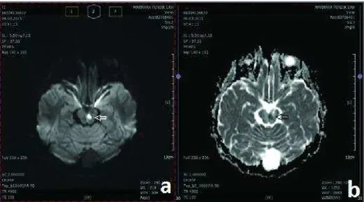
with vertigo and difficulty in talking and wal king. He had recurrent oral (24-30/year) and genital ulcers on his history. On examination, he had active oral ulcers, acneiform lesions on the torso of the body, genital ul-cer scar, dysarthria, left-sided hypoesthesia and ata xia. Magnetic resonance imaging (MRI) of the brain showed contrast-enhanced lesion at the right ponto-cerebellar peduncle and a haemorrhagic lesion in the right basal ganglia (Figure 1). Diffusion-weighted MRI and MR venography excluded acute infarction and du-ral sinus thrombosis. With the exclusion of infectious causes by examination of the CSF, the patient was di-agnosed as parenchymal NBD according to Interna-tional Consensus Recommendation (ICR) criteria for NBD diagnosis. Treatment with pulse methylpred-nisolone (1g/day, iv for 3 days) and CyP were initia ted. With the pulse steroid treatment, neurologic findings dramatically improved. After 8 months of CyP (1g/month iv) treatment, the therapy was continued with azathioprine (AZA) (150mg/day) and oral methylprednisolone (initial dose of 1mg/kg/day with gradual tapering until 4 mg/day). The patient was in remission with the maintenance therapy for 4 years. However, while on maintenance therapy, 2 years ago, he was admitted with nausea and loss of vision in the right eye. He had no signs of uveitis but MRI revealed recurrence of the brain stem lesions. This relapse was treated with 3 days of iv 1g pulse methylprednisolone. The maintenance therapy was set as AZA (150mg/day) and methylprednisolone (tapered to 4mg/day), and colchicine. However, at the end of the first year of this
treatment, due to clinical remission, the patient volun-tarily stopped taking medications. Ten months after cessation of the drugs, he was admitted to another hos-pital with diplopia, absent gag reflex, dysarthria, right he mihypoesthesia and quadriparesis. Diffusion--weighted MRI revealed hyper intense lesions at left mesencephalon and central pons with hypo-isointense signal changes on apparent diffusion coefficient maps. The lesions were considered as acute ischemia due to a vasculitic process, which was consistent with parenchymal NBD. CSF examination (35 white blood cells/µl with normal glucose and protein) was not sugges tive of infection and the lesions were regarded as relapse of NBD. For the acute NBD attack, considering the severity of the neurologic findings, treatment with pulse methylprednisolone (1g/day iv) was started. Se ven days of pulse steroid therapy did not result in signi -ficant neurological improvement and the patient was referred to our institution.
On neurologic examination, the patient was alert, partially cooperative and oriented. He had dysarthria, horizontal gaze palsy (left one-and-a-half syndrome), mild tetraparesis and right hemihypoesthesia. The find-ings were in favour of aggressive NBD. The patient was treated with 2 pulses of high dose iv CyP 500mg/2 weeks, methylprednisolone 60 mg/day iv and adali-mumab 40 mg/14 days subcutaneously. He had no pre-vious tuberculosis (TB) contact, the Quantiferon TB gold test was negative, and chest X-ray was normal. Nevertheless, prophylactic isoniazid (INH) was ad-ministered due to the aggressive immunosuppressive treatment. Neurological examination after 2 weeks of treatment revealed nystagmus, dysarthria, 4/5 motor strength and wide based ataxic gait. Computed to-mography (CT) of thorax was performed to assess pul-monary vascular involvement of Behcet’s disease 4 weeks later. Thorax CT showed 2.5 cm pleural effu-sion and atelectasis on right hemithorax without any vascular changes. Bronchoscopy and pleural sampling were performed to exclude infectious and other caus-es. Pleural effusion was exudative with lactate dehy-drogenase of 715U/L, and total protein of 3.1g/dL. To-tal cell count was 6110/ µl with a predominance of polymorphonuclear cells (78%). Cultures for bacteria and acid-fast stain of the effusion were all negative. Bro-choalveolar lavage fluid polymerase chain reaction (PCR) was positive for Mycobacterium tuberculosis and culture was positive for methicillin resistant Staphylo-coccus auerus(MRSA). As the clinical presentation was not pneumonic, culture positivity for MRSA was
con-sidered as colonization. Since the patient was severely immunosuppressed, vancomycin was initiated. For TB, quadruple antiTB treatment (INH, rifampicin, pyrazi -namide, and ethambutol) was started. Due to the ele-vation of liver enzymes, quadruple anti-TB treatment was switched to the combination of ethambutol and moxifloxacin. Methylprednisolone dose was decreased to 32 mg/day. Therapy for MRSA was continued with trimethoprim-sulfamethoxazole (two months). When levels of liver enzymes returned to normal, INH was added to anti-TB treatment (with a plan of 2 months quadriple anti-TB and 7 months INH and rifampin combination). One month later, the patient was dis-charged with 5/5 muscle strength in 4 extremities, without any respiratory symptoms.
DIscUssION
NBD is a serious complication of BD, which has no proven curative treatment yet. Patients with NBD have a poor long-term outcome. In a study of the 820 BD pa-tients, 275 had neurologic symptoms and 68 (24.7%) were diagnosed as having nonparenchymal CNS in-volvement. Of these with NBD, 25% became depen-dent (were unable to perform activities of daily living) or died during follow-up, and the mortality rate was 10.4%3.
Considering the high disability, morbidity and mor-tality rates of NBD, prompt introduction of immuno-suppressive treatment is crucial. Despite treatment, se-vere relapses can occur during the disease course. In this report, we presented a patient with severe relaps-ing NBD, which caused quadriparesis, was unrespon-sive to high-dose glucocorticoids and finally necessi-tated an uncommon aggressive immunosuppressive treatment. So far, concomitant use of TNFi and CyP with high dose glucocorticoids has never been report-ed for NBD. This combination was only usreport-ed in a phase 2 trial of adalimumab in severe ANCA-associated vas-culitis5. Although that study reported similar rates of
adverse events with adalimumab and CyP combination compared to standard therapy, the follow-up duration, and sample size were small5. Regarding the severe
course of our patient, despite all the risks, this aggres-sive combination therapy was started as other treat-ment options failed.
been reported to be efficacious in controlling NBD. Addi tionally there are positive studies with infliximab and adalimumab which are TNFi mainly used for ocu -lar BD6-8. In a case by Leccese et al ocular BD treated
with infliximab and cyclosporine progressed to NBD and after 2 months of adalimumab therapy, there was an unexpected improvement of the neurological symp-toms with complete regression of the active lesions in MRI8. In a pediatric case, treatment with adalimumab
resulted in resolution of systemic and neurological signs, along with improvement of MRI abnormalities9.
In another NBD case report by Belzunegui et al, after insufficient responses to sequential pulse CyP, gluco-corticoids and infliximab, lesions disappeared with adalimumab10. Case notifications with tocilizumab and
etanercept have also been reported11,12. In our case,
con-sidering the severity of the relapses, inability of previ-ous maintenance AZA therapy in full suppression of relapses and unresponsiveness to high-dose glucocor-ticoids, instead of starting CyP alone, other immuno-suppressives, a relatively rapid-acting agent, adali-mumab, was added to the treatment regimen. Inflam-matory reaction in BD arises from disruption of home-ostasis resulting in altered innate and adaptive immune responses, pathogenic T cell activation in the peri pheral blood, and in inflammatory sites. Researches have shown that both Th1 and Th17 expansions were pre-sent, while regulatory T cell response was suppressed13.
Although the exact immunological and molecular path-ways in NBD have not been fully understood yet, it is suggested that release of interleukin (IL)-1, -6 and -8, and TNF-aand IFN-ginto CSF were increased in NBD
patients14. This reflects a nonspecific inflammatory
pattern compatible with autoinflammatory disease pa thways. The potential beneficial effects observed with TNFi, anti-IL6 and anti-IL1 also support these findings. Recently, encouraging responses with anti-IL1 agents, anakinra, canakinumab and, gevakizumab, especially in difficult and multi-resistant cases of BD have also been reported15, 16. Cantarini et al. showed rapid
reso-lution of disease activity with anakinra in eight of nine drug-resistant BD patients17. Only one of those cases
had neurologic involvement and anakinra improved ocular involvement of that patient17. Although there are
no data about effects of anti-IL1 agents in NBD, in dif-ficult relapsing cases like ours, anti-IL1 agents may also be an alternative after failure of standard therapy or TNFi. However, there are currently no randomized controlled trials (RCT) in BD with these biologic agents, therefore in order to understand benefits and risks or
steroid sparing effects of these drugs, well-designed multi-center RCTs are needed.
Another noteworthy issue related to the treatment applied in this patient is infections. It is well known that biologic agents, especially TNFi, are associated with reactivation of latent TB and serious infections. In this case although the tests for latent TB was negative and prophylactic INH was started, the patient deve -loped pulmonary TB. A recent multicenter Turkish study reported 73 new TB cases among 10.434 TNFi--exposed patients with rheumatic diseases (0.69%). In this research, the frequency of TB was the highest among TNFi-exposed BD patients (5 of 124; 4%). TB was more frequent in patients exposed to adalimumab compared with etanercept, but the difference was sta-tistically insignificant (p = 0.08). The median time for occurrence of TB since the initiation of infliximab, ada -limumab, or etanercept was 13 months (range 1–96), 13 months (range 3–36), and 7 months (range 4–60), respectively18. However, in our case the interval
be-tween the initiation of CyP and adalimumab combina-tion therapy and development of TB was relatively low. In this case, concomitant high-dose, long-term gluco-corticoid therapy could probably have a significant role in the increased risk of infection with the treatment mentioned earlier. Although the risk of infections with TNFi or CyP has not been well examined yet in BD as in other rheumatic diseases, the literature from RA and systemic lupus suggest that concomitant use of gluco-corticoids either with TNFi or CyP significantly in-creases the risk of serious infections, including TB19-20.
Therefore in our case, each of the medications proba-bly contributed to the development of infections.
In conclusion, we describe a patient with serious NBD refractory to conventional immunosuppressive therapy. In this case, due to serious clinical course, CyP and adalimumab were given together. Although this treatment led to a good clinical response, TB developed as a complication of immunosuppressive treatment. In cases of severe clinical course, with close follow-up, adalimumab can be considered for the treatment of NBD. However, a high awareness is required for oppor -tunistic infections.
cOrrEsPONDENcE tO
Korkmanz FN
Department of Internal Medicine, Marmara University School of Medicine Ba ıbüyük Campus
Ba ıbüyük Mah. Maltepe Ba ıbüyük Yolu Sok. No:9/1 Maltepe – Istanbul
rEFErENcEs
1. Akman-Demir G, Saip S, Siva A. Behçet’s disease. Curr Treat Options Neurol 2011; 13:290–310.
2. Kalra S, Silman A, Akman-Demir G. Diagnosis and manage-ment of Neuro-Behçet’s disease: international consensus re-commendations. J Neurol 2014; 261:1662-1676.
3. Noel N, Bernard R, Wechsler B, et al. Long-term outcome of neuro-Behçet’s disease. Arthritis Rheumatol 2014; 66:1306-14. 4. Kural-Seyahi E, Fresko I, Seyahi N, et al. The long-term morta-lity and morbidity of Behcet syndrome: a 2-decade outcome survey of 387 patients followed at a dedicated center. Medici-ne 2003; 82:60–76.
5. Laurino S, Chaudhry A, Booth A, Conte G, Jayne D. Prospecti-ve study of TNFalpha blockade with in ANCA-associated sys-temic vasculitis with renal involvement. Nephrol Dial Trans-plant 2010 25:3307-3314.
6. Al-Araji A, Siva A, Saip S, et al. Treatment of NeuroBehcet’s di-sease with infliximab. An international multi-centre case-series of 18 patients. Clin Exp Rheumatol 2010; 28: S119.
7. Olivieri I, Leccese P, D’Angelo S, et al. Efficacy of adalimumab in patients with Behcet’s disease unsuccessfully treated with in-fliximab. Clin Exp Rheumatol 2011; 29:S54–S57.
8. Leccese P, D’Angelo S, Angela P, Coniglio G, Olivieri I. Swit-ching to adalimumab is effective in a case of neuro-Behcet’s di-sease refractory to infliximab Clin Exp Rheumatol 2010; 28:S102.
9. Robinson AB, Gallentine WB, Rabinovich CE. Pediatric neuro--Behçet’s disease responsive to adalimumab. Pediatr Neurol 2010; 43:291-293.
10. Belzunegui J, López L, Paniagua I, Intxausti JJ, Maíz O. Effica-cy of infliximab and adalimumab in the treatment of a patient with severe neuro-Behçet’s disease. Clin Exp Rheumatol 2008; 26:S133-134.
11. Alty JE, Monaghan TM, Bamford JM. A patient with neuro--Beçhet’s disease is successfully treated with etanercept: further evidence for the value of TNF alpha blockade. Clin Neurol Neu-rosurg 2007; 109:279–281.
12. Shapiro LS, Farrell J, Borhani Haghighi A. Tocilizumab treat-ment for neuro-Behcet’s disease, the first report. Neurosurg 2012; 114:297-298.
13. Houman MH, Bel Feki N. Pathophysiology of Behçet’s disease. Rev Med Interne 2014; 35:90-96.
14. Saruhan-Direskeneli G, Yentür SP, Akman-Demir G, I ik N, Ser-daro lu P. Cytokines and chemokines in neuro-Behçet’s disease compared to multiple sclerosis and other neurological diseases. J Neuroimmunol 2003; 145:127–134.
15. Cantarini L, Lopalco G. Effectiveness and tuberculosis-related safety profile of interleukin-1 blocking agents in the manage-ment of Behçet’s disease. Autoimmun Rev 2015; 14:1-9. 16. Caso F, Costa L, Rigante D. Biological treatments in Behçet’s
di-sease: beyond anti-TNF therapy. Mediators Inflamm 2014; 2014:107421.
17. Cantarini L, Vitale A, Scalini P, et al. Anakinra treatment in drug--resistant Behcet’s disease: a case series. Clin Rheumatol 2015; 34:1293-301.
18. Kisacik B, Pamuk ON, Onat AM, et al. Characteristics Predic-ting Tuberculosis Risk under Tumor Necrosis Factor-a Inhibi-tors: Report from a Large Multicenter Cohort with High Back-ground Prevalence. J Rheumatol 2016;43(3):524-529. 19. Lahiri M, Dixon WG.Risk of infection with biologic
antirheu-matic therapies in patients with rheumatoid arthritis. Best Pract Res Clin Rheumatol 2015;29:290-305.