Harboring a Gammaretroviral Long Terminal Repeat at
the
Nras
/
Csde1
Locus
Borja Balları´n-Gonza´lez., Louise Berkhoudt Lassen., Randi Jessen, Annette Fu¨chtbauer, Ernst-Martin Fu¨chtbauer", Finn Skou Pedersen*"
Department of Molecular Biology and Genetics, Aarhus University, Aarhus, Denmark
Abstract
To investigate mechanisms and phenotypic effects of insertional mutagenesis by gammaretroviruses, we have developed mouse lines containing a single Akv 1-99 long terminal repeat (LTR) and a floxed PGK/Tn5 neomycin cassette at theNras proto-oncogene at positions previously identified as viral integration sites in Akv 1-99 induced tumors. The insert did not compromise the embryonic development, however, the cassette had an effect onNrasexpression in all tissues analyzed. Cre-mediated excision of the PGK/Tn5 neomycin cassette in two of the lines caused upregulation ofNras. Altogether, the knock-in alleles are characterized by modulation of expression of the target gene from more than ten-fold upregulation to three-fold downregulation and exemplify various mechanisms of deregulation by insertional mutagenesis. LTR knock-in mice may serve as a tool to investigate mechanisms of retroviral insertional mutagenesis and as a way of constitutive or induced modulation of expression of a target gene.
Citation:Balları´n-Gonza´lez B, Lassen LB, Jessen R, Fu¨chtbauer A, Fu¨chtbauer E-M, et al. (2013) DeregulatedNrasExpression in Knock-In Animals Harboring a Gammaretroviral Long Terminal Repeat at theNras/Csde1Locus. PLoS ONE 8(2): e56029. doi:10.1371/journal.pone.0056029
Editor:Christine A. Kozak, National Institute of Allergy and Infectious Diseases, United States of America
ReceivedOctober 3, 2012;AcceptedJanuary 4, 2013;PublishedFebruary 13, 2013
Copyright:ß2013 Balları´n-Gonza´lez et al. This is an open-access article distributed under the terms of the Creative Commons Attribution License, which permits unrestricted use, distribution, and reproduction in any medium, provided the original author and source are credited.
Funding:This study was supported in part by grants from the Danish Cancer Society, the Danish Medical Research Council and the Danish Genetically Modified Animal Resource (DAGMAR) funded by the Danish Agency for Science, Technology, and Innovation. No additional external funding received for this study. The funders had no role in study design, data collection and analysis, decision to publish, or preparation of the manuscript.
Competing Interests:The authors have declared that no competing interests exist. * E-mail: fsp@mb.au.dk
.These authors contributed equally to this work.
"These authors also contributed equally to this work.
Introduction
Retroviruses insert a double-stranded DNA copy of their genome non-specifically into the genome of the host and thereby act as insertional mutagens that can disrupt gene regulation or cause the production of an altered gene product [1]. The integrated retrovirus, termed the provirus, contains strong transcription-regulatory signals that can induce or enhance the expression of nearby genes. When such affected genes are involved in cell survival and proliferation, their deregulated expression may contribute to tumorigenesis [2]. In tumors caused by retroviral insertions, the proviruses constitute a tag allowing the identifica-tion of candidate genes with a possible role in tumorigenesis. By this approach several recent studies have contributed to the discovery of new proto-oncogenes and also, when performed in genetically modified mice expanded our knowledge on oncogene cooperativity [3–4]. Moreover, insertional mutagenesis is a concern in gene therapy by retroviral vectors [5]. By another development, evidence is also emerging that endogenous retrovi-ruses of mice and humans may contribute to oncogenesis by the activation of nearby genes without any need for new retroviral insertions in somatic cells [6].
Based on the analysis of somatic integrations selected during malignant transformation various types of virus-induced gene activation have been proposed. By the process known as enhancer
insertion a provirus increases the production of a normal transcript of an adjacent target gene [2]. In these cases, proviruses are often found outside the transcription unit of the target gene and in many cases upstream of the target gene and in the opposite transcrip-tional orientation. Other types of insertranscrip-tional mutagenesis result in the formation of chimeric RNA species containing viral and host sequences. One example of this is promoter insertion in which proviruses are integrated in the same transcriptional orientation as the proto-oncogene, either upstream or within its 59end [2].
The work reported by Martin-Hernandez et al. [7] represents an example of gene over-expression caused by promoter insertion. Three out of 13 murine B-cell lymphomas induced by the leukemogenic Akv1-99 virus had retroviral integrations into the
Nras/Csde1locus [7]. In all three cases viral-Nraschimeric RNAs were detected and the overall level of mRNA with NRAS-encoding potential significantly increased, whereas the retroviral integrations did not influence the expression ofCsde1. Since no activating mutations of Nras were detected, the sole over-expression of the wild type gene seems to constitute an important factor in the development of B-cell lymphomas in this experimen-tal setting.
mouse knock-in models using retroviral sequences. These animals contain a single Akv1-99 LTR integrated at the exact same position as a previously identified retroviral integration in a tumor, placed in either the same or opposite transcriptional orientation relative toNrasand with or without a flanking floxed PGK/Tn5 neo cassette. We here report that the different alleles up or down-regulate Nras expression to various degrees dependent upon the orientation and position of the LTR. Mice of this series have already proven valuable in the analysis of mechanisms of deregulation of host genes by insertional mutagenesis [8] as well as investigation of phenotypic effects ofNrasover-expression [9].
Results
Generation of Alleles with Targeted Knock-in of an LTR and a Floxed PGK/Tn5 Neomycin Cassette
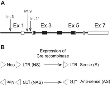
The positions chosen for targeted insertion of an LTR-containing cassette corresponded to the three retroviral integra-tions identified in B-cell lymphomas by Martin-Hernandez et al. [7] within an 800 bp window upstream of the coding region for NRAS (Figure 1A). Integration 3 was located in the 39untranslated region of Csde1 upstream of the Nras promoter, whereas integrations 9 and 11 were both located in intron 1 ofNras. All three integrations had the same transcriptional orientation asNras. To address the role of the orientation of the LTR, targeted insertions were made with the LTR in the same as well as the opposite transcriptional orientation asNras. The knock-in plasmids harbored an Akv1-99 LTR and, to allow selection, a floxed PGK-neomycin-resistance expression cassette placed in the same transcriptional orientation as the LTR. The alleles with the neomycin selection marker (neo) and an LTR in sense orientation relative toNras are termed LTR3NS, LTR9NS, and LTR11NS for the three positions, respectively, whereas the alleles with neo and an LTR in antisense orientation relative toNrasare termed LTR3NAS, LTR9NAS, and LTR11NAS (Figure 1B). As seen in Figure 1B, neo was placed upstream of the LTR relative to the
transcriptional orientation of Nras in all cases. G418 resistant colonies of CJ7 ES cells [10] with the desired inserts were identified by Southern blotting.
The LTR Knock-in Cassette Affects Nras Expression in ES Cells
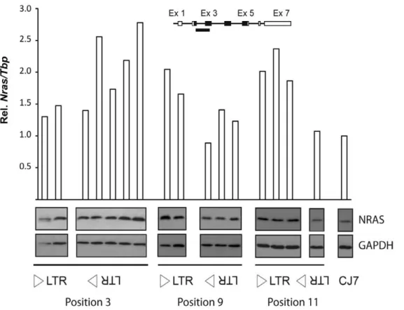
To address the effect of the modified alleles onNrasexpression, quantitative real-time PCR (qPCR) analysis was done using an amplicon spanning the exon 2-exon 3 junction ofNras. Analysis of the CJ7-derived clones (Figure 2) showed that the position 3 knock-in alleles had only a minor effect in sense orientation and a pronounced effect in antisense orientation in four out of five clones analyzed. On the other hand, for positions 9 and 11, the CJ7-derived clones showed a pronounced upregulation of Nras for knock-in alleles in sense orientation and only a minor effect in case of anti-sense orientated alleles. Western blotting analysis using an NRAS-specific antibody confirmed that the knock-in alleles also had an effect on the levels of NRAS (Figure 2).
Nras Transcription is Deregulated in Animals with a Cassette in Intron 1
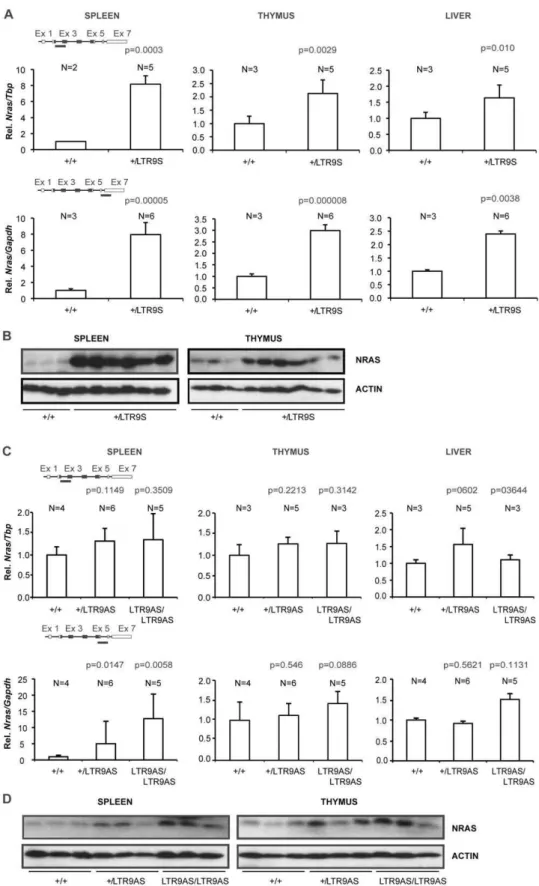
The effect of the knock-in alleles was first analyzed in animals targeted in intron 1 using position 9 as the example. Mice heterozygous or homozygous for the two position 9 alleles, LTR9NS and LTR9NAS, were both born at the expected ratios and phenotypically normal. To assess the influence of the knock-in cassettes on Nras transcription, we employed qPCR using two amplicons covering parts of exon 2 and exon 3 and parts of exon 6 and exon 7, respectively. Introduction of the targeting cassette with the LTR in the same orientation as the Nras gene (the LTR9NS allele) caused a clear increase ofNras mRNA levels in spleen, thymus and liver (Figure 3A). The measured increase in mRNA levels was similar for the two amplicons. In all cases the heterozygous+/LTR9NS animals hadNrasmRNA levels between the wild type (+/+) and homozygous knock-in (LTR9NS/ LTR9NS) animals. The effect onNras mRNA levels was highest in the spleen, where homozygous knock-in animals showed a four-fold increase inNrasmRNA relative to wild type (wt).
Western blotting using an NRAS specific antibody detected higher protein levels in knock-in than in wt animals, again more pronounced in spleen than in thymus (Figure 3B). The liver samples were excluded from the Western analysis due to a low signal to noise ratio.
Expression of the LTR-Nras chimeric transcripts previously identified in the tumor harboring a provirus at position 9 [7] was verified by the RT-PCR using LTR andNrasspecific primers, and it was confirmed that the generation of these transcripts does not abolish transcription from the normalNraspromoter (Figure 3C). Analysis ofNrasmRNA levels in mice harboring the LTR9NAS allele with the LTR placed in the opposite transcriptional orientation of Nras revealed downregulation of Nras mRNA in spleen, thymus, and liver when analyzed with the amplicon spanning exons 2 and 3 (Figure 4a, upper panels). The mRNA levels in heterozygotes were intermediate between those of wt and of LTR9NAS/LTR9NAS animals. The largest effect was an about two-fold reduction observed in spleen tissue. In contrast, analysis using the amplicon spanning exons 6 and 7 detected an upregulation in animals harboring the LTR9NAS allele in spleen, but not in thymus and liver tissues. Western blotting analysis (Figure 4B) showed a decrease in NRAS protein in mice harboring the LTR9NAS allele in spleen and thymus in consistency with the mRNA levels detected with the exon2-exon3 probe.
Figure 1. Overview of knock-in alleles.(A). Schematic represen-tation of Nras. Arrows indicate the identified Akv 1-99 proviral integrations (integration 3, 9 and 11) [7]. Boxes represent exons and the coding region is depicted in black. (B). Representation of the ‘‘targeting cassettes’’ introduced in the sense (S) and antisense (AS) knock-in models. Upon expression of Cre recombinase a LoxP sequence (triangle) and the neomycin selection marker (Neo) can be removed from the construct. LTR = long terminal repeat.
doi:10.1371/journal.pone.0056029.g001
The discrepancy between the RNA levels detected in spleen using the two different qPCR probes indicated that alternative RNA species might be induced in the LTR9NAS allele. One of the possible explanations could be the formation of RNA from transcription initiation sites downstream of exon 3. To investigate this, 59RACE analysis ofNrasRNA was done on samples from+/
+and LTR9NAS/LTR9NAS mice (Figure 4C).
As expected, in wild type spleen, all the detected RNA species (28) clustered around the canonical transcription start site forNras
mRNA. On the other hand, when spleen from knock-in homozygotes animals was analyzed, a unique cluster of 84 initiation sites was identified at the intron 3/exon 4 boundary. Hence, transcriptional initiation around the intron 3/exon 4 boundary may contribute to the discrepancy between the qPCR data from LTR9NAS/LTR9NAS spleens using the two different qPCR amplicons. The failure to detect the canonical transcription start site most probably results from the selection during the process for the identification of short RNA species and the high expression of these alternative transcripts. The over-representation of these alternative transcripts in 5`RACE analysis was confirmed through the investigation of an LTR9NAS/LTR9NAS thymus. In this tissue, where the same tendency in Nras mRNA expression could be observed irrespectively of the utilized qPCR amplicon (Figure 4A), more RNA 59ends were detected at the alternative than at the canonical promoter. We previously reported that the LTR9NAS allele also expressesNrasRNA species initiated at an antisense promoter in the LTR [8] and containing exons 2 and 3 of Nras. These data indicate that in LTR9NAS/LTR9NAS animals, Nras transcription is deregulated, quantitatively with
respect to RNA levels and qualitatively with respect to transcrip-tional initiation sites.
Removal of the PGK/Tn5 Neomycin Cassette Leads to More Pronounced Deregulation of Nras Expression
We next wanted to investigate the effect of removal of the floxed PGK/Tn5 neomycin cassette. Mice harboring the LTR9NS or LTR9NAS alleles were mated with EIIa-Cre transgenic mice and the loss of the floxed cassette verified by PCR. This generated the alleles LTR9S and LTR9AS.Nras mRNA levels were measured using the same qPCR amplicons as used in Figures 3 and 4. In spleen,+/LTR9S heterozygotes showed about eight fold higher levels than+/+animals (Figure 5A). The levels of Nras mRNA in adult LTR9S/LTR9S homozygotes could not be analyzed since these animals had an early lethality phenotype [9]. The results show that LTR9S causes higherNrasmRNA levels than LTR9NS in thymus, liver, and spleen. Altogether, the results demonstrate that removal of the PGK/Tn5 neomycin cassette from the allele with the LTR in sense orientation leads to upregulation ofNras
mRNA, possibly because the LTR and the Nras promoter are brought in closer proximity and/or the loss of an inhibitory effect on transcription caused by the neomycin cassette [11]. The Western blot analyses of NRAS protein levels reveal strong upregulation in heterozygous animals relative to wt in agreement with the mRNA levels (Figure 5B).
Comparing mouse strains with alleles LTR9NAS and LTR9AS revealed that removal of the PGK/Tn5 neomycin cassette caused either an upregulation or had no effect onNras mRNA levels.
Figure 2. Analysis ofNrasexpression in knock-in clones of mouse ES cells.The analysis included two LTR3NS clones, five LTR3NAS clones, two LTR9NS clones, three LTR9NAS clones, three LTR11NS clones, one LTR11NAS clones as well as parental CJ7 cells.NrasmRNA was quantified by qPCR employing an amplicon covering part of exon 2 and exon 3 (insert). Expression was normalized to that ofTbpand represented as relative to the parental CJ7 ES cell line. The panels below the histogram present Western blot analysis of NRAS in protein extracts from the listed ES cell clones. GAPDH was used as reference.
Using the amplicon spanning exons 2 and 3, animals carrying the LTR9AS allele gave higherNrasmRNA values than+/+in spleen and thymus (Figure 5C). The levels detected with the exon 6-exon
7 amplicon were strongly increased in spleen, presumably caused by intragenic transcriptional initiation as observed for the LTR9NAS allele. Western blotting analysis showed that excision
Figure 3. Analysis of knock-in animals harboring the LTR integrated in the sense orientation at position 9.(A).Nrasexpression was quantified by qPCR employing two different methods, SYBR green (amplicon covering part of exon 2 and 3) or a TaqMan hydrolysis probe (amplicon covering part of exon 6 and 7). Expression was normalized to that ofTbporGapdhdepending on the employed strategy (SYBR green or TaqMan probe, respectively) and represented as relative to that of wild type animals. N represents the number of animals in the different groups. Paired Student’s t test was used to determine p-values relative to+/+animals. (B). Western blot analyses of spleen and thymus samples using antibodies against NRAS or GAPDH. C) PCR analysis of mRNA from spleen of homozygous LTR9NS (samples 1 and 2) and wild type animals (samples 3 and 4). Two distinct chimeric mRNAs can be detected by an LTR and anNrasspecific primer in combination (left half of gel). These transcripts depicted at the bottom of the figure contain viral as well as cellular sequences and differ in length due to splicing or not from a cellular splice donor at the firstNras intron. LTR initiated transcription does not seem to suppress the activity of the normalNraspromoter, as the putativeNrastranscript could be detected in both wild type and homozygous LTR9NS animals employing the appropriateNrasspecific primers (right half of gel).
doi:10.1371/journal.pone.0056029.g003
of the PGK/neo cassette also caused upregulation at the protein level (Figure 5D) of NRAS.
Nras Expression is Deregulated in Animals with a Cassette Inserted Upstream of the Promoter
To analyze the effect of insertion of an LTR upstream of the
Nraspromoter, we investigated tissues of adult animals heterozy-gous or homozyheterozy-gous for LTR3NS and LTR3NAS. These animals were phenotypically normal. We used the amplicon spanning exons 2 and 3 previously shown to correlate with protein levels as well as the amplicon spanning exons 6 and 7. The data (Figure 6) show thatNrasexpression is increased regardless of the orientation of the cassette, that heterozygous animals are intermediate between wt and homozygous knock-in animals, and that the LTR3NAS allele gives higherNrasexpression than the LTR3NS allele. The two amplicons gave similar results. Hence, neither the LTR3NAS locus nor the LTR3NS locus cause significant activation of the cryptic promoter at the intron 3-exon 4 boundary as did LTR9NAS and LTR9AS. Since the PGK/Tn5 cassette in these strains is located further upstream from theNras promoter, we did not investigate the effect of Cre-mediated cassette excision uponNrasexpression.
Discussion
To address how retroviral insertional mutagenesis in the germ line or in somatic tissues may deregulate host genes and cause disease we have generated a series of novel mouse strains which harbor an LTR inserted at theNras locus at positions previously identified as targets for retroviral insertions in B-cell lymphomas [7]. None of the knock-in alleles cause embryonic lethality neither as homozygotes or heterozygotes. However, mice homozygous for the allele causing the highest over-expression ofNrasin the spleen, manifest with a phenotype of granulocytosis, T-cell expansion, and decease within three weeks after birth [9].
The knock-in alleles showed deregulation ofNrasranging from more than ten-fold upregulation to a downregulation of three fold. Expression levels in heterozygotes were intermediates between wild type and homozygous knock-in animals. In spleen, the order of expression of mRNA including the coding exons ofNrasamong the different alleles was: LTR9S.LTR3NAS.LTR9NS.LTR3NS.
LTR9AS.wt.LTR9NAS. The values observed in adult tissues roughly corresponded to those of the engineered embryonic stem cells used to generate the mouse lines, when considering that the ES cells are heterozygous for the knock-in allele. In the present study as well as in a recent publication [9], we have used the knock-in alleles for constitutive deregulation only. However, since we observed an increased level of
NrasmRNA in adult tissues following germ-line excision of the PGK/ neo, the alleles can also be used to address questions of the effect of tissue-specific or induced over-expression of wtNras. A number of tools for tissue specific or inducible activation of Cre recombinase can be used for such studies [12–13].
For position 3, upstream of the Nras promoter, both cassette orientations gave rise to an increase inNrasexpression, however, the antisense orientation to a higher level than did the sense
orientation, originally detected in the B-cell lymphoma [7]. The antisense orientation upstream of a promoter is a configuration of insertional mutagenesis most commonly found for lymphoma induction by MLVs. However, in the present case the LTR is located close to the promoter, which might explain the upregula-tion observed by the LTR3NS as well.
For position 9, on the other hand, the cassette in sense orientation (LTR9NS) stimulated expression of Nras mRNA whereas the antisense orientation LTR9NAS reduced it. In case of LTR9NS we detected the normal Nras mRNA in which the LTR was excised as part of intron 1 as well as two types of LTR initiated mRNAs lacking exon 1 ofNras. The two LTR-initiated mRNAs corresponded to those observed in the original tumor 9 harboring a provirus at this position, indicating that the inserted solo-LTR functions similarly to the inserted provirus. LTR9NAS gave rise to RNA species initiated at several sites at the locus, including an antisense promoter in the LTR [8] as well as the normalNraspromoter and a cryptic promoter at the intron 3/exon 4 boundary ofNras. The enhancer of the inserted LTR activated transcription start sites in a window of about 250 bp at the cryptic promoter whereas the transcription start sites at the normal promoter are confined to a much smaller window irrespective of the presence or absence of the LTR cassette. Such enhancer activation of cryptic promoters has previously been reported to use scattered transcription start sites [14].
Endogenous retroviruses are known to be targets for epigenetic silencing of transcription in the early embryo and mouse retroviruses transferred to embryonic stem cells may be subject to such silencing mechanisms [15]. In humans, failure to sustain such silencing in adult tissues has been linked to LTR-driven expression of a neighboring gene as an oncogenic mechanism [6]. A major target for silencing of murine leukemia viruses such as Akv is overlapping with the primer binding site for proline tRNA [16], but there is also evidence of other determinants in the viral genome including the LTR [17]. In the present study the knock-in cassette contains only the LTR, and not the proline primer binding site. Only one of the knock-in alleles, LTR9NAS did result in reducedNrasexpression. However, this reduction was only 2–3 fold and therefore relatively minor compared to the strong repression observed for some endogenous retrovirus. Whether this reduction involves epigenetic mechanisms or altered promoter configurations is not clear. We note, however, that Cre-mediated excision of the PGK/neo cassette, previously found to downreg-ulate gene expression [11] causes upregulation of Nras mRNA relative to wt, suggesting that the LTR does not contribute to the reduction of expression ofNrasmRNA in LTR9NAS. Moreover, a cryptic promoter further downstream in Nras is induced in LTR9NAS as well as in LTR9AS indicating that the LTR does not cause a general reduction in transcriptional activity of the target locus. The results therefore indicate that the downregulation observed in LTR9AS is unrelated to epigenetic repression targeted to the LTR.
In conclusion, we have shown that a gammaretroviral LTR inserted into the mouse germ line is transcriptionally active and mimics a number of features of retroviral insertional mutagenesis
Figure 4. Analysis of knock-in animals harboring the LTR integrated in the antisense orientation at position 9.(A).Nrasexpression was quantified by qPCR employing two different methods, SYBR green (amplicon covering part of exon 2 and 3) or a TaqMan hydrolysis probe (amplicon covering part of exon 6 and 7). Expression was normalized to that ofTbporGapdhdepending on the employed strategy (SYBR green or TaqMan probe, respectively) and represented as relative to that of wild type animals. N represents number of animals in the different groups. Paired Student’s t test was used to determine p-values relative to+/+animals. (B). Western blot analyses of spleen and thymus samples using antibodies against NRAS or GAPDH. (C). Rapid amplification of cDNA ends: Initiation sites of alternative transcripts within theNrasgene or viral LTR were identified by the usage of the GeneRacerTM kit (Invitrogen). Position of the detected transcription start sites are depicted with respect to the first nucleotide of exon 1 or 4. Height of the bars indicates the frequency of the detected transcripts.
doi:10.1371/journal.pone.0056029.g004
Figure 5.Nrasexpression in knock-in animals with and without the neomycin selection marker.Nrasexpression was quantified by qPCR employing two different methods, SYBR green (amplicon covering part of exon 2 and 3) or a TaqMan hydrolysis probe (amplicon covering part of exon 6 and 7). Expression was normalized to that ofTbporGapdhdepending on the employed strategy (SYBR green or TaqMan probe, respectively) and represented as relative to that of wild type animals. Panels A and B: qPCR and Western analysis of the LTR9S allele. Only+/+and+/LTR9S animals are included since LTR9S/LTR9S animal die within three weeks. Panels C and D: qPCR and Western Blot analysis of the LTR9AS allele. Paired Student’s t test was used to determine p-values relative to+/+animals.
in somatic tissues such as promoter insertion, alternative splicing, enhancer insertion, activation of a cryptic promoter [18] [8] [19], and the formation of chimeric RNA initiated at retroviral antisense promoters [8]. This type of knock-in mice provides novel models for the analysis of phenotypic consequences of deregulation of target genes for retroviral insertional mutagenesis [9].
Materials and Methods
Knock-in, ES Cells, Animals
Homology arms for the targeting vectors were retrieved by recombineering in bacteria [20]. Linearized targeting vector DNA was electroporated into CJ7 ES cells [21]. Successful targeting was verified by Southern blot and positive ES cell clones were injected into B6D2F2 blastocysts [22]. Chimeric mice were mated with C57Bl/6J, offspring was genotyped by PCR with primers flanking the individual insertion sites. In ES cells, the PGK-TN5-neo cassette was removed by transient transfection with an expression vector coding for Cre recombinase. In mice, the PGK-TN5-neo cassette was removed by mating knock-in mice with transgenic mice expressing Cre recombinase under the control of the EIIa promoter [23].
RNA Isolation and cDNA Synthesis
RNA was isolated from frozen tissues or cultured cells with the TRIzol Reagent (Invitrogen) using the protocol provided by the manufacturer. Random primers were used to reverse-transcribe 2.5mg RNA of each RNA sample following the recommendations included in the Fermentas cDNA synthesis kit or the M-MLV reverse transcriptase kit (Invitrogen).
Polymerase Chain Reaction
All reagents employed in the PCR reactions were purchased from Invitrogen except primers, which were acquired from DNA Technology. The PCR reaction mix commonly used consists of the following solutions: 5mL 10x buffer; 8mL 1.25 mM dNTP (deoxynucleoside triphosphate mix); 1.5mL 50 mM MgCl2;
1.5mL forward primer (10 pmol/mL); 1.5mL reverse primer (10 pmol/mL); 0,25mL Taq polymerase (5 U/mL); 31.25mL ddH20; 1.5mL template (100–500 ng).
Quantitative-real time-PCR
qPCR analyses were performed in the Stratagene Mx3005pTM Real-time PCR machine. Two standard curves, one for theNras
and the other for the reference gene, composed by serial dilutions of cDNA from ‘‘wild type’’ tissue (spleen, thymus or liver) were included in each run. In order to determine an adequate reference gene, a pilot experiment with a few samples was conducted in which Nras expression was normalized against several house keeping genes.Tbp(TATA-box binding protein),Gapdh, andHprt
all produced equivalent results.
For the N-terminal detection the Nras (Mm00477878_g1) taqman probe was used with the referenceGapdh (4352932E) or
Hprt (Mm00446968_m1) probes used as internal standard. C-terminal detection ofNras was done with Platinum SYBR Green qPCR SuperMix-UDG (Invitrogen) with primers forNras:
[5’ - ACTGGTCTCTCATGGCACTGTACT - 3’];
[5’ - TACAAACTGGTGGTGGTTGGAGCA - 3’] and primers forTbp:
[5’ -AGAGAGCCACGGACAACTG - 3’]; [5’ - ACTCTAGCATATTTTCTTGCTGCT - 3’]
Rapid Amplification of cDNA Ends
Initiation sites of alternative transcripts within theNrasgene or viral LTR were identified by the usage of the GeneRacerTM kit (Invitrogen). The sequential 59 dephosphorylation/decapping steps included in this kit ensure the ligation of a specific adaptor RNA oligonucleotide only to full-length (previously capped) mRNA, validating the identified sequences as putative initiation site and not artifacts originated by RNA truncation. cDNA synthesis was performed following the manufacturer’s recommen-dations from 2mg of RNA and utilizing the random primers provided in the kit. PCR products amplified with a DNA oligonucleotide complementary to the adaptor oligonucleotide and a gene specific primer were subsequently TOPO cloned (TOPOHTA CloningHKit for Sequencing, Invitrogen) in order to detect both common and rare initiation sites.
Protein Extraction and Western Blot Analyses
Proteins were extracted from tissue or cultivated cells cultures by homogenization in RIPA buffer (10 mM Tris-HCl (pH 8.0), 150 mM NaCl, 1% Triton X-100, 0.1% SDS, 0.5% sodium deoxycholate) supplemented with proteinase inhibitors (0.2 mM PMSF, 20mg/mL aprotinin). For detecting NRAS, 15–20mg protein per lane was electrophoresed through 12.5% or 16% polyacrylamide gels and immunodetected with monoclonal anti-NRAS antibody (dilution 1:300, sc-31; Santa Cruz Biotechnology) followed by visualization using the ECL Plus Western Blotting Detection system (GE Healthcare) and medical films (Konica Minolta Medical and Graphic Inc.). To confirm equal loading, membranes were stripped and re-hybridized with either an anti-GAPDH antibody (dilution 1:300, sc-20357) or an anti-Beta-actin antibody (dilution 1:300, sc-1616, Santa Cruz Biotechnology).
Acknowledgments
The authors thank Lisbeth Ahm Hansen for injection of ES cells in blastocysts and Lone Højgaard Nielsen, Zane Binate, and Tine Birch for technical assistance.
Author Contributions
Performed the experiments: BBG LBL. Conceived and designed the experiments: BBG EMF FSP. Analyzed the data: BBG LBL. Wrote the paper: FSP. Contributed reagents/materials/analysis tools: RJ ACF.
Figure 6. Analysis of knock-in animals harboring the LTR inserted at position 3.Nrasexpression was quantified by qPCR employing an amplicon employing two different methods, SYBR green (amplicon covering part of exon 2 and 3) or a TaqMan hydrolysis probe (amplicon covering part of exon 6 and 7). Expression was normalized to that ofTbporHprtdepending on the employed strategy (SYBR green or TaqMan probe, respectively) and represented as relative to that of wild type animals. N represents the number of animals in the different groups. Alleles with the cassette in sense (panel A) or antisense (panel B) orientation were analyzed. Paired Student’s t test was used to determine p-values relative to+/+ animals.
References
1. Mitchell RS, Beitzel BF, Schroder AR, Shinn P, Chen H, et al. (2004) Retroviral DNA integration: ASLV, HIV, and MLV show distinct target site preferences. PLoS Biol 2: E234.
2. Uren AG, Kool J, Berns A, van Lohuizen M (2005) Retroviral insertional mutagenesis: past, present and future. Oncogene 24: 7656–7672.
3. Blyth K, Vaillant F, Hanlon L, Mackay N, Bell M, et al. (2006) Runx2 and MYC collaborate in lymphoma development by suppressing apoptotic and growth arrest pathways in vivo. Cancer Res 66: 2195–2201.
4. Kool J, Uren AG, Martins CP, Sie D, de Ridder J, et al. (2010) Insertional mutagenesis in mice deficient for p15Ink4b, p16Ink4a, p21Cip1, and p27Kip1 reveals cancer gene interactions and correlations with tumor phenotypes. Cancer Res 70: 520–531.
5. Biasco L, Baricordi C, Aiuti A (2012) Retroviral integrations in gene therapy trials. Mol Ther 20: 709–716.
6. Lamprecht B, Walter K, Kreher S, Kumar R, Hummel M, et al. (2010) Derepression of an endogenous long terminal repeat activates the CSF1R proto-oncogene in human lymphoma. Nat Med 16: 571–579, 571p following 579. 7. Martin-Hernandez J, Sorensen AB, Pedersen FS (2001) Murine leukemia virus
proviral insertions between the N-ras and unr genes in B-cell lymphoma DNA affect the expression of N-ras only. J Virol 75: 11907–11912.
8. Rasmussen MH, Ballarin-Gonzalez B, Liu J, Lassen LB, Fuchtbauer A, et al. (2010) Antisense transcription in gammaretroviruses as a mechanism of insertional activation of host genes. J Virol 84: 3780–3788.
9. Lassen LB, Ballarin-Gonzalez B, Schmitz A, Fuchtbauer A, Pedersen FS, et al. (2012) Nras overexpression results in granulocytosis, T-cell expansion and early lethality in mice. PLoS One 7: e42216.
10. Swiatek PJ, Gridley T (1993) Perinatal lethality and defects in hindbrain development in mice homozygous for a targeted mutation of the zinc finger gene Krox20. Genes Dev 7: 2071–2084.
11. Haldar M, Karan G, Tvrdik P, Capecchi MR (2008) Two cell lineages, myf5 and myf5-independent, participate in mouse skeletal myogenesis. Dev Cell 14: 437–445.
12. Orban PC, Chui D, Marth JD (1992) Tissue- and site-specific DNA recombination in transgenic mice. Proc Natl Acad Sci U S A 89: 6861–6865. 13. Kuhn R, Schwenk F, Aguet M, Rajewsky K (1995) Inducible gene targeting in
mice. Science 269: 1427–1429.
14. Rasmussen MH, Wang B, Wabl M, Nielsen AL, Pedersen FS (2009) Activation of alternative Jdp2 promoters and functional protein isoforms in T-cell lymphomas by retroviral insertion mutagenesis. Nucleic Acids Res 37: 4657– 4671.
15. Rowe HM, Trono D (2011) Dynamic control of endogenous retroviruses during development. Virology 411: 273–287.
16. Wolf D, Goff SP (2009) Embryonic stem cells use ZFP809 to silence retroviral DNAs. Nature 458: 1201–1204.
17. Matsui T, Leung D, Miyashita H, Maksakova IA, Miyachi H, et al. (2010) Proviral silencing in embryonic stem cells requires the histone methyltransferase ESET. Nature 464: 927–931.
18. Liu J, Sorensen AB, Wang B, Wabl M, Nielsen AL, et al. (2009) Identification of novel Bach2 transcripts and protein isoforms through tagging analysis of retroviral integrations in B-cell lymphomas. BMC Mol Biol 10: 2.
19. Pyrz M, Wang B, Wabl M, Pedersen FS (2010) A retroviral mutagenesis screen identifies Cd74 as a common insertion site in murine B-lymphomas and reveals the existence of a novel IFNgamma-inducible Cd74 isoform. Mol Cancer 9: 86. 20. Lee EC, Yu D, Martinez de Velasco J, Tessarollo L, Swing DA, et al. (2001) A highly efficient Escherichia coli-based chromosome engineering system adapted for recombinogenic targeting and subcloning of BAC DNA. Genomics 73: 56– 65.
21. Swiatek PJ, Lindsell CE, del Amo FF, Weinmaster G, Gridley T (1994) Notch1 is essential for postimplantation development in mice. Genes Dev 8: 707–719. 22. Wertz K, Fu¨chtbauer EM (1994) B6D2F1 - an improved mouse hybrid strain for
the production of ES cell germ line chimeras. Transgenics 1: 277–280. 23. Lakso M, Pichel JG, Gorman JR, Sauer B, Okamoto Y, et al. (1996) Efficient
in vivo manipulation of mouse genomic sequences at the zygote stage. Proc Natl Acad Sci U S A 93: 5860–5865.