Neural Bases of Unconscious Error Detection
in a Chinese Anagram Solution Task:
Evidence from ERP Study
Hua-zhan Yin1☯*, Dan Li1☯, Junyi- Yang2, Wei Li2, Jiang Qiu2*, Ying-yu Chen1
1Department of Psychology, Chongqing Normal University, Chongqing, 401331, China,2School of Psychology, Southwest University (SWU), Chongqing, 400715, China
☯These authors contributed equally to this work. *[email protected](HZY);[email protected](JQ)
Abstract
In everyday life, error monitoring and processing are important for improving ongoing perfor-mance in response to a changing environment. However, detecting an error is not always a conscious process. The temporal activation patterns of brain areas related to cognitive con-trol in the absence of conscious awareness of an error remain unknown. In the present study, event-related potentials (ERPs) in the brain were used to explore the neural bases of unconscious error detection when subjects solved a Chinese anagram task. Our ERP data showed that the unconscious error detection (UED) response elicited a more negative ERP component (N2) than did no error (NE) and detect error (DE) responses in the 300–400-ms time window, and the DE elicited a greater late positive component (LPC) than did the UED and NE in the 900–1200-ms time window after the onset of the anagram stimuli. Taken together with the results of dipole source analysis, the N2 (anterior cingulate cortex) might reflect unconscious/automatic conflict monitoring, and the LPC (superior/medial frontal gyrus) might reflect conscious error recognition.
Introduction
The brain needs to continuously monitor behavior in real time to accomplish specific tasks. When the results of behavior are not on target, namely as erroneous behavior occurs, the brain detects the error and makes adjustments to subsequent behavior in order to reach the expected goal and thus fit the environment better[1–4]. The error monitoring system helps to correct immediate behavioral faults[5].
Many researchers have been suggesting that error processing might be executed at different levels of consciousness, and the processing of error detection and error awareness occurs at dif-ferent levels in the error monitoring system. Specifically, error detection occurs when an error is detected by the brain but the subject is unaware of the error, whereas error awareness is defined as the ability to explicitly report the error verbally or press a key to indicate the error [4,6–8]. Typically, we make a mistake but are not aware of it, so researchers pay more attention to error detection.
a11111
OPEN ACCESS
Citation:Yin H-z, Li D, Yang J, Li W, Qiu J, Chen Y-y (2016) Neural Bases of Unconscious Error Detection in a Chinese Anagram Solution Task: Evidence from ERP Study. PLoS ONE 11(5): e0154379. doi:10.1371/journal.pone.0154379
Editor:Wenbo Luo, Liaoning Normal University, CHINA
Received:August 28, 2015
Accepted:April 12, 2016
Published:May 5, 2016
Copyright:© 2016 Yin et al. This is an open access article distributed under the terms of theCreative Commons Attribution License, which permits unrestricted use, distribution, and reproduction in any medium, provided the original author and source are credited.
Data Availability Statement:All relevant data are within the paper and its Supporting Information files.
Funding:This research was supported by the Ministry of Education of Humanities and Social Science project of China (12YJC190035), and the China Postdoctoral Science Foundation (2013M530759). The funders had no role in study design, data collection and analysis, decision to publish, or preparation of the manuscript.
Event-related potentials (ERPs) have made it possible to precisely record the spatiotemporal cortical activation patterns associated with error detection. Most commonly, the ERP methods employed for this purpose analyze response-locked and stimulus-locked electrophysiological activity. In response-locked electrophysiological activity, for example, previous ERP studies have indicated that error negativity (Ne/ERN) and error positivity (Pe) might be associated with error monitoring and error detection [4,7,9–12]. Specifically, the ERN is a negative deflec-tion with a fronto-central maximum that peaks 50–100 ms after an erroneous response in choice reaction time tasks [13–16]. The Pe is a positive deflection that has a centro-parietal maximum peaking at 200–400 ms, and frequently follows the ERN after an erroneous response [17,4]. Some studies have also suggested that the ERN is elicited irrespective of whether or not the subject is aware of the error [18–21], whereas the Pe is associated with conscious error rec-ognition [20,22]. In stimulus-locked electrophysiological activity, Britz and Michel investigated the temporal dynamics of the scalp topography of stimulus-locked high-density ERPs elicited by errors and correct trials in the Stroop task, and showed amplitude differences between error and correct trials in two time windows around 200 ms and 400 ms after stimulus onset, but these differences in source strength did not reach statistical significance[23].
Previous ERP studies have mainly focused on response-locked electrophysiological activity in error processing, and have found that ERN and Pe were related to error processing. How-ever, in our study, we have tried to explore stimulus-locked electrophysiological activity instead of response-locked brain activity.
With the development of brain imaging technology, research has increasingly focused on the activation patterns of brain areas associated with error processing. For example, some stud-ies have provided direct evidence that the mPFC/ACC are activated when subjects make an erroneous response without being consciously aware of it [3–4,24]. Others studies have found that the anterior cingulate cortex (ACC) might be involved in the conflict monitoring mecha-nism during information processing [25–28]. A number of studies have found that activation of the medial frontal gyrus is related to conscious cognitive control [29–30].
conditions, and the average LPC amplitude and activation intensity of the superior/medial frontal gyrus in the DE condition are higher than those in the UED and NE conditions [36–
38].
Method
Participants
Twenty undergraduates (11 women, 9 men) aged 19 to 24 (mean age 20.8 years,SD= 1.58) at Southwest University in China participated as paid volunteers. They were right-handed, had normal or corrected-to-normal vision, and no current or past neurological or psychiatric disor-ders. This study was approved by the IRB at Southwest University. Subjects all gave their writ-ten informed consent before the experiment started.
Materials
In a logographic language such as Chinese, characters are composed of radicals, which in turn, are composed of strokes. Chinese characters are normally written using a fixed stroke sequence (e.g., which stroke is drawn first, and which second stroke is prescribed). Radicals usually con-sist of several strokes and are arranged or combined in a prescribed way to form a Chinese character. Generally, Chinese characters can be broken down into components or radicals. For example, the Chinese character“xiang (think)”(想) is made up of three radicals,“mu”(木),
“mu”(目) and“xin”(心). In the formal experiment, Chinese anagrams were presented for sub-jects to solve. The anagram was created from a character by scrambling or rearranging the radi-cals that make up the character. Two types of anagrams were used,lexicalandnonlexical anagrams. The radicals of a lexical anagram were intact radicals from a character that can be used as building blocks to reconstruct the original character. A nonlexical anagram contained one radical that was altered from the radical in the original character, for example, by adding one stroke to, or removing one stroke from the original radical. This makes it impossible to reconstruct the anagram into a lexical character since one“building block”is altered. In our study, there were no significant differences in operating frequency and strokes of the Chinese characters described as nonlexical and lexical. The changes were very minor and not easily noticeable (For more details, seeFig 1). Subjects were presented with either a lexical or a non-lexical anagram and required to determine as fast as they could whether they could reconstruct the anagram into a lexical character. It was the Chinese counterpart of solving, for example, the English anagram of“oxmia”by rearranging the letter into“axiom”[31].
Design and Procedure
were instructed to press the“1”key (“Yes”response) if they thought that the Chinese anagram could be reconstructed into a correct (i.e., lexical) character, or press the“2”key (“No” response) if they thought that the Chinese anagram could not be reconstructed into a correct character, or to make no response if they could not decide whether the Chinese anagram could be reconstructed into a correct character. In half of the trials, the anagram was a nonlexical anagram. In the other half of the trials, the anagram was a lexical anagram. After the response was made, a blank screen appeared for 3000 ms, unless the subject reversed the selection and pressed the response key. After the blank screen, a target probe or a distracter probe (visual angle of 2.86°) would remain until the subject pressed a key within 4000 ms. A target probe was a reconstructed“character”version of the earlier displayed anagram keeping the original radicals in the anagram intact regardless of whether the anagram was lexical or nonlexical (in which one of the radicals was altered). A distractor probe was one in which one radical in the anagram was changed from its original form, either from a correct to an incorrect or from an incorrect to a correct one. If the subjects responded“yes”to the anagram and did not change their response, they needed to respond to the probe to indicate whether their reconstructed character was the same as the probe by pressing“1”for“yes”or“2”for“no”. If they responded “no”or did not respond to the anagram, they needed to indicate whether the probe was a Fig 1. Flow of stimuli in each trial of the ERP experiment.There were two different trial types: lexical Chinese anagrams and nonlexical Chinese anagrams. In the formal experiment, a lexical Chinese anagram or nonlexical Chinese anagram was presented to the subjects; and subjects then decided whether the anagram could be reconstructed into a correct Chinese character. A correct or incorrect Chinese character was subsequently presented as a probe to determine the anagram solution of the subject. Subjects were required to judge whether the character they generated was the same as the probe character. No error (NE) condition means that there was no error in the stimulus, and the subjects responded correctly, so no error was detected. Unconscious error detection (UED) means that subjects were not aware of having generated an incorrect character when a nonlexical Chinese anagram appeared. By contrast, the detect error (DE) condition meant that subjects detected that the nonlexical Chinese anagram could not be reconstructed into a correct Chinese character.
lexical or nonlexical character by pressing“1”for“yes”or“2”key for“no”(for more details,
seeFig 1). In half of the trials, the probes were target probes. In the other half of the trials, the
probes were distractor probes. To illustrate the test procedure, suppose a lexical anagram was displayed and a subject correctly judged that it could be reconstructed into a lexical character and did not correct the response. In that case, when the probe was displayed, the subject should have responded“yes”to a target probe, and“no”to a distractor probe. Now, suppose a nonlexi-cal anagram was displayed and the subject failed to detect the altered radinonlexi-cal in the anagram and did not correct the response. In that case, when the probe was displayed, he or she should respond“yes”to a distractor probe (showing the corrected radical instead of the incorrect radi-cal in the anagram) and“no”to a target probe (showing the original incorrect radical). Half of the subjects were instructed to press“1”for“yes”and press“2”for“no”. The other half of sub-jects were instructed to press“2”for“yes”and press“1”for“no”.
In our study, we were interested in no error (NE), unconscious error detection (UED), and detect error (DE) response types. The UED response was an unconsciousness error response to a nonlexical anagram stimulus, but the DE and NE were correct responses to nonlexical and lexical anagram stimuli. Our aim was to study the neural bases of unconsciousness error detec-tion, so we could distinguish the difference between unconsciousness error responses and cor-rect responses elicited by different stimuli or cognitive processing when we compared UED with DE and NE responses. Specifically, the NE response meant that subjects responded“yes” to a lexical anagram and did not change their response, and subsequently responded“yes”to a target probe (a correct Chinese character) or“no”to a distractor probe (an incorrect character). Thus, in this response type, there was no error in the stimulus, and the subjects’response was consistent with the stimulus. By contrast, the UED response meant that subjects responded “yes”to a nonlexical Chinese anagram, were apparently unaware of the nonlexical feature (the altered radical) in the anagram, and did not change their response. In other words, they believed that they had generated a correct character from the radicals of the nonlexical Chinese anagram and did not correct their response. Thus, this response would be classified as a UED if subjects responded“yes”to the nonlexical anagram and did not correct the response, and sub-sequently responded“yes”to the distractor probe (a correct character), or responded“no”to the target probe (an incorrect Chinese character). The DE response type meant that subjects responded“no”to a nonlexical anagram and did not change their responses, and subsequently responded“no”to a target probe (an incorrect Chinese character) or“yes”to a distractor probe (a correct character).
Electrophysiological recording and analysis
Brain electrical activity was recorded from 64 scalp sites using tin electrodes mounted in an elastic cap (Brain Products, Gilching, Germany), with the average reference electrode on the left and right mastoids and a ground electrode on the medial frontal aspect. The vertical elec-trooculograms (EOGs) were recorded supra- and infra-orbitally at the left eye. The horizontal EOG was recorded from the left versus right orbital rim. All interelectrode impedance was maintained below 5 kO. The EEGs and EOGs were amplified using a 0.05–80 Hz bandpass and continuously sampled at 500 Hz/channel for offline analysis. Artifacts from eye-blink move-ments were rejected offline. Trials with EOG artifacts (where the mean EOG voltage
exceeded ± 100μV) and those contaminated with artifacts due to amplifier clipping, bursts of electromyographic activity, or peak-to-peak deflections exceeding ± 100μV were excluded from averaging.
including 1400 ms post-stimulus and 200 ms pre-stimulus. As observed in the grand averaged waveforms, the ERPs elicited by the UED, DE, and NE responses were clearly distinct from each other. All these differences were prominent over the front-central and central-parietal regions of the scalp. Thus, the following six electrode points were chosen for statistical analysis within the 300–400-ms time window: F1, Fz, F2, FC1, FCz, and FC2. The following four elec-trode points were chosen for statistical analysis within the 900–1200-ms time window: P1, P2, CP1, and CP2. Mean amplitudes were analyzed using two-way repeated-measures Analysis of variance (ANOVA). The factors were response type (UED, DE, and NE) and electrode site. For all analyses, thepvalue was corrected for deviations based on the Greenhouse–Geisser
procedure.
Dipole source analysis
The Brain Electrical Source Analysis program (BESA Version 5.0 Software; BESA, Gräfelfing, Germany) was used to perform dipole source analysis, using the four-shell ellipsoidal head model. In order to focus on scalp electrical activity related to the processing of unconscious error detection, the averaged ERPs evoked by the DE were subtracted from the ERPs evoked by the UED in the 300–400-ms and 900–1200-ms time ranges. When the dipole points were determined, the software automatically determined the location of the dipoles. The relevant residual variance criterion was used.
Results
Behavioral data
The mean number of trials for NE, UED, and DE responses were 81 ± 5, 38 ± 6, and 40 ± 6, respectively. We recorded the reaction time (RT) from when the anagram appeared until a response was made. Mean RTs for the NE, UED, and DE were 1410 ± 66 ms, 1433 ± 75 ms, and 1498 ± 67 ms, respectively. A repeated-measures ANOVA of mean RTs showed that the main effect of response type was significant,F(2, 38) = 13.764,p<0.001. Post-hoc testing showed that the mean RT of the UED response was significantly shorter than that of the DE response (p<0.01) and the mean RT of the NE response was also significantly shorter than that of the DE response (p<0.001). The nonlexical anagram that subjects thought could not be reconstructed into a correct character imposed an increasing load on the working memory in the DE response, so the RTs of the DE response were significantly longer than those of the UED and NE. However, there was no significant difference between the mean RTs of UED and NE responses (p= 0.092).
Electrophysiological scalp data
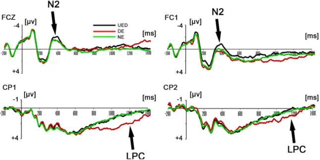
Results for two-way repeated-measures ANOVAs revealed that there was a significant main effect of response type for the main amplitude in the 300–400-ms time window,F(2, 38) = 4.121,p<0.05. Post-hoc tests showed that the UED elicited a more negative ERP component than did the DE (p<0.05) and the NE (p<0.05) (seeFig 2). In addition, the results of the ANOVAs showed that the main effect of response type for the main amplitude in the 900– 1200-ms time window was also significant,F(2, 38) = 9.22,p<0.01. Post-hoc tests showed the DE elicited a significantly greater late positive component (LPC) than did the UED (p<0.05) and the NE (p<0.001; seeFig 2andFig 3).
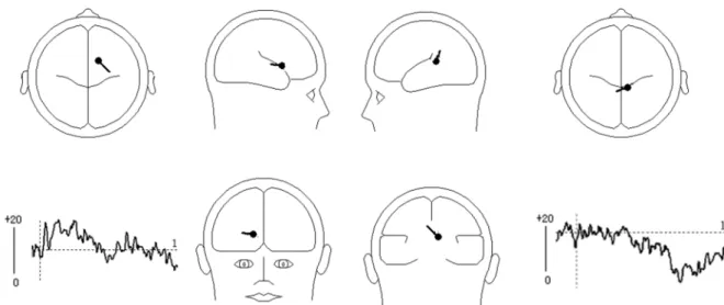
component was needed to explain 100% of the variance in the data between 300–400 ms. Therefore, a dipole was fitted with no restriction on the direction and location. The result indi-cated that the dipole was loindi-cated in the ACC (BA32: x = 14.4, y = 23.5, z = 42.8). This model best explained the data and accounted for most of the variance, with a residual variance (RV) of 18.7% (seeFig 4). Secondly, PCA indicated that one component was needed to explain 100% of the variance in the data between 900–1200 ms. Therefore, the dipole was fitted with no restriction on the direction and location. The result indicated that the first dipole was located Fig 2. Grand-mean ERP waveforms of the N2 and LPC components. Top:Grand average ERPs FCZ, FC1 for the UED, DE and NE response types in the time window of 300-400ms.Bottom:Grand average ERPs CP1, CP2 for the UED, DE and NE response types in the time window of 900-1200ms.
doi:10.1371/journal.pone.0154379.g002
Fig 3. Grand-mean ERP difference waveforms and topographies of the N2 and LPC components. Top:Grand average ERPs FCz for the UED and DE conditions and difference waves (UED minus DE) in the time window of 300-400ms and CP1 for the UED and DE conditions and difference waves (DE minus UED) in the time window of 900-1200ms.Bottom: Topographical maps of the voltage amplitudes for the difference waves (UED minus DE) from 300 ms to 400ms and the voltage amplitudes for the difference waves (DE minus UED) from 900 ms to 1200 ms.
near the middle frontal gyrus (GFm)/gyrus frontalis medialis (GFd) (BA6: x = 9.2, y = -15.1, z = 51.3). This model explained the data best and accounted for most of the variance, with a RV of 19.4% (seeFig 4).
Discussion
In the present study, we were interested in brain electrical activity related to error detection that occurred during the solution of an anagram before erroneous responses were executed. That is to say, our ERP results focused on the stimulus-locked electrical activity. We showed that there were two ERP components (N300-400 and LPC) that were different in UED, DE, and NE responses.
At present, there are two possible interpretations of N300-400: one is semantic integration related to the N400, the other is the negative ERP component N2. First, the UED elicited a greater negative component (N2) in the 300–400-ms time window than did the NE and DE after the onset of the anagram. The N400 is a negative ERP component that peaks around 400 ms after word onset [32,39], so we could define the negative component in the 300–400-ms time window in our study as the N400. The N400 appears to be an index of semantic integra-tion processes (Sitnikova et al., 2003)[40]. For example, the N400 is more negative in response to semantically incongruous words than congruous ones [39,41]. The N400 also has been described as reflecting how easily the word can be integrated into the current context) [39,42–
43]. Perhaps the most widely accepted account of the N400 is that its amplitude is associated with the difficulty or mental effort required to integrate an item into the semantic context [40].
The N300–400 was actually the second negative component in the waveforms. Therefore, it might be an N2 component. The N2 is a negative ERP component peaking between 200 and 400 ms after stimulus onset and maximal at the fronto-central scalp location [32,44]. The N2 reflects the activity of the conflict-monitoring mechanism [44–46]. The N2 is typically larger after incongruent compared with congruent trials [47]and is larger in trials that include conflict between competing response representations, such as incongruent trials in the Stroop task [44,48]. However, some research has indicated that the N2 is increased in trials involving com-peting response activations but not in trials involving conflict in stimulus categorization Fig 4. Results of dipole source analysis of the difference waves (UED minus DE) in the N2 and LPC components.The left-bottom and right-bottom images show the source activity waveforms; the figures display the mean locations of the dipoles. Left:In the time range of 300–400 ms, the dipole was located near the cingulate gyrus (BA32: x = 14.4, y = 23.5, z = 42.8); Right: In the time range of 900–1200 ms, the dipole was located near the middle frontal gyrus (BA6: x = 9.2, y = -15.1, z = 51.3)
[46,49], The N2 has also been proven to reflect conflict monitoring for correct responses[50]. Moreover, there is evidence that the N2 is also sensitive to the frequency of particular trial types [44], with the N2 amplitude being greater for low frequency trials [44,51–52]. The con-flict monitoring function is essential for regulating many forms of everyday behavior [44] and the ACC is the crucial brain area that responds to conflict in information processing [53–54]. The ACC is thought to be associated with the selection and coupling of perceptual information to motor actions [34]. The amplitude of the N2 component related to response conflict is thought to be detectable by the ACC (Bartholow et al., 2009; Liotti et al., 2000; Van Veen & Carter, 2002)[55–56,48]. Based on these findings, we can infer from our study that the
enhanced fronto-central N2 (conflict monitoring) component in the 300–400-ms time window located near the ACC indicated that conflict occurred in the UED response. Moreover, the UED response was an unconsciously incorrect response that elicited a more negative ERP com-ponent (N2) than did the correct NE and DE responses, so we can infer that the enhanced N2 in our study is also a signal of unconscious error detection.
Second, we also found that the DE elicited a greater LPC magnitude than did the NE and UED between 900–1200 ms. Previous studies have indicated that slow waves in the ERP are correlated with rehearsal/retention operations in the working memory [37–38]. Moreover, Berti et al. found that the larger the processing demands needed to keep object information in the working memory [57], the higher the slow wave activity (King & Kutas, 1995; Mecklin-ger & Pfeifer, 1996; Ruchkin et al., 1992)[37–38,58]. Similarly, Helenius et al. (2010) recorded stimulus-locked electrophysiological activity in a Go/NoGo task, and found that the NoGo correct trials elicited a prominent LPC after stimulus onset, as well as a positive deflection after the NoGo error trial peak, which was approximately 50 ms later than the LPC for the NoGo correct trials [59]. Increased perceptual effort was required in parietal association areas to encode the word characteristics of degraded words in nonverbal processes[36]. In our study, dipole analysis localized the generator of the LPC to the medial/superior frontal gyrus. A medial superior frontal gyrus (mSFG) region centered on the pre-supplementary motor area (pre-SMA) is thought to contribute to the higher cognitive function involved in task control, selection of actions [60], attention deployment[61], and recognition memory [62].
However, because the task for subjects was to judge whether a Chinese anagram could form a correct character, they tended to expect that the Chinese anagram would form a correct char-acter. Subjects were required to analyze lexical Chinese anagrams and nonlexical Chinese ana-grams in the NE and UED conditions, respectively, and then judge whether the characters were correct. In the DE condition, subjects were required to analyze a nonlexical Chinese anagram, and then judge whether or not it was a correct character, so they would once again evaluate and confirm their choice based on this tendency. Therefore, the increased LPC in the DE response type in our study might be related to re-evaluation and confirmation processes in the working memory when a stimulus did not form a correct character.
Conclusions
Supporting Information
S1 File. The Average mapping of ERP for each subject in this study. (RAR)
S2 File. The Grand Average mapping of ERPs for all subjects in this study. (RAR)
Author Contributions
Conceived and designed the experiments: HZY JQ. Performed the experiments: JYY WL DL. Analyzed the data: JYY WL DL YYC. Contributed reagents/materials/analysis tools: JYY DL YYC. Wrote the paper: DL JYY HZY JQ.
References
1. Dockree PM, Tarleton YM, Carton S, Fitzgerald MC. Connecting Self-Awareness and Error-Awareness in Patients with Traumatic Brain Injury.[J]. Journal of the International Neuropsychological Society, 2015, 21(7):1–10.
2. Harty S, Robertson IH, Miniussi C, Sheehy OC, Devine CA, McCreery S, et al. Transcranial direct cur-rent stimulation over right dorsolateral prefrontal cortex enhances error awareness in older age.[J]. Journal of Neuroscience the Official Journal of the Society for Neuroscience, 2014, 34(10):3646–3652. 3. Hester R, Foxe JJ, Molholm S, Shpaner M, Garavan H. Neural mechanisms involved in error
process-ing: a comparison of errors made with and without awareness.[J]. Neuroimage, 2005, 27(3):602–608. PMID:16024258
4. Klein TA, Endrass T, Kathmann N, Neumann J, von Cramon DY, Ullsperger M. Neural correlates of error awareness.[J]. Neuroimage, 2007, 34(4):1774–1781.
5. Rabbitt PM. Errors and error correction in choice-response tasks.[J]. Journal of Experimental Psychol-ogy, 1966, 71(2):264–272. PMID:5948188
6. Navarro-Cebrian A, Knight RT, Kayser AS. Error-monitoring and post-error compensations: dissocia-tion between perceptual failures and motor errors with and without awareness.[J]. Journal of Neurosci-ence the Official Journal of the Society for NeurosciNeurosci-ence, 2013, 33(30):12375–12383.
7. Ralph JW, Goodin DS, Long JM, Aminoff MJ. Electrical brain events associated with awareness of errors[J]. Brain Research, 2009, 1251:213–222. doi:10.1016/j.brainres.2008.11.024PMID:19046953 8. van Gaal S, Lamme VA. Unconscious high-level information processing: implication for neurobiological
theories of consciousness.[J]. Neuroscientist A Review Journal Bringing Neurobiology Neurology & Psychiatry, 2012, 18(3):287–301.
9. Falkenstein M, Hohnsbein J, Hoormann J, Blanke L. Effects of crossmodal divided attention on late ERP components. II. Error processing in choice reaction tasks.[J]. Electroencephalography & Clinical Neurophysiology, 1991, 78(6):447–455.
10. Gehring WJ. A Neural System for Error Detection and Compensation[J]. Psychological Science, 1993, 4(6):385–390.
11. Vidal F, Hasbroucq T, Grapperon J, Bonnet M. Is the‘error negativity’specific to errors?[J]. Biological Psychology, 2000, 51(2–3):109–128. PMID:10686362
12. Wiersema JR, Meere JJVD, Roeyers H. Developmental changes in error monitoring: An event-related potential study[J]. Neuropsychologia, 2007, 45(8):1649–1657. PMID:17303199
13. Carter CS, Braver T, Barch DM, Botvinick M, Noll D, Cohen J D, et al. Anterior cingulate cortex, error detection, and the online monitoring of performance.[J]. Science, 1998, 280(5364):747–749. PMID: 9563953
14. Dehaene S, Posner MI, Tucker DM. Localization of a Neural System for Error Detection and Compen-sation[J]. Psychological Science, 1994, 5(5):303–305.
15. Kiehl KA, Liddle PF, Hopfinger JB. Error processing and the rostral anterior cingulate: An event-related fMRI study[J]. Psychophysiology, 2000, 37(2):216–223. PMID:10731771
16. Nieuwenhuis S, Yeung N, Cohen JD. Stimulus modality, perceptual overlap, and the go/no‐go N2[J]. Psychophysiology, 2004, 41:157–160. PMID:14693011
18. Endrass T, Franke C, Kathmann N. Error Awareness in a Saccade Countermanding Task[J]. Journal of Psychophysiology, 2005, 19(4):275–280.
19. Tanja E, Benedikt R, Norbert K. ERP correlates of conscious error recognition: aware and unaware errors in an antisaccade task.[J]. European Journal of Neuroscience, 2007, 26(6):1714–1720. PMID: 17880402
20. Nieuwenhuis S, Ridderinkhof KR, Blom J, Band GPH, Kok A. Error-related brain potentials are differen-tially related to awareness of response errors: Evidence from an antisaccade task[J]. Psychophysiol-ogy, 2001, 38(5):752–760. PMID:11577898
21. O'Connell RG, Dockree PM, Bellgrove MA, Kelly SP, Hester R, Garavan H, et al. The role of cingulate cortex in the detection of errors with and without awareness: a high-density electrical mapping study. [J]. European Journal of Neuroscience, 2007, 25(8):2571–2579. PMID:17445253
22. Belopolsky AV, Kramer AF, Jan T. The role of awareness in processing of oculomotor capture: evi-dence from event-related potentials.[J]. Journal of Cognitive Neuroscience, 2008, 20(12):2285–2297. doi:10.1162/jocn.2008.20161PMID:18457508
23. Britz J, Michel CM. Errors can be related to pre-stimulus differences in ERP topography and their con-comitant sources. Neuroimage,2010, 49(3):2774–2782. doi:10.1016/j.neuroimage.2009.10.033PMID: 19850140
24. Ursu S, Clark KH, Stenger V, Carter C. Conflict-related activity in the caudal anterior cingulate cortex in the absence of awareness.[J]. Biological Psychology, 2009, 80(3):279–286. doi:10.1016/j.biopsycho. 2008.10.008PMID:19026710
25. Abutalebi J, Della Rosa PA, Green DW, Hernandez M, Scifo P, Keim R, et al. Bilingualism tunes the anterior cingulate cortex for conflict monitoring.[J]. Cerebral Cortex, 2012, 22(9):2076–86. doi:10. 1093/cercor/bhr287PMID:22038906
26. Barch D, Edu RW, Braver T, Akbudak E, Conturo T, Ollinger J, et al. Anterior cingulate cortex and response conflict: effects of response modality and processing domain.[J]. Cerebral Cortex, 2001, 11 (9):837–48. PMID:11532889
27. Botvinick M, Nystrom LE, Fissell K, Carter CS, Cohen JD. Conflict monitoring versus selection-for-action in anterior cingulate cortex.[J]. Nature International Weekly Journal of Science, 1999, 402 (6758): 179–181.
28. Forster SE, Carter CS, Cohen JD, Cho RY. Parametric manipulation of the conflict signal and control-state adaptation.[J]. Journal of Cognitive Neuroscience, 2011, 23(4):923–935. doi:10.1162/jocn.2010. 21458PMID:20146615
29. Jiang J, Zhang Q, Gaal SV. Conflict awareness dissociates theta-band neural dynamics of the medial frontal and lateral frontal cortex during trial-by-trial cognitive control[J]. Neuroimage, 2015, 116:102–
111. doi:10.1016/j.neuroimage.2015.04.062PMID:25957992
30. Welander-Vatn A, Jensen J, Otnaess MK, Agartz I, Server A, Melle I, et al. The neural correlates of cog-nitive control in bipolar I disorder: An fMRI study of medial frontal cortex activation during a Go/No-go task[J]. Neuroscience Letters, 2013, 549(33):51–56.
31. Novick LR, Sherman SJ. On the nature of insight solutions: evidence from skill differences in anagram solution.[J]. Quarterly Journal of Experimental Psychology A, 2003, 56(56):351–382.
32. Folstein JR, Petten CV. Influence of cognitive control and mismatch on the N2 component of the ERP: A review[J]. Psychophysiology, 2008, 45(1):152–170. PMID:17850238
33. Folstein JR, Petten CV, Rose SA. Novelty and conflict in the categorization of complex stimuli[J]. Psychophysiology, 2008, 45(3):467–479. PMID:18047482
34. Gajewski P, Stoerig PM. ERP-correlates of response selection in a response conflict paradigm.[J]. Brain Research, 2008, 1189(2):127–134.
35. Holmes AJ, Pizzagalli DA. Response conflict and frontocingulate dysfunction in unmedicated partici-pants with major depression.[J]. Neuropsychologia, 2008, 46(12):2904–2913. doi:10.1016/j. neuropsychologia.2008.05.028PMID:18577391
36. Juottonen K, Revonsuo A, Lang H. Dissimilar age influences on two ERP waveforms (LPC and N400) reflecting semantic context effect[J]. Brain Research Cognitive Brain Research, 1996, 4(2):99–107. PMID:8883923
37. Mecklinger A, Pfeifer E. Event-related potentials reveal topographical and temporal distinct neuronal activation patterns for spatial and object working memory[J]. Cognitive Brain Research, 1996, 4 (3):211–224. PMID:8924049
39. Nan VDM, Kolk HHJ, Chwilla DJ, Vissers CTWM. Monitoring in Language Perception.[J]. Language & Linguistics Compass, 2009, 3(5):1211–1224.
40. Tatiana S, Gina K, Holcomb PJ. Semantic integration in videos of real-world events: an electrophysio-logical investigation[J]. Psychophysiology, 2003, 40(1):160–164. PMID:12751813
41. Kutas M, Hillyard SA. Event-related brain potentials to semantically inappropriate and surprisingly large words.[J]. Biological Psychology, 1980, 11(2):99–116. PMID:7272388
42. van Berkum JJ, Hagoort P, Brown CM. Semantic integration in sentences and discourse: evidence from the N400.[J]. Journal of Cognitive Neuroscience, 1999, 11(6):657–671. PMID:10601747 43. Chwilla DJ, Hagoort P, Brown CM. The Mechanism Underlying Backward Priming in a Lexical Decision
Task: Spreading Activation versus Semantic Matching[J]. Quarterly Journal of Experimental Psychol-ogy A, 1998, august 1(3):531–560.
44. Dickter CL, Bartholow BD. Ingroup categorization and response conflict: Interactive effects of target race, flanker compatibility, and infrequency on N2 amplitude. Psychophysiology, 2010, 47(3):596–601. doi:10.1111/j.1469-8986.2010.00963.xPMID:20136734
45. Botvinick MM, Braver TS, Barch DM, Carter CS, Cohen JD. Conflict monitoring and cognitive control. Psychological review, 2001, 108(3):624–652. PMID:11488380
46. Vincent VV, Carter CS. The timing of action-monitoring processes in the anterior cingulate cortex.[J]. Journal of Cognitive Neuroscience, 2002, 14(4):593–602. PMID:12126500
47. Ridderinkhof KR, de Vlugt Y, Bramlage A, Spaan M, Elton M, Snel J, et al. Alcohol consumption impairs detection of performance errors in mediofrontal cortex.[J]. Science, 2003, 298(5601):2209–2211. 48. Liotti M, Woldorff MG, Iii RP, Mayberg HS. An ERP study of the temporal course of the Stroop
color-word interference effect [J]. Neuropsychologia, 2000, 38(5):701–711. PMID:10689046
49. Veen VV, Cohen JD, Botvinick MM, Stenger VA, Carter CS. Anterior Cingulate Cortex, Conflict Monitor-ing, and Levels of Processing[J]. Neuroimage, 2002, 14(6):1302–1308.
50. Yeung N, Botvinick MM, Cohen JD. The neural basis of error detection: conflict monitoring and the error-related negativity[J]. Psychological review,2004, 111(4):931–959. PMID:15482068
51. Bartholow B, Pearson MC, Sher K, Fabiani M, Gratton G. Strategic control and medial frontal negativity: Beyond errors and response conflict[J]. Psychophysiology, 2005, 42(1):33–42. PMID:15720579 52. Nieuwenhuis S, Yeung N, Wildenberg WVD, Ridderinkhof KR. Electrophysiological correlates of
ante-rior cingulate function in a go/no-go task: Effects of response conflict and trial type frequency[J]. Cogni-tive AffecCogni-tive & Behavioral Neuroscience, 2003, 3(1):17–26.
53. Chris B, Serje R, Risko EF, Derek B. Item-specific adaptation and the conflict-monitoring hypothesis: a computational model.[J]. Psychological Review, 2007, 114(4):1076–1086. PMID:17907873 54. Mennes M, Wouters H, Van DBB, Lagae L, Stiers P. ERP correlates of complex human decision
mak-ing in a gamblmak-ing paradigm: detection and resolution of conflict.[J]. Psychophysiology, 2008, 45 (5):714–720. doi:10.1111/j.1469-8986.2008.00678.xPMID:18665870
55. Bartholow BD, Riordan MA, Saults JS, Lust SA. Psychophysiological evidence of response conflict and strategic control of responses in affective priming[J]. Journal of Experimental Social Psychology, 2009, 45(4):655–666.
56. Van VV, Carter CS. The anterior cingulate as a conflict monitor: fMRI and ERP studies.[J]. Physiology & Behavior, 2003, 77(4–5):477–482.
57. Berti S, Geissler HG, Lachmann T, Mecklinger A. Event-related brain potentials dissociate visual work-ing memory processes under categorial and identical comparison conditions[J]. Brain Research Cogni-tive Brain Research, 2000, 9(2):147–155. PMID:10729698
58. King JW, Kutas M. Who Did What and When? Using Word- and Clause-Level ERPs to Monitor Working Memory Usage in Reading.[J]. Journal of Cognitive Neuroscience, 1995, 7(3):376–95. doi:10.1162/ jocn.1995.7.3.376PMID:23961867
59. Helenius P, Marja L, Laura H, Ritva P, Markku N. Neural correlates of late positivities associated with infrequent visual events and response errors.[J]. Neuroimage, 2010, 53(53):619–28.
60. Rushworth MFS, Walton ME, Kennerley SW, Bannerman DM. Action sets and decisions in the medial frontal cortex.[J]. Trends in Cognitive Sciences, 2004, 8(9):410–417. PMID:15350242
61. Foucaud DB, Richard L, Emmanuelle V, Magali S, Hughes D, Serge K, et al. Functions of the left supe-rior frontal gyrus in humans: a lesion study.[J]. Brain A Journal of Neurology, 2007, 129(12):3315–
3128.